. 2022 Feb 12;43(7):683-697.
doi: 10.1093/eurheartj/ehab757.

Calcific aortic valve disease: from molecular and cellular mechanisms to medical therapy

Affiliations

Calcific aortic valve disease: from molecular and cellular mechanisms to medical therapy

Simon Kraler et al. Eur Heart J. .

Abstract

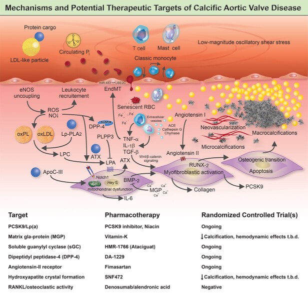
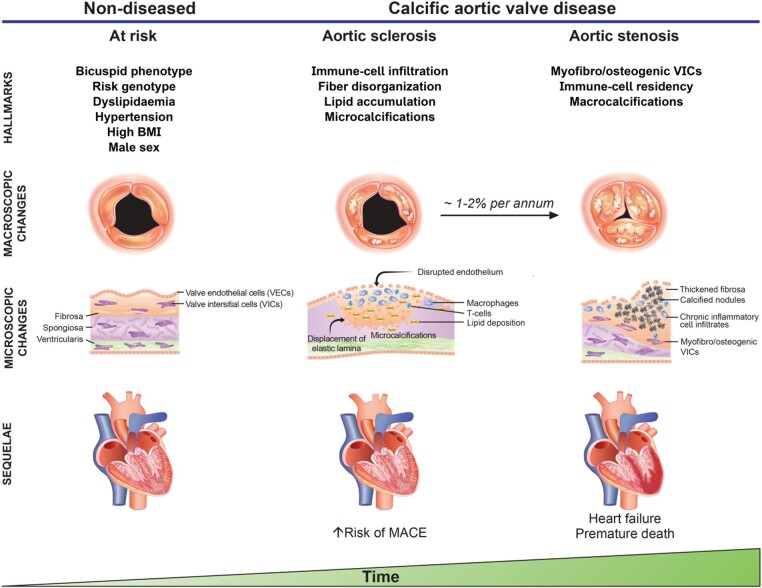
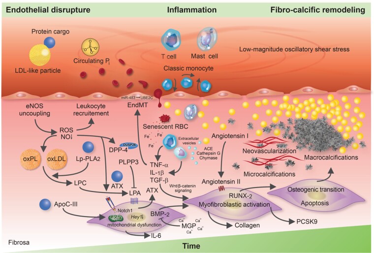
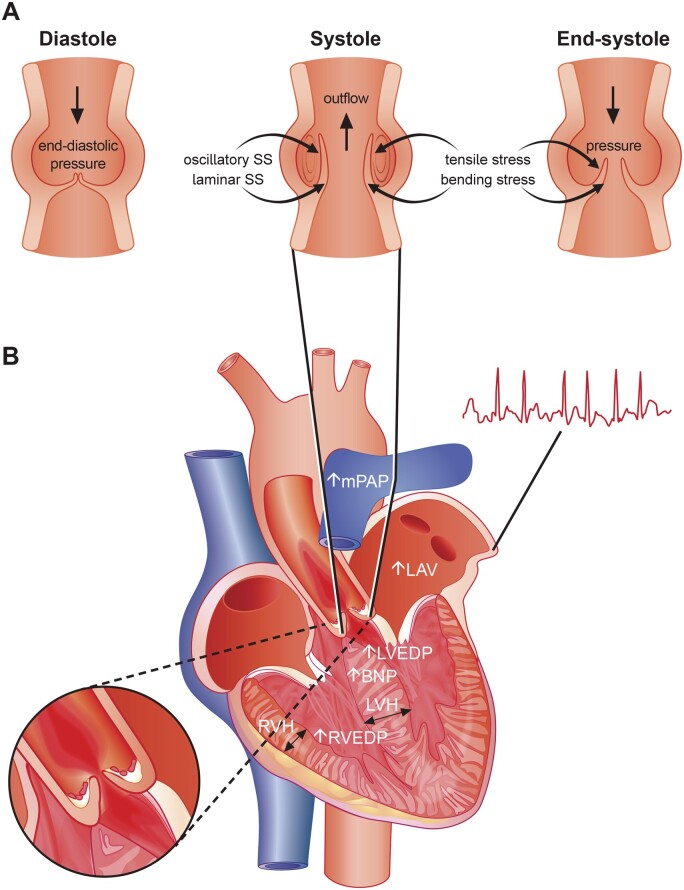
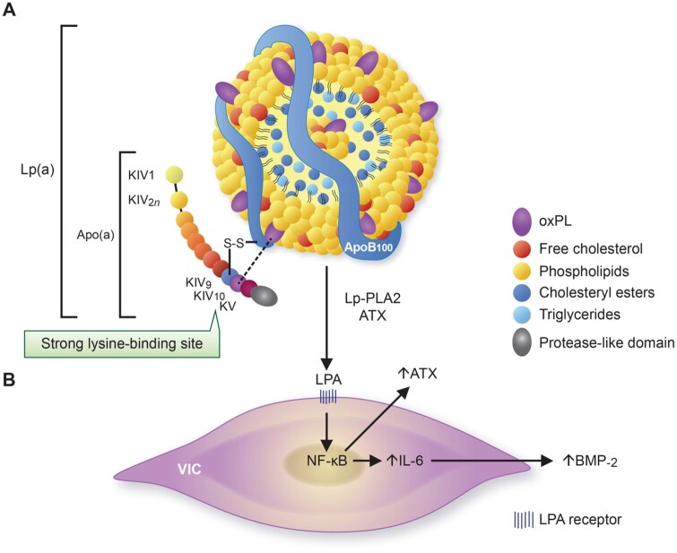
Calcific aortic valve disease (CAVD) is a highly prevalent condition that comprises a disease continuum, ranging from microscopic changes to profound fibro-calcific leaflet remodelling, culminating in aortic stenosis, heart failure, and ultimately premature death. Traditional risk factors, such as hypercholesterolaemia and (systolic) hypertension, are shared among atherosclerotic cardiovascular disease and CAVD, yet the molecular and cellular mechanisms differ markedly. Statin-induced low-density lipoprotein cholesterol lowering, a remedy highly effective for secondary prevention of atherosclerotic cardiovascular disease, consistently failed to impact CAVD progression or to improve patient outcomes. However, recently completed phase II trials provide hope that pharmaceutical tactics directed at other targets implicated in CAVD pathogenesis offer an avenue to alter the course of the disease non-invasively. Herein, we delineate key players of CAVD pathobiology, outline mechanisms that entail compromised endothelial barrier function, and promote lipid homing, immune-cell infiltration, and deranged phospho-calcium metabolism that collectively perpetuate a pro-inflammatory/pro-osteogenic milieu in which valvular interstitial cells increasingly adopt myofibro-/osteoblast-like properties, thereby fostering fibro-calcific leaflet remodelling and eventually resulting in left ventricular outflow obstruction. We provide a glimpse into the most promising targets on the horizon, including lipoprotein(a), mineral-binding matrix Gla protein, soluble guanylate cyclase, dipeptidyl peptidase-4 as well as candidates involved in regulating phospho-calcium metabolism and valvular angiotensin II synthesis and ultimately discuss their potential for a future therapy of this insidious disease.

Keywords: Ageing; Calcific aortic valve disease; Lipoprotein(a); Medical therapy; Nitric oxide; Notch1.


Figures

Graphical Abstract
Graphical Abstract
A complex network of cellular and molecular mechanisms underpins the pathobiology of calcific aortic valve disease. According to the current concept, disrupture of the endothelial layer covering the fibrosa promotes the uptake of oxidatively modified lipids (along with the protein-cargo they carry), red blood cells, and immune-cells, thereby promoting an inflammation-calcification feedback loop that results in fibro-calcific remodelling, leaflet stiffening and ultimately narrowing of the left ventricular outflow tract, with its dreadful clinical sequelae such as aortic stenosis, heart failure and premature death. Beyond LDL-C lowering by statins, other previously identified molecules, including PCSK9/Lp(a), mineral-binding matrix Gla protein, soluble guanylate cyclase, dipeptidyl peptidase-4 as well as candidates involved in regulating valvular angiotensin II synthesis and phosphocalcium metabolism, have been targeted pharmacologically in randomized controlled trials. While in some of these studies an attenuation of calcification burden could be observed, effects of target modulation on haemodynamic disease progression, a clinically much more relevant surrogate of disease burden, are uncertain and need to be rigorously assessed in future trials.
Figure 1
Figure 1
Risk factors, structural changes and sequelae of calcific aortic valve disease at different disease stages. A variety of risk factors, including a bicuspid phenotype, dyslipidaemia, hypertension, diabetes, and increased body mass index enhance the risk to develop calcific aortic valve disease. Endothelial disruption, lipid accumulation, immune-cell infiltration and collagen fibre disorganization occur early in the disease process, with fine-stippled mineralisations being a hallmark of early disease stages. Of note, patients without left ventricular outflow obstruction but sclerotic changes of the aortic valve are at increased risk for major adverse cardiovascular events, likely mediated by the frequent co-existence of coronary atherosclerosis. While the rate of transition from aortic sclerosis to symptomatic aortic valve stenosis varies considerably between patients, findings from the population-based Cardiovascular Health Study suggest that 1–2% of patients with aortic sclerosis progress to aortic stenosis annually, of which three-quarter develop heart failure, undergo valve replacement or die within 2 to 5 years of follow-up.
Figure 2
Figure 2
Molecular and cellular mechanisms involved in calcific aortic valve disease pathogenesis. The injured endothelium covering the fibrosa fosters the uptake of immune-cells, red blood cells as well as low-density lipoprotein-like particles and their protein cargo, such as autotaxin and lipoprotein-associated phospholipase A2. Reactive oxygen species formation, enhanced by nitric oxide synthase uncoupling, aggravates the oxidative modification of lipids, promotes endothelial immune-cell trafficking and induces valvular interstitial cell apoptosis—yielding apoptotic bodies which may form additional nidi for the deposition of calcium and phosphorus crystals. While lipoprotein-associated phospholipase A2 hydrolyses the ester bond of oxidized phospholipids, autotaxin—which is secreted by valvular interstitial cells—catalyzes lysophosphatidic acid synthesis by choline group removal. Importantly, apoC-III colocalizes with calcific regions, promotes mitochondrial stress and increases interleukin-6 and bone morphogenetic protein-2 expression in human valvular interstitial cells. Matrix metalloproteinase/tissue inhibitors of matrix metalloproteinases imbalances disrupt extracellular matrix homeostasis and promote leaflet stiffening, while bone morphogenetic protein-2 drives osteogenic transition of valvular interstitial cells through increased expression of pro-osteogenic transcription factors, such as RUNX2. Infiltrated mast cells release chymase which facilitates angiotensin II synthesis, thereby promoting valvular interstitial cell-mediated collagen production and thus stiffening of aortic valve leaflets—a potent promoter of osteogenic valvular interstitial cell differentiation. Neovascularization, fuelled by vascular endothelial growth factor secretion, exacerbates immune-cell recruitment and cytokine secretion, which in turn boosts the fibro-calcific response.
Figure 3
Figure 3
Hemodynamic flow across the aortic valve and myocardial alterations occurring with advanced calcific aortic valve disease. (A) The hemodynamic forces aortic leaflets are exposed to are shown. Note that disturbed hemodynamic flow can perturb tissue homeostasis by acting on pro-inflammatory and pro-fibrotic signalling, thereby promoting calcific aortic valve disease progression and eventually the development of aortic stenosis. (B) As calcific aortic valve disease progresses and impediments in left ventricular outflow occur, left ventricular hypertrophy and myocardial fibrosis evolves leading to reduced left ventricular longitudinal function, although left ventricular ejection fraction typically remains unchanged in the majority of patients. If left untreated, the left atrium enlarges, enhancing the susceptibility to atrial fibrillation. Due to left ventricular hypertrophy and the reduced diastolic pressure gradient, coronary flow reserve can substantially decrease leading to cardiomyocyte loss further perpetuating processes underlying myocardial fibrosis. At late disease stages, secondary pulmonary hypertension and right-ventricular dysfunction evolves.
Figure 4
Figure 4
Structure of lipoprotein(a) and its pro-osteogenic effects on valvular interstitial cells. (A) Lipoprotein(a) is characterized by a low-density lipoprotein-like particle (note the single apoB100 molecule) that is covalently linked to the unique apolipoprotein(a) glycoprotein which is encoded by the LPA gene. While its lipid core consists mainly of cholesteryl esters and (some) triglycerides, its outer shell is mainly composed of phospholipids and free cholesterol. Although the majority of oxidized phospholipid is bound to apolipoprotein(a), lipids can also be covalently linked to apoB100 or even found freely in the lipid-shell. Twelve domains form apolipoprotein(a), with 10 (i.e. KIV1–KIV10) being homologous to plasminogen kringle-IV and one representing a kringle-V-like domain (i.e. KV) which is followed by an inactive protease-like domain. Different functions have been ascribed to each, with KIV10 being characterized by a strong lysine-binding site crucial for oxidized phospholipid binding. (B) Lipoprotein-associated phospholipase A2 and autotaxin can transform oxidized phospholipid to lysophosphatidylcholine and lysophosphatidic acid, respectively, thereby promoting endogenous interleukin-6 and autotaxin production through NF-κB activation. Interleukin-6 can induce increased bone morphogenetic protein-2 expression in a paracrine manner resulting in osteogenic transition of adjacent valvular interstitial cells and eventually aortic valve calcification.

References

    1. Novaro GM, Katz R, Aviles RJ et al. Clinical factors, but not C-reactive protein, predict progression of calcific aortic-valve disease. The Cardiovascular Health Study. J Am Coll Cardiol 2007;50:1992–1998. - PubMed
    1. Sverdlov AL, Ngo DTM, Chan WPA et al. Determinants of aortic sclerosis progression: implications regarding impairment of nitric oxide signalling and potential therapeutics. Eur Heart J 2012;33:2419–2425. - PubMed
    1. Faggiano P, Antonini-Canterin F, Erlicher A et al. Progression of aortic valve sclerosis to aortic stenosis. Am J Cardiol 2003;91:99–101. - PubMed
    1. Otto CM, Lind BK, Kitzman DW, Gersh BJ, Siscovick DS. Association of aortic-valve sclerosis with cardiovascular mortality and morbidity in the elderly. N Engl J Med 1999;341:142–147. - PubMed
    1. Coffey S, Cox B, Williams MJA. The prevalence, incidence, progression, and risks of aortic valve sclerosis: a systematic review and meta-analysis. J Am Coll Cardiol 2014;63:2852–2861. - PubMed

Publication types