. 2021 Oct 19;11(11):jkab297.
doi: 10.1093/g3journal/jkab297.

Vitamin A deficiency affects gene expression in the Drosophila melanogaster head

Affiliations

Vitamin A deficiency affects gene expression in the Drosophila melanogaster head

Deepshe Dewett et al. G3 (Bethesda). .

Abstract

Insufficient dietary intake of vitamin A causes various human diseases. For instance, chronic vitamin A deprivation causes blindness, slow growth, impaired immunity, and an increased risk of mortality in children. In contrast to these diverse effects of vitamin A deficiency (VAD) in mammals, chronic VAD in flies neither causes obvious developmental defects nor lethality. As in mammals, VAD in flies severely affects the visual system: it impairs the synthesis of the retinal chromophore, disrupts the formation of the visual pigments (Rhodopsins), and damages the photoreceptors. However, the molecular mechanisms that respond to VAD remain poorly understood. To identify genes and signaling pathways that are affected by VAD, we performed RNA-sequencing and differential gene expression analysis in Drosophila melanogaster. We found an upregulation of genes that are essential for the synthesis of the retinal chromophore, specific aminoacyl-tRNA synthetases, and major nutrient reservoir proteins. We also discovered that VAD affects several genes that are required for the termination of the light response: for instance, we found a downregulation of both arrestin genes that are essential for the inactivation of Rhodopsin. A comparison of the VAD-responsive genes with previously identified blue light stress-responsive genes revealed that the two types of environmental stress trigger largely nonoverlapping transcriptome responses. Yet, both stresses increase the expression of seven genes with poorly understood functions. Taken together, our transcriptome analysis offers insights into the molecular mechanisms that respond to environmental stresses.

Keywords: Drosophila; carotene; chromophore; photoreceptor; phototransduction; retinoic acid; rhabdomere; rhodopsin; transcriptome; vision; visual pigment; vitamin A.


Figures

Figure 1
Figure 1
Vitamin A deprivation affects Drosophila photoreceptor structure and Rhodopsin expression. (A) The schematic depicts the key steps of phototransduction. Dietary β-carotene is converted by NinaB and NinaG to the retinal chromophore that binds to opsin to form the Rhodopsin pigment. Activation of Rhodopsin triggers the phototransduction cascade and results in the opening of two types of cation channels, Trp and Trpl. The termination of the light response is mediated by two Arrestins (Arr1 and Arr2), which inactivate Rhodopsin, and several downstream factors (Stops, InaC, and Culd). The factors that terminate phototransduction are highlighted by a red outline. For details, see text. (B) Flies were raised on minimal medium with β-carotene (vitA+, left) or without β-carotene (vitA−, right). The images show that vitamin A deprivation had no obvious effect on the external morphology of the head or the eye. Total RNA was extracted from heads of adult flies for sequencing and differential gene expression (DEG) analysis. (C) Vitamin A replete (vitA+) wild-type adult eye. The rhabdomeres (green) have a round shape and the inner photoreceptors express Rh5 (blue) or Rh6 (red). (C’) Chronic vitamin A deprivation (vitA−) causes small rhabdomeres (green, compare to C) and affects Rhodopsin expression in the adult eye: Rh6 (red) is abnormally accumulated (arrows) outside of the rhabdomeres (green) and Rh5 is not detectable. (D) The vitamin A replete (vitA+) wild-type retina expresses mature Rh1 (blue). (D’) Vitamin A deprivation (vitA−) impairs Rh1 (blue) maturation and results in an abnormal localization (compare to D). Scale bars, 10 µm.
Figure 2
Figure 2
Vitamin A deprivation affects gene expression in the adult Drosophila head. (A) The volcano plot shows the profiles of DEGs that respond to vitamin A deprivation (vitA−). Genes with significant differential expression in the adult head are highlighted in blue or yellow color; 18 genes are significantly downregulated upon vitamin A deprivation (blue, left) and 50 genes are upregulated (yellow, right). The fold change is plotted for each gene relative to its P-value with a cut-off of abs(logFC) > 1.5-fold and a false discovery rate of FDR < 0.05. (B) Heat map of all DEGs. Three replicates are shown for vitamin A replete (vitA+) and deprived (vitA−) conditions, respectively. Shades of blue represent different levels of downregulation and shades of yellow represent different levels of upregulation.
Figure 3
Figure 3
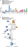
Enriched GO terms for genes that respond to vitamin A deprivation. (A) The bar graph shows the fold change of DEGs that respond to vitamin A deprivation and are associated with the GO terms phototransduction (dark blue), Rhodopsin metabolic process (light blue), retinoid metabolic process (orange), tRNA aminoacylation (magenta), and nutrient reservoir activity (green). Positive values indicate upregulation upon vitamin A deprivation, negative values indicate downregulation. (B) The schematic highlights phototransduction-, Rhodopsin metabolism-, and retinoid metabolism-related genes that respond to vitamin A deprivation. Color code corresponds to (A), white indicates no significant transcriptional response to vitamin A deprivation. Note that the vitamin A deprivation-responsive Arr1, Arr2, Culd, stops, and inaC all play a role in the deactivation of the light response (emphasized by red outline).
Figure 4
Figure 4
RT-qPCR validates vitamin A deprivation-responsive genes that were identified by total RNA-seq. The bar graph shows the fold change as detected by total RNA-seq (gray) or RT-qPCR (brown) for DEGs that respond to VAD. Three biological replicates were analyzed. Note that the shown genes are associated with different GO term categories such as Rhodopsin metabolic process or retinoid metabolism (ninaB and ninaG), phototransduction (ninaB, Arr1, Arr2, and CG11426), and tRNA aminoacylation (LeuRS and LysRS).
Figure 5
Figure 5
Summary of the effects of vitamin A deprivation in the Drosophila head. Vitamin A deprivation causes structural and functional defects in the eye; moreover, it affects gene expression in the adult head (18 genes downregulated, 50 genes upregulated).

References

    1. Ahmad ST, Joyce MV, Boggess B, O'Tousa JE.. 2006. The role of drosophila ninaG oxidoreductase in visual pigment chromophore biogenesis. J Biol Chem. 281:9205–9209. - PubMed
    1. Bahner M, Frechter S, Da Silva N, Minke B, Paulsen R, et al.2002. Light-regulated subcellular translocation of Drosophila TRPL channels induces long-term adaptation and modifies the light-induced current. Neuron. 34:83–93. - PubMed
    1. Baker EK, Colley NJ, Zuker CS.. 1994. The cyclophilin homolog ninaA functions as a chaperone, forming a stable complex in vivo with its protein target rhodopsin. EMBO J. 13:4886–4895. - PMC - PubMed
    1. Ballinger DG, Xue N, Harshman KD.. 1993. A Drosophila photoreceptor cell-specific protein, calphotin, binds calcium and contains a leucine zipper. Proc Natl Acad Sci USA. 90:1536–1540. - PMC - PubMed
    1. Barrett JR. 2005. Focusing on vision through an environmental lens. Environ Health Perspect. 113:A822–A827. - PMC - PubMed

Publication types