. 2021 Dec 2;49(21):12502-12516.
doi: 10.1093/nar/gkab1096.

Inhibition of SARS-CoV-2 coronavirus proliferation by designer antisense-circRNAs

Affiliations

Inhibition of SARS-CoV-2 coronavirus proliferation by designer antisense-circRNAs

Christina Pfafenrot et al. Nucleic Acids Res. .

Abstract

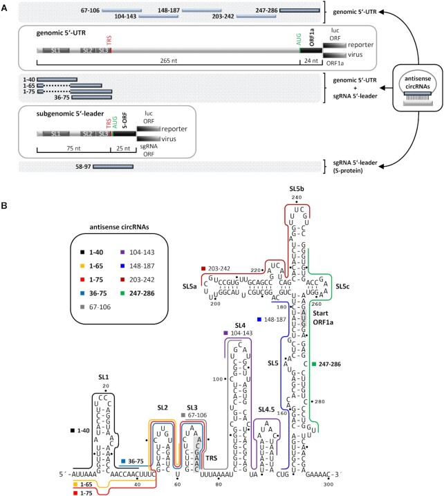
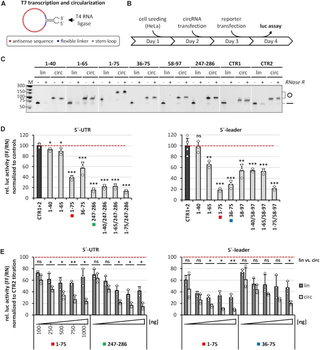
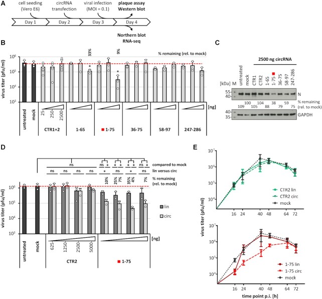
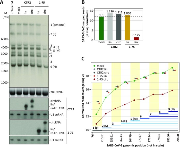
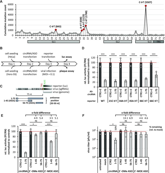
Circular RNAs (circRNAs) are noncoding RNAs that exist in all eukaryotes investigated and are derived from back-splicing of certain pre-mRNA exons. Here, we report the application of artificial circRNAs designed to act as antisense-RNAs. We systematically tested a series of antisense-circRNAs targeted to the SARS-CoV-2 genome RNA, in particular its structurally conserved 5'-untranslated region. Functional assays with both reporter transfections as well as with SARS-CoV-2 infections revealed that specific segments of the SARS-CoV-2 5'-untranslated region can be efficiently accessed by specific antisense-circRNAs, resulting in up to 90% reduction of virus proliferation in cell culture, and with a durability of at least 48 h. Presenting the antisense sequence within a circRNA clearly proved more efficient than in the corresponding linear configuration and is superior to modified antisense oligonucleotides. The activity of the antisense-circRNA is surprisingly robust towards point mutations in the target sequence. This strategy opens up novel applications for designer circRNAs and promising therapeutic strategies in molecular medicine.


Figures

Figure 1.
Figure 1.
Design of AS-circRNA targeting SARS-CoV-2 RNA. (A) Schematic representation of the 5′-UTR (top, nts 1–265) and 5′-leader (bottom; nts 1–75) sequences, targeted by AS-circRNAs, either in a luciferase-reporter (luc), or in the viral SARS-CoV-2 context (ORF1a or S). Target regions of individual AS-circRNAs are represented as blue bars with nucleotide coordinates. The conserved 5′-terminal stem-loop elements (SL1-3) are indicated, as well as the transcription regulatory sequence (TRS) of the 5′-leader. Note that the target regions of AS_1–65 and AS_1–75 circRNAs omit the first stem–loop (SL1), represented as a dashed line. (B) Target regions of AS-circRNAs within the 5′-leader (nts 1–75) and 5′-UTR of SARS-CoV-2 (nts 1–265), represented in the context of the secondary structure model of this region [nts 1–300; (3,4)]. For a schematic representation, see panel A. The core TRS element (nts 70–75) and the AUG start codon of ORF1a (nts 266–268) are shaded in grey. Note that AS_58–97 circRNA is not included, since it overlaps with the ORF of the S protein.
Figure 2.
Figure 2.
Screening of active AS-circRNAs: 5′-UTR and 5′-leader reporter assays. (A) Design of synthetic AS-circRNAs. CircRNAs were produced by in vitro T7 transcription and subsequent circularization by T4 RNA ligase. Each circular RNA is composed of a stem–loop with overhangs for efficient ligation (grey), a short stretch of unrelated nucleotides for enhanced flexibility (blue), and the antisense sequence (red). (B) Experimental workflow for luciferase reporter assays in HeLa cells transfected with synthetic circRNAs. (C) AS-circRNA synthesis. RNase R treatment and aberrant electrophoretic migration confirm the circularity of the produced circRNAs. Gel-purified linear and circular RNAs (lin/circ) were treated with RNase R, or left untreated (–/+), and analyzed by denaturing polyacrylamide electrophoresis and ethidium bromide staining. Mobilities of circular (o) and linear (–) forms are marked. M, DNA markers (sizes in bp). (D) Luciferase reporter assays reveal regions accessible to AS-circRNAs, based on the repression of luciferase activity by specific SARS-CoV-2 5′-UTR (left) and 5′-leader reporter constructs (right). HeLa cells were transfected with the respective circRNA (as indicated below the diagram) or a combination thereof (e.g. AS_1–40/247–286). The color code highlights those AS-circRNAs that were analyzed in more detail in panel E. After 24 h, the respective reporter was transfected (5′-UTR or 5′-leader), and relative luciferase activities (ratio of Firefly and Renilla expression) were measured, normalized to control circRNAs CTR1 and 2 (mean and standard deviations of three replicates, *P < 0.05, **P < 0.005, ***P < 0.001, ns = not significant, two-sided t-test). (E) Dose dependence and comparison of circular versus linear configuration of selected antisense-RNA regions. HeLa cells were transfected with increasing amounts (100–1000 ng per assay) of circRNAs (light gray), or their linear counterparts (dark grey; as indicated below the diagram). After 24 h, the respective reporter constructs were transfected (5′-UTR or 5′-leader), and relative luciferase activities (Firefly/Renilla expression ratios) were measured, normalized to control circRNA CTR2 (mean and standard deviations of three replicates, *P < 0.05, **P < 0.005, ***P < 0.001, ns = not significant, two-sided t-test).
Figure 3.
Figure 3.
Inhibition of SARS-CoV-2 proliferation by AS-circRNAs: viral infection assays. (A) Experimental workflow for viral infection assays using Vero E6 cells. (B) Screening of AS-circRNAs by virus titer assays identifies AS_1–75 as the most effective antiviral circRNA. Vero E6 cells were transfected with increasing quantities of circRNAs (25, 250 and 2500 ng per assay; light gray; as indicated below the diagram). After 24 h, cells were infected with SARS-CoV-2 (MOI = 0.1 pfu/cell). The effects on virus titers were measured by virus plaque assays using cell cutlure supernatants collected at 24 h post-infection (mean and standard deviations of three experiments, *P < 0.05, ns = not significant, two-sided t-test). Untreated (without RNA and transfection reagent) and mock-treated cells (without RNA, but with transfection reagent) served as controls. Residual virus titers of significantly affected samples are indicated as ‘percent remaining’ relative to mock treatment. (C) Viral protein synthesis assays: Western blot analysis of the viral nucleocapsid protein (N) confirms reduction of viral protein accumulation in cells treated with specific AS-circRNAs. Vero E6 cells transfected with 2500 ng of respective circRNAs per assay were harvested, lysed, and equal amounts of protein were analyzed by Western blotting, using the nucleocapsid protein as a marker for viral protein accumulation (quantitation of protein levels relative to mock); GAPDH was used as loading control. M, protein markers (sizes in kDa). (D) AS_1–75 circRNA: dose dependence of antiviral effect, in comparison to its linear counterpart. Vero E6 cells were transfected with increasing amounts (625, 1250, 2500 and 5000 ng) of AS_1–75 circRNA (light gray), or of its linear counterpart (dark gray), followed by viral infection (MOI = 0.1 pfu/cell) after 24 h. Plaque assays were used to determine virus titers in culture supernatants collected at 24 h post-infection (mean and standard deviations of three experiments, *P < 0.05, ns = not significant, two-sided t-test). As controls, untreated (without RNA and transfection reagent) and mock-treated cells (without RNA, but with transfection reagent) were used, as well as transfections with linear or circular control RNA (CTR2). The virus titer of significantly affected samples is indicated as ‘percent remaining’, relative to mock treatment. (E) Durability of antiviral activity of AS_1–75 circRNA. Vero E6 cells were transfected with AS_1–75 circRNA or its linear counterpart (bottom panel; in red), followed by viral infection (MOI = 0.1 pfu/cell) after 24 h (mean and SEM of three experiments). Plaque assays were performed to determine virus titers in culture supernatants collected at the indicated time points (16–72 h post-infection). As controls, mock-treated cells (without RNA, but with transfection reagent) were used (top and bottom panels; in black), as well as transfections with linear or circular control RNA (CTR2; top panel; in green).
Figure 4.
Figure 4.
Inhibition of viral RNA synthesis and processing by AS_1–75 circRNA. (A) Northern blot analysis of genomic and subgenomic viral RNAs. Vero E6 cells were transfected with AS_1–75 circRNA or its linear counterpart; as controls, untreated (without RNA and transfection reagent) and mock-treated cells (without RNA, but with transfection reagent) were used, as well as cells transfected with linear or circular control RNA (CTR2). At 24 h post-transfection, cells were infected with SARS-CoV-2 (MOI = 0.1 pfu/cell). At 24 h post-infection, total RNA was prepared and subjected to glyoxal-Northern blot analysis, to detect genomic and all subgenomic viral RNA species. M, RiboRuler High Range RNA Ladder (Thermo Fisher Scientific). As input control, 28S rRNA was detected by ethidium bromide staining, and CTR2 and AS_1–75 RNAs by specific Northern probes. (B) RNA-seq analysis of total viral RNA synthesis. The total numbers of SARS-CoV-2-mapped reads (in mio; normalized to total read number) were compared for RNAs isolated from virus-infected Vero E6 cells that were mock-transfected or transfected with CTR2 control and AS_1–75 RNA, each in linear or circular form, with ratios of read numbers relative to mock conditions indicated. (C) Effect of AS_1–75 circRNA on viral genome (g) and subgenomic RNA production in infected cells. Cumulative read coverages (in log2; normalized to total read number) are plotted for mock-treated, and CTR2 control RNA (lin/circ) or AS_1–75 RNA (lin/circ)-transfected cells. The SARS-CoV-2 genome was divided in nine sections with boundaries defined by the body-TRS sites of the eight subgenomic RNAs (sections used for cumulative read numbers are marked by thick lines; genomic positions are indicated below and drawn not in scale).
Figure 5.
Figure 5.
AS-circRNAs exhibit robust activity against SARS-CoV-2 mutant sequences and are superior to modified ASOs. (A) Summary of naturally occurring point mutations within the viral 5′-leader of SARS-CoV-2. All annotated mutations are indicated per nucleotide position [cumulative mutation count, as deposited in the ViGTK database (https://www.biosino.org/ViGTK/); as at 30 April 2021). The five most frequent single-point mutations in the 5′-leader region (positions 1–75) are highlighted in red (occurrences given in parentheses) and were selected for mutational analysis. Secondary structures and regulatory elements are marked (SL1–3, TRS). (B) Experimental workflow for luciferase reporter assays in HeLa cells, and for viral infection assays using Vero E6 cells, transfected with synthetic circRNAs or modified antisense oligonucleotides (ASOs). (C) Schematic representation of the 5′-leader (nts 1–75) sequence, targeted by a AS-circRNA (1–75) or two antisense oligonucleotides (ASOs, 1–45 and 56–75), either in a luciferase-reporter (luc), or in the viral SARS-CoV-2 context (sgRNAs/genome). Target regions of individual AS-circRNA or ASOs are represented as blue bars with nucleotide coordinates. Secondary structures and regulatory elements are marked (SL1–3, TRS, AUG). Note that the targeting regions of AS_1–75 circRNA and 1–45 ASO omit the first stem-loop (SL1), represented as a dashed line. (D) Activity of AS_1–75 circRNA in presence of single point mutations within the 5′-leader: luciferase reporter assays. HeLa cells were transfected with 1–75 AS-circRNA or with control circRNAs. After 24 h, the respective 5′-leader reporter plasmids, either without (WT) or with the indicated point mutations, were transfected, and relative luciferase activities (ratio of Firefly and Renilla expression) were measured, normalized to control circRNAs CTR1 and 2 (mean and standard deviations of three replicates, P < 0.001***, two-sided t-test). (E) Activity of 2′-OMe or 2′-MOE modified ASOs: luciferase reporter assays. HeLa cells were transfected with the AS_1–75 circRNA (1 μg) or ASOs (molar equivalents), respectively. After 24 h, the 5′-leader reporter was transfected, and relative luciferase activities (ratio of Firefly and Renilla expression) were measured, normalized to control circRNAs CTR1 and 2, or correspondingly modified control ASOs CTR1 and 2 (mean and standard deviations of three replicates, P < 0.05*, P < 0.001***, ns = not significant, two-sided t-test). Fold differences in translational repression between AS_1–75 circRNA and ASO treatments are indicated. (F) Antiviral activity of 2′-OMe or 2′-MOE modified ASOs: virus infection assays. Vero E6 cells were transfected with AS_1–75 circRNA (2500 ng per assay) or with ASOs (molar equivalents). After 24 h, cells were infected with SARS-CoV-2 (MOI = 0.1 pfu/cell). The antiviral effects were measured by virus plaque assays at 24 h post-infection (mean and standard deviations of three experiments, *P < 0.05, ns = not significant, two-sided t-test). Untreated (without RNA and transfection reagent) and mock-treated cells (without RNA, but with transfection reagent) served as controls. In addition, control circRNA CTR2 and the correspondingly modified control ASO CTR2 were used. Residual virus titers of significantly affected samples are indicated as ‘percent remaining’ relative to mock treatment, as well as fold differences between circRNA and ASO treatments.

Similar articles

Cited by

References

    1. Perlman S., Netland J.. Coronaviruses post-SARS: update on replication and pathogenesis. Nat. Rev. Microbiol. 2009; 7:439–450. - PMC - PubMed
    1. Perlman S., Masters P.S.. Howley P.M., Knipe D.M., Whelan S.. Coronaviridae: the viruses and their replication. Fields Virology. 2021; I:Philadelphia, PA: Wolters Kluwer; 410–448.
    1. Madhugiri R., Karl N., Petersen D., Lamkiewicz K., Fricke M., Wend U., Scheuer R., Marz M., Ziebuhr J.. Structural and functional conservation of cis-acting RNA elements in coronavirus 5′-terminal genome regions. Virology. 2018; 517:44–55. - PMC - PubMed
    1. Miao Z., Tidu A., Eriani G., Martin F.. Secondary structure of the SARS-CoV-2 5′-UTR. RNA Biol. 2021; 18:447–456. - PMC - PubMed
    1. Sola I., Almazán F., Zúñiga S., Enjuanes L.. Continuous and discontinuous RNA synthesis in coronaviruses. Annu. Rev. Virol. 2015; 2:265–288. - PMC - PubMed

Publication types

MeSH terms