. 2021 Dec 2;49(21):12377-12393.
doi: 10.1093/nar/gkab1123.

TRF2 promotes dynamic and stepwise looping of POT1 bound telomeric overhang

Affiliations

TRF2 promotes dynamic and stepwise looping of POT1 bound telomeric overhang

Tapas Paul et al. Nucleic Acids Res. .

Abstract

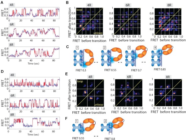
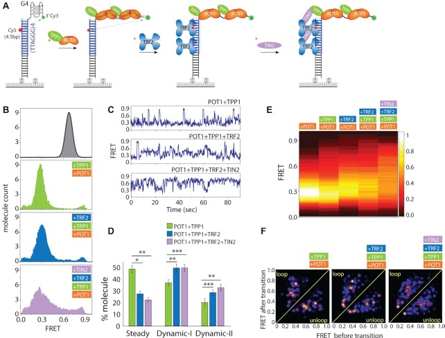
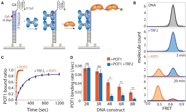
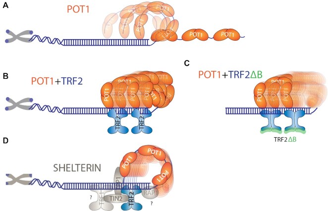
Human telomeres are protected by shelterin proteins, but how telomeres maintain a dynamic structure remains elusive. Here, we report an unexpected activity of POT1 in imparting conformational dynamics of the telomere overhang, even at a monomer level. Strikingly, such POT1-induced overhang dynamics is greatly enhanced when TRF2 engages with the telomere duplex. Interestingly, TRF2, but not TRF2ΔB, recruits POT1-bound overhangs to the telomere ds/ss junction and induces a discrete stepwise movement up and down the axis of telomere duplex. The same steps are observed regardless of the length of the POT1-bound overhang, suggesting a tightly regulated conformational dynamic coordinated by TRF2 and POT1. TPP1 and TIN2 which physically connect POT1 and TRF2 act to generate a smooth movement along the axis of the telomere duplex. Our results suggest a plausible mechanism wherein telomeres maintain a dynamic structure orchestrated by shelterin.


Figures

Figure 1.
Figure 1.
POT1 is stably bound to telomeric G4/4R. (A) Schematic smFRET model of before and after POT1 (two orange lobes represent OB1 and OB2 domains of a single POT1 molecule) binding to telomeric G4/4R DNA (Top4.5 construct). (B) The FRET histograms of 4R before and after POT1 (25 nM) binding. In y-axis, molecule count means the normalized number of molecules. (C) FRET histograms after 30 min incubation with 4R only (top), 100 mM KCl (center) and C4 (bottom) to the POT1 bound G4. (D) The representative real-time smFRET trace of POT1 binding to 4R (protein flow at ∼10 s). (E) The heatmap (n > 100), generated by synchronizing the POT1 bound state. (F) Single-exponential fitting of POT1 bound fraction to 4R overhang at different POT1 concentrations. (G) Linearly fitted binding rate represents the corresponding POT1 binding rate to 4R overhang at different concentration. (H) Determination of the apparent dissociation constant (KD-app) of POT1 to 4R/G4.
Figure 2.
Figure 2.
POT1 bound telomeric overhang shows dynamics. (A) The representative smFRET traces of POT1 bound to telomeric 4R after wash of free protein show steady (FRET ∼0.3, top), Dynamic-I (FRET ∼0.2–0.6, second from top), and Dynamic-II (FRET ∼0.2–0.9, bottom two) traces. (B, C) Quantification of molecular behavior of POT1 bound G4 (steady versus two types of dynamic) at 25 nM after wash of free protein (B) and the protein concentrations ranging from 50 to 1 nM before wash of free protein (C). (D) Dwell time of dynamic FRET traces, δt1 for Dynamic-I and δt2 for Dynamic–II respectively. (E, F) Model cartoon of Top0 (E) and Top7.5 (F) 4R construct, based on the acceptor dye position from the top of the duplex and beside the representative POT1 bound smFRET traces respectively. (G) FRET heatmap histogram of three constructs generated from the dynamic traces (keeping the bin size 0.2). White asterisk denotes level of highest FRET values observed in traces. (H) Schematic dynamic model of POT1 bound telomeric G4/4R overhang. (I) Schematic smFRET model of monomer POT1 bound to telomere overhang. (J) The representative smFRET steady (top) and dynamic (bottom) traces of POT1 bound to telomeric 2R overhang. (K) Quantification of molecular behavior of POT1 bound 2R (steady vs dynamic). (L) Dwell time (δt1) of dynamic FRET traces of 2R overhang. (M) Schematic dynamic model of POT1 bound telomeric 2R overhang.
Figure 3.
Figure 3.
Telomeric duplex bound TRF2 enhances POT1-overhang dynamics. (A) Schematic model of telomeric duplex (four repeats of TTAGGG tract) with G4 overhang sequentially binding POT1 at the overhang followed by TRF2 at the duplex. The basic domain of TRF2 can bind to ss-ds junction, for simplicity isn’t shown here. (B) The FRET histograms of telomeric duplex with G4 (∼0.65 FRET, black solid line), POT1 bound condition (∼0.3 FRET, orange solid line), and POT1, TRF2 together (∼0.3 FRET, solid blue filled). (C) POT1 and TRF2 bound representative smFRET traces of Dynamic-I (top) and Dynamic-II (bottom) states. (D) Quantification of molecular behavior (steady vs two types of dynamics) of 4R bound POT1 in presence and absence of TRF2. All statistics for this figure are calculated using a two-tailed two-sample Student's t test (*P < 0.05; **P < 0.01). (E) Representative smFRET trace of real time TRF2 binding at telomeric duplex (flow at ∼10 s) contain POT1 pre-bound overhang. (F) The heatmap (n > 100) of POT1 bound state in absence (top) and presence (bottom) of TRF2 (flow at ∼10 s).
Figure 4.
Figure 4.
Enhanced dynamics also observed in longer telomeric overhang. (A, B) The representative real time smFRET traces of TRF2 binding at telomeric duplex (flow at ∼10 s) to the pre-bound POT1 of 6R (A) and 8R (B) overhangs. (C, D) The heatmap (n > 100) of POT1 bound state in absence (top) and presence (bottom) of TRF2 (flow at ∼10 s) of 6R (C) and 8R (D) overhangs. (E) FRET heatmap histograms generated from the dynamic traces of POT1 only and POT1 followed by TRF2 addition to 2R, 4R, 6R and 8R overhangs containing telomeric duplex (keeping the bin size 0.2). (F) Proposed model of TRF2 induced POT1 overhang dynamics of the respective construct.
Figure 5.
Figure 5.
POT1 bound overhangs show discrete steps moving up and down along the TRF2 bound duplex. (A, D) Representative smFRET traces (blue) fitted by HMM (red), (BE) transition density plot (TDP) and, (CF) the proposed models of POT1 bound 4R, 6R and 8R telomeric overhangs moving up and down to the TRF2 bound four (A–C) and two (D–F) repeats of TTAGGG containing duplex.
Figure 6.
Figure 6.
TPP1 and TIN2 generates smaller substeps within the TRF2-POT1 induced movement. (A) Experimental schematics of four shelterin components added in succession to telomeric duplex (four repeats of TTAGGG tract) with G4/4R overhang. First, premixed POT1–TPP1 is applied followed by TRF2 and TIN2 which interact with TRF2 and TPP1. (B) FRET histogram of telomeric duplex with 4R (top), with POT1-TPP1 (second from top), with TRF2 (third from top) and TIN2 bound condition (bottom). (C) Representative smFRET traces. (D) Quantification of steady and dynamic patterns. All statistics for this figure are calculated using a two-tailed two-sample Student's t test (*P < 0.05; **P < 0.01, ***P < 0.001). (E) FRET heatmap histograms generated from the dynamic traces. (F) Transition density plot (TDP) of the corresponding protein conditions.
Figure 7.
Figure 7.
Telomeric duplex bound TRF2 delays POT1 binding to telomeric overhangs. (A) Schematic model of telomeric duplex (four repeats of TTAGGG tract) with the G4/4R overhang, sequentially binding TRF2 at the duplex followed by POT1 at the overhang. (B) The FRET histograms of telomeric duplex with 4R overhang (top), TRF2 bound condition (second from top), and POT1 binding at 3 and 20 min, respectively (bottom two). (C) Single-exponential fitting of POT1 bound fraction in the absence and presence of TRF2. (D) The bar graph represents the corresponding POT1 binding rate in the absence and presence of TRF2 of different overhang. All statistics for this figure are calculated using a two-tailed two-sample Student's t test (ns, not significant; *P < 0.05; **P < 0.01).
Figure 8.
Figure 8.
Proposed dynamic model of POT1 and POT1 with TRF2 bound to telomeric DNA. (A) POT1 bound telomeric overhang showing dynamics. (B) TRF2 recruit POT1 bound 3′ end towards the junction and moving up and down along the duplex axis. (C) TRF2ΔB doesn’t recruit POT1 bound 3′ end towards the junction. (D) Shelterin components bridging interactions within the proteins bound at telomeric DNA.

Similar articles

Cited by

References

    1. De Lange T. Shelterin: the protein complex that shapes and safeguards human telomeres. Genes Dev. 2005; 19:2100–2110. - PubMed
    1. Sfeir A., De Lange T.. Removal of shelterin reveals the telomere end-protection problem. Science. 2012; 336:593–597. - PMC - PubMed
    1. Bandaria J.N., Qin P., Berk V., Chu S., Yildiz A.. Shelterin protects chromosome ends by compacting telomeric chromatin. Cell. 2016; 164:735–746. - PMC - PubMed
    1. Farr C., Fantes J., Goodfellow P., Cooke H.. Functional reintroduction of human telomeres into mammalian cells. Proc. Natl Acad. Sci. U.S.A. 1991; 88:7006–7010. - PMC - PubMed
    1. Reddel R.R. Alternative lengthening of telomeres, telomerase, and cancer. Cancer Lett. 2003; 194:155–162. - PubMed

Publication types

MeSH terms