. 2021 Dec 16;49(22):13019-13030.
doi: 10.1093/nar/gkab1160.

The SARS-CoV-2 RNA polymerase is a viral RNA capping enzyme

Affiliations

The SARS-CoV-2 RNA polymerase is a viral RNA capping enzyme

Alexander P Walker et al. Nucleic Acids Res. .

Abstract

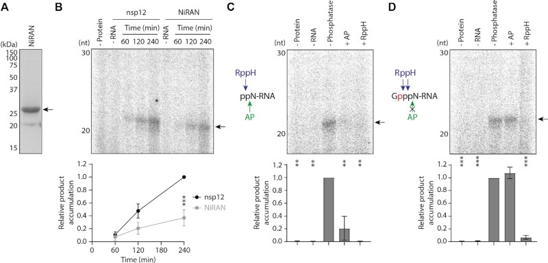
SARS-CoV-2 is a positive-sense RNA virus responsible for the Coronavirus Disease 2019 (COVID-19) pandemic, which continues to cause significant morbidity, mortality and economic strain. SARS-CoV-2 can cause severe respiratory disease and death in humans, highlighting the need for effective antiviral therapies. The RNA synthesis machinery of SARS-CoV-2 is an ideal drug target and consists of non-structural protein 12 (nsp12), which is directly responsible for RNA synthesis, and numerous co-factors involved in RNA proofreading and 5' capping of viral RNAs. The formation of the 5' 7-methylguanosine (m7G) cap structure is known to require a guanylyltransferase (GTase) as well as a 5' triphosphatase and methyltransferases; however, the mechanism of SARS-CoV-2 RNA capping remains poorly understood. Here we find that SARS-CoV-2 nsp12 is involved in viral RNA capping as a GTase, carrying out the addition of a GTP nucleotide to the 5' end of viral RNA via a 5' to 5' triphosphate linkage. We further show that the nsp12 NiRAN (nidovirus RdRp-associated nucleotidyltransferase) domain performs this reaction, and can be inhibited by remdesivir triphosphate, the active form of the antiviral drug remdesivir. These findings improve understanding of coronavirus RNA synthesis and highlight a new target for novel or repurposed antiviral drugs against SARS-CoV-2.


Figures

Figure 1.
Figure 1.
SARS-CoV-2 viral RNAs are 7-methylguanosine capped. (A) Schematic of the full-length SARS-CoV-2 genome with RT-qPCR primer binding sites indicated with arrows. (B) RT-qPCR was performed on total RNA from mock infected cells and cells 12 hours post-infection with SARS-CoV-2. Reaction products were resolved by agarose gel electrophoresis. Off-target amplicons from uninfected cells are visible for 8 and N sgRNA primers at high cycle numbers. (C) Vero cells were infected with SARS-CoV-2 and viral RNA accumulation was measured by RT-qPCR. Quantification of RNA levels is from n = 2 independent infections, data are mean ± s.e.m. (D) Schematic of RNA IP using an anti-m7G antibody, with an anti-his tag antibody as a negative control. (E) m7G capped RNA was immunoprecipitated from the total RNA of SARS-CoV-2 infected cells and quantified by RT-qPCR. RNA levels were normalised to uncapped 5S rRNA and the input was set to 0, such that a positive value indicates enrichment. Quantification of RNA enrichment is from n = 2 independent infections, data are mean ± s.e.m. Anti-his and anti-m7G values were compared by unpaired two-tailed t-test. *P < 0.05, **P < 0.01.
Figure 2.
Figure 2.
Nsp12 has guanylyltransferase activity. (A) Schematic of nsp13 5′ triphosphatase activity followed by a canonical GTase reaction mechanism, which consists of nucleotidylation of the enzyme (E) with GTP (Gppp), then transfer of GMP (Gp) to a diphosphorylated RNA substrate (ppN-RNA). The 32P isotope of α-32P-GTP is indicated in red. (B) Purified SARS-CoV-2 nsp13 was visualised by SDS PAGE, the arrow indicates the His6-ZBasic-tagged nsp13 band. (C) 5′ triphosphatase activity of purified nsp13 was tested by incubation with γ-32P-ATP for the indicated amount of time. The γ-32P-ATP substrate and inorganic phosphate (Pi) product were visualised by denaturing PAGE and autoradiography, arrows indicate the anticipated bands (left). AP was used as a positive control, and as a negative control nsp13 was inactivated by heating to 70°C for 5 min prior to the reaction, then was incubated with the γ-32P-ATP substrate for 5 mins. Quantification of γ-32P-ATP substrate and Pi product is from n = 2 independent reactions, data are mean ± s.e.m. (right). Pi present in the untreated γ-32P-ATP stock was subtracted from all samples during quantification. (D) SARS-CoV-2 nsp7 and nsp8 were expressed and purified from E. coli cells and visualised by SDS PAGE. Arrows indicate bands corresponding to the proteins of interest. (E) SARS-CoV-2 nsp12 was expressed and purified from Sf9 cells and visualised by SDS PAGE. The arrow indicates the purified nsp12 band. (F) The indicated SARS-CoV-2 proteins were incubated with α-32P-GTP and diphosphorylated RNA, then radiolabelled RNA products were visualised by denaturing PAGE and autoradiography. The arrow indicates the anticipated product, and the asterisk denotes a faster mobility product which could result from contaminating RNA kinase activity in some protein preparations, or decapping of the anticipated product by a phosphatase such as residual nsp13. (G) Nsp12 was incubated with diphosphorylated RNA and α-32P-UTP or α-32P-GTP, then radiolabelled RNA products were visualised by denaturing PAGE and autoradiography. The arrow indicates the anticipated product (top). Quantification of the product is from n = 3 independent reactions (bottom). Data are mean ± s.e.m., analysed by one-way ANOVA. ***P<0.01. (H) Nsp12 or vaccinia capping enzyme were incubated with α-32P-GTP and diphosphorylated RNA for the indicated amount of time. Radiolabelled RNA products were visualised by denaturing PAGE and autoradiography (left), the arrow indicates the anticipated product. Quantification of the product is from n = 3 independent reactions, data are mean ± s.e.m. (right). (I) Schematic of AP and RppH activity on the diphosphorylated RNA substrate (left). Diphosphorylated RNA was treated with AP or RppH, then incubated with nsp12 or vaccinia capping enzyme and α-32P-GTP (top). The arrow indicates the anticipated product. Quantification of the product is from n = 2 independent reactions (bottom). Data are mean ± s.e.m., analysed by one-way ANOVA. ***P < 0.001. (J) Schematic of AP and RppH activity on the capped RNA product (left). Diphosphorylated RNA was incubated with nsp12 or vaccinia capping enzyme and α-32P-GTP, then reaction products were treated with AP and RppH (top). The arrow indicates the anticipated product. Quantification of the product is from n = 2 independent reactions (bottom). Data are mean ± s.e.m., analysed by one-way ANOVA. **P < 0.01.
Figure 3.
Figure 3.
Mutations in the nsp12 NiRAN domain disrupt guanylyltransferase activity. (A) Structure of the SARS-CoV-2 nsp7/8/12 complex bound to RNA product, nsp9 and GDP (PDB: 7CYQ). Nsp12 (grey) is shown as a surface with the thumb and fingers subdomains highlighted in blue and green respectively. In this structure the NiRAN domain (cyan) at the nsp12 N-terminus is bound to GDP (orange), while nsp7 and nsp8 (turquoise and green ribbons) facilitate RNA product (orange/red) binding. (B) Close-up view of GDP bound to the nsp12 NiRAN domain with key amino acid residues highlighted, including D218 which coordinates an Mg2+ ion (silver). (C) Mutant nsp12 proteins purified from Sf9 cells were visualised by SDS PAGE. The arrow indicates the purified nsp12 band. (D) Schematic of the 40nt RNA template (LS1) and 20nt radiolabelled RNA primer (LS2). (E) Wild type or D760A/D761A mutant nsp12 was incubated with nsp7 and nsp8, then tested for RNA polymerase activity on the LS1/LS2 RNA template. Reaction products were resolved by denaturing PAGE and autoradiography (top). The arrow indicates the anticipated 40nt RNA product, and the asterisk denotes incompletely denatured RNA which has slower mobility on the gel. Quantification of the 40nt RNA product is from n = 3 independent reactions (bottom). Data are mean ± s.e.m., analysed by two-way ANOVA. ***P < 0.001. (F) Mutant nsp12 proteins were tested for RNA polymerase activity in the presence of nsp7, nsp8 and LS1/LS2 RNA template (top). The arrow indicates the anticipated 40nt RNA product, and the asterisk denotes incompletely denatured RNA which has slower mobility on the gel. Quantification of the 40nt product is from n = 3 independent reactions (bottom). Data are mean ± s.e.m., analysed by one-way ANOVA. ***P < 0.001. (G) Mutant nsp12 proteins were tested for GTase activity using α-32P-GTP and a diphosphorylated RNA substrate, the arrow indicates the anticipated product (top). Quantification of the product is from n = 3 independent reactions (bottom). Data are mean ± s.e.m., analysed by one-way ANOVA. ***P < 0.001.
Figure 4.
Figure 4.
The nsp12 NiRAN domain alone is a guanylyltransferase enzyme. (A) The NiRAN domain from SARS-CoV-2 nsp12 was expressed and purified from E. coli, then visualised by SDS PAGE. The arrow indicates the purified NiRAN domain band. (B) Full-length nsp12 and the purified NiRAN domain were tested for GTase activity using α-32P-GTP and a diphosphorylated RNA substrate, the arrow indicates the anticipated product (top). Quantification of the product is from n = 3 independent reactions (bottom). Data are mean ± s.e.m., analysed by two-way ANOVA. ***P < 0.001. (C) Schematic of AP and RppH activity on the diphosphorylated RNA substrate (left). Diphosphorylated RNA was treated with AP or RppH, then incubated with purified NiRAN domain and α-32P-GTP (right). The arrow indicates the anticipated product. Quantification of the product is from n = 2 independent reactions (bottom). Data are mean ± s.e.m., analysed by one-way ANOVA. **P < 0.01. (D) Schematic of AP and RppH activity on the capped RNA product (left). Diphosphorylated RNA was incubated with purified NiRAN domain and α-32P-GTP, then reaction products were treated with AP and RppH (right). The arrow indicates the anticipated product. Quantification of the product is from n = 2 independent reactions (bottom). Data are mean ± s.e.m., analysed by one-way ANOVA. ***P < 0.001.
Figure 5.
Figure 5.
In vitro reconstitution of 7-methylguanosine cap synthesis. (A) SARS-CoV-2 nsp14 was expressed and purified from E. coli, then visualised by SDS PAGE. The arrow indicates the purified nsp14 band. (B) Nsp12 GTase reactions were treated with nsp14 and SAM, then reaction products were visualised by dot-blotting using an anti-m7G antibody (top). Quantification of the product is from n = 3 independent reactions (bottom). Data are mean ± s.e.m., analysed by one-way ANOVA. ***P < 0.001.
Figure 6.
Figure 6.
Inhibition of SARS-CoV-2 RNA polymerase and guanylyltransferase functions by remdesivir triphosphate and a model for coronavirus RNA capping. (A) Structure of remdesivir triphosphate, active metabolite of remdesivir. (B) Remdesivir triphosphate was titrated into RNA polymerase reactions containing nsp7/8/12 and LS1/LS2 RNA template. The black arrow indicates the anticipated 40nt RNA product, red arrows indicate major premature termination products, and the asterisk denotes incompletely denatured RNA which has slower mobility on the gel. (C) Schematic of the LS1/LS2 RNA template suggesting possible identities of major premature termination products. Incorporated bases are shown in bold, with remdesivir triphosphate residues in red. Red arrows indicate the point of termination. (D) Remdesivir triphosphate was titrated into nsp12 GTase reactions with α-32P-GTP and a diphosphorylated RNA substrate, the arrow indicates the anticipated product. (E) Remdesivir triphosphate was titrated into NiRAN domain GTase reactions, the arrow indicates the anticipated product. (F) Inhibition curves for remdesivir triphosphate in SARS-CoV-2 RNA polymerase and GTase reactions. Quantifications of the 40nt RNA product, nsp12 GTase product and NiRAN GTase product are each from n = 3 independent reactions. Data are mean ± s.e.m., fit to dose-response inhibition curves by nonlinear regression in GraphPad Prism 9. (G) Model for coronavirus RNA capping. The γ-phosphate of triphosphorylated viral RNA is removed by the 5′ triphosphatase activity of nsp13. The nsp12 NiRAN domain GTase links diphosphorylated viral RNA to GTP (purple), which involves a GMP-enzyme intermediate. Nsp14 methylates GTP at the N7 position using SAM (blue) as a methyl donor, and producing SAH. Nsp16 methylates the 2’ hydroxyl group of the nucleotide at position 1 of the RNA, generating viral RNA with a cap-1 structure as the final product.

References

    1. Gorbalenya A.E., Baker S.C., Baric R.S., de Groot R.J., Drosten C., Gulyaeva A.A., Haagmans B.L., Lauber C., Leontovich A.M., Neuman B.W.et al. .. The species Severe acute respiratory syndrome-related coronavirus: classifying 2019-nCoV and naming it SARS-CoV-2. Nat. Microbiol. 2020; 5:536–544. - PMC - PubMed
    1. Wu A., Peng Y., Huang B., Ding X., Wang X., Niu P., Meng J., Zhu Z., Zhang Z., Wang J.et al. .. Genome composition and divergence of the novel coronavirus (2019-nCoV) originating in China. Cell Host Microbe. 2020; 27:325–328. - PMC - PubMed
    1. Gorbalenya A.E., Enjuanes L., Ziebuhr J., Snijder E.J.. Nidovirales: evolving the largest RNA virus genome. Virus Res. 2006; 117:17–37. - PMC - PubMed
    1. Snijder E.J., Decroly E., Ziebuhr J.. The nonstructural directing coronavirus RNA synthesis and processing. Adv. Virus Res. 2016; 96:59–126. - PMC - PubMed
    1. V’kovski P., Kratzel A., Steiner S., Stalder H., Thiel V.. Coronavirus biology and replication: implications for SARS-CoV-2. Nat. Rev. Microbiol. 2021; 19:155–170. - PMC - PubMed

Publication types

MeSH terms