. 2022 Feb 3;34(2):889-909.
doi: 10.1093/plcell/koab289.

Phosphatidic acid modulates MPK3- and MPK6-mediated hypoxia signaling in Arabidopsis

Affiliations

Phosphatidic acid modulates MPK3- and MPK6-mediated hypoxia signaling in Arabidopsis

Ying Zhou et al. Plant Cell. .

Abstract

Phosphatidic acid (PA) is an important lipid essential for several aspects of plant development and biotic and abiotic stress responses. We previously suggested that submergence induces PA accumulation in Arabidopsis thaliana; however, the molecular mechanism underlying PA-mediated regulation of submergence-induced hypoxia signaling remains unknown. Here, we showed that in Arabidopsis, loss of the phospholipase D (PLD) proteins PLDα1 and PLDδ leads to hypersensitivity to hypoxia, but increased tolerance to submergence. This enhanced tolerance is likely due to improvement of PA-mediated membrane integrity. PA bound to the mitogen-activated protein kinase 3 (MPK3) and MPK6 in vitro and contributed to hypoxia-induced phosphorylation of MPK3 and MPK6 in vivo. Moreover, mpk3 and mpk6 mutants were more sensitive to hypoxia and submergence stress compared with wild type, and fully suppressed the submergence-tolerant phenotypes of pldα1 and pldδ mutants. MPK3 and MPK6 interacted with and phosphorylated RELATED TO AP2.12, a master transcription factor in the hypoxia signaling pathway, and modulated its activity. In addition, MPK3 and MPK6 formed a regulatory feedback loop with PLDα1 and/or PLDδ to regulate PLD stability and submergence-induced PA production. Thus, our findings demonstrate that PA modulates plant tolerance to submergence via both membrane integrity and MPK3/6-mediated hypoxia signaling in Arabidopsis.


Figures

Figure 1
Figure 1
Submergence induces PLDα1- and PLDδ-derived PA accumulation, which triggers the nuclear localization of RAP2.12. A, Amounts of membrane lipids (PG, PC, PE, PI, PS, and PA) in the rosettes of 4-week-old WT Col-0 plants under light submergence treatment (Sub) and after recovery (R) for the indicated times. B, Various PA and PE species in the rosettes of 4-week-old WT, pldα1, pldδ, and pldα1 pldδ plants before light submergence treatment (air) and after 2 days of submergence treatment (submergence). C, Exogenous application of PA, but not PC, PE, or PS, induces the translocation of RAP2.12-GFP from the plasma membrane to the nucleus. Detached leaves of 3-week-old RAP2.12-GFP transgenic plants were treated with 50-µM liposomes prepared from PA, PC, PE, or PS (natural lipid mixtures purified from soy, Avanti Polar Lipids) for 3 h. Leaves similarly treated with dilution buffer were set as mock controls (Mock). The GFP fluorescence was detected by confocal microscopy. Red arrows indicate nuclear signal induced by PA application. Bars, 20 μm. All experiments were performed on three biological replicates with similar results. Values represent means ± sd (n = 4) of four independent technical replicates, and each replicate was collected from the rosettes of at least seven plants. Asterisks with “H” or “L” indicate significantly higher or lower levels than in control plants (A) or in WT (B) at each time point (*P < 0.05, **P < 0.01 by Student’s t test).
Figure 2
Figure 2
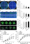
Knockouts in PLDα1 and PLDδ result in increased sensitivity to hypoxia, but higher tolerance to submergence. A and B, Phenotypes (A) and percentage of seedlings with green cotyledons (B) for WT, pldα1, pldδ, and pldα1 pldδ seeds germinated under normoxia (air, 21% O2) or hypoxia (3% O2) conditions for 12 days. C and D, Phenotypes (C) and survival rates (D) of WT, pldα1, pldδ, and pldα1 pldδ seedlings grown under hypoxia (0.1% O2) conditions. Six-day-old WT, pldα1, pldδ, and pldα1 pldδ seedlings grown on half-strength MS solid medium were transferred to normoxia (air) or hypoxia (0.1% O2) for 5 days followed by recovery for 3 days under normal growth conditions. E and F, Phenotypes (E) and dry weights (F) of 4-week-old submerged WT, pldα1, pldδ, and pldα1 pldδ plants under submergence treatment. Plants were photographed before submergence (air) and after submergence treatment for 8 days, followed by 3 days recovery (submergence) under normal growth conditions. Dry weights were determined after recovery for 7 days. Bars, 1.5 cm. G and H, Ion leakage (G) and water loss (H) in 4-week-old WT, pldα1, pldδ, and pldα1 pldδ plants under light submergence treatment or after reoxygenation for the indicated times. All experiments were performed on three biological replicates. Data are means ± sd of three biological replicates. Asterisks indicate significant differences from WT (*P < 0.05, **P < 0.01 by Student’s t test).
Figure 3
Figure 3
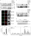
PA binds to MPK3 and MPK6 and enhances submergence-induced MPK3 and MPK6 activity. A, Lipid binding specificity of recombinant MPK3 and MPK6 proteins on membrane filters. About 50 μM of various lipids (PA, PC, PE, PI, PG, and PS dissolved in chloroform; natural lipid mixtures purified from soy, Avanti Polar Lipids) were spotted onto nitrocellulose membrane and incubated with 10 μg of purified GST-MPK3, GST-MPK6, or GST protein. Binding was detected by immunoblotting using an anti-GST antibody. Equal volume of chloroform was spotted as negative control (Blank). B, Pull-down assay showing the physical interaction between PA and recombinant MPK3 and MPK6 proteins. Recombinant proteins were incubated with PA beads, and the precipitated GST-MPK3 and GST-MPK6 were detected with anti-GST antibody. GST-PYL4 was used as a negative control. C, Dissociation constant (Kd) for the binding of recombinant MPK3 and MPK6 proteins to liposomes of PA, PC, PI, and PS (natural lipid mixtures purified from soy, Avanti Polar Lipids). A serial dilution of various liposomes ranging from 1.5 nM to 50 μM was prepared for mixing with the labeled proteins, and their binding affinities were measured by MST analysis. Kd, dissociation constant. ND, not detected. D and E, MPK3 and MPK6 are activated by submergence. Ten-day-old WT seedlings were exposed to light submergence (LS, D) or in the dark submergence (DS, E). MPK3/MPK6 kinase activities were detected with anti-pTEpY antibody. MPK3 and MPK6 proteins were detected with anti-MPK3 and anti-MPK6 antibodies, respectively. Actin, detected with an anti-actin antibody, was used as loading control. F, Quantification of MPK phosphorylation activity shown in (D) and (E). Data were calculated according to relative intensity from three independent experiments and the average values ± sd are shown. G, MPK3 and MPK6 kinase activities in WT and pldα1 pldδ plants and following submergence for 0.5, 1, 3, and 6 h. Total proteins were extracted and immunoblotting assays were performed using anti-pTEpY, anti-MPK3, and anti-MPK6 antibodies, with Ponceau S-stained total protein as loading control. hpt, hour posttreatment. Relative intensity of each p-MPK3 or pMPK6 band normalized to the loading control is shown below. H, PA induces MPK3 and MPK6 activity in planta. Ten-day-old WT seedlings were treated without (Mock) or with 50 μM PA or PS liposomes (natural lipid mixtures purified from soy, Avanti Polar Lipids) for 0.5, 1, and 3 h, and immunoblotting assays were performed using anti-pTEpY, anti-MPK3, anti-MPK6, and anti-actin antibodies, with actin as loading control. Relative intensity of each p-MPK3 or pMPK6 band normalized to the loading control is shown below. All experiments were performed on three biological replicates with similar results. Data in (C) and (F) are means ± sd of three biological replicates. Asterisks indicate significant differences from WT at 0 h (*P < 0.05 by Student’s t test).
Figure 4
Figure 4
MPK3 and MPK6 are required for the regulation of plant tolerance to hypoxia and submergence. A and B, Phenotypes (A) and ratios of seedlings with green cotyledons (B) for WT, mpk3, mpk6, and mpk3 mpk6-es (mpk3,6-es) seedlings grown under 3% O2 conditions. Seeds of WT, mpk3, mpk6, and mpk3,6-es were sown on half-strength MS medium with 10 μM β-estradiol and germinated under normoxia (air) or hypoxia (3% O2) for 10 days. C and D, Phenotypes (C) and survival rates (D) for WT, mpk3, mpk6, and mpk3,6-es seedlings grown under 0.1% O2 exposure. Six-day-old WT, mpk3, mpk6, and mpk3,6-es seedlings grown on MS solid medium were transferred to MS medium containing 10 μM estradiol, and continued to grow under normoxia (air) or hypoxia (0.1% O2) for 5 days followed by recovery for 3 days. E and F, Phenotypes (E) and dry weights (F) of 4-week-old submergence-treated WT, mpk3, mpk6, and mpk3,6-es plants. Plants were photographed before submergence (air) and after submergence for 8 days, followed by 3 days recovery (submergence). Dry weights were calculated after recovery for 7 days. Bars: 1.5 cm. G and H, Ion leakage (G) and H2O2 levels (H) in 4-week-old WT, mpk3, mpk6, and mpk3,6-es plants before submergence (Day 0) and after LS treatment followed by recovery (R) for the indicated times. All experiments were performed on three biological replicates with similar results, and average data calculated from three biological replicates are shown. Data are means ± sd of three independent biological replicates. Asterisks indicate significant differences from WT (*P < 0.05, **P < 0.01 by Student’s t test).
Figure 5
Figure 5
PA enhances MPK3- and MPK6-mediated phosphorylation of RAP2.12 to activate its transcriptional activity. A, Co-IP assay showing the interaction between MPK3/MPK6 and RAP2.12. Constructs encoding MPK3-FLAG and MPK6-FLAG, and RAP2.12-HA were transiently transfected in WT Arabidopsis protoplasts and immunoprecipitated with anti-FLAG beads. B, Immunoblot analyses showing RAP2.12 protein levels when co-expressed with MPK3 or MPK6. RAP2.12-HA was co-transfected with or without MPK3-FLAG or MPK6-FLAG in WT Arabidopsis protoplasts overnight. pUC119-eGFP-HA was co-transfected to determine transfection efficiency for each sample. Anti-HA and anti-FLAG antibodies were used for immunoblotting. Relative intensity of each protein band normalized to the GFP-HA control is shown below. C, Dual-LUC reporter assay showing RAP2.12-activated transcription of ADH1 in the absence (mock) or presence of PA. When indicated, protoplasts were treated with 10-μM PA liposomes (natural lipid mixture purified from soy, Avanti Polar Lipids) for 16 h. “a” indicates significantly higher or lower levels than in control; “b” indicates significantly higher or lower levels than with RAP2.12 alone; “c” indicates significantly higher or lower levels than in mock-treated protoplasts. D, Dual-LUC reporter assays showing RAP2.12-activated transcription of ADH1 in WT and pldα1 pldδ protoplasts. “a” indicates significantly higher or lower levels than controls not co-transfected with RAP2.12 (CK), “b” indicates significantly higher or lower levels than RAP2.12 in WT. E, Activated MPK3 and MPK6 phosphorylate RAP2.12 in vitro. Phosphorylated recombinant MPK3, MPK6, MKK5DD, as well as RAP2.12 were detected with anti-thiophosphate ester rabbit monoclonal antibodies after gel electrophoresis (top), recombinant MKK5DD, MPK3, and MPK6 were detected with anti-His antibody (middle), and recombinant RAP2.12 was detected with anti-GST antibody (bottom). Reactions lacking the specified components (‒) were used as controls. Recombinant proteins were separated by 10% SDS–PAGE after incubation in protein kinase buffer containing ATPγS and PNBM. F, MPK6-mediated RAP2.12 phosphorylation is enhanced by the application of PA. Phosphorylated recombinant MPK6 and MKK5DD, as well as RAP2.12 were detected with anti-thiophosphate ester rabbit monoclonal antibodies after gel electrophoresis (top), recombinant MKK5DD and MPK6 were detected with anti-His antibody (middle), and recombinant RAP2.12 was detected with anti-GST antibody (bottom). Reactions lacking the specified components (‒) were used as controls. Recombinant proteins were separated by 10% SDS–PAGE after incubation in protein kinase buffer containing ATPγS and PNBM. Relative intensity of phosphorylated proteins normalized to the control is shown below. G, Phosphorylation of RAP2.12 by MPK6 in vivo. Constructs encoding MKK5DD-HA and MPK6-HA, and RAP2.12-FLAG were transiently transfected in Arabidopsis protoplasts. Proteins were extracted 16 h after incubationto allow protein accumulation. The phosphorylation of RAP2.12 was confirmed by incubation with phosphatase and phosphatase inhibitor, and the immunoblots were probed with anti-HA and anti-FLAG antibodies. All experiments were performed on three biological replicates with similar results. For all blots, numbers on the left indicate the molecular weight (kDa) of each band. For the LUC reporter assay, data are means ± sd of three independent experiments. Asterisks indicate significant differences from WT (**P < 0.01 by Student’s t test).
Figure 6
Figure 6
MPK3 and MPK6 feedback-regulate PLDα1 and PLDδ to modulate submergence-induced PA accumulation. A, Co-IP assay showing the interaction between MPKs (MPK3 and MPK6) and PLDs (PLDα1 and PLDδ) in vivo. Constructs encoding MPK3-HA and MPK6-HA and FLAG-PLDα1 and FLAG-PLDδ were transiently co-transfected in WT Arabidopsis protoplasts and immunoprecipitated with anti-FLAG beads. B, BiFC assay of MPKs (MPK3 and MPK6) and PLDs (PLDα1 and PLDδ) in Arabidopsis. Constructs encoding split YFP, nYFP and cYFP fusions MPKs-nYFP (MPK3-nYFP and MPK6-nYFP) and PLDs-cYFP (PLDα1-cYFP and PLDδ-cYFP) were co-transfected in WT Arabidopsis protoplasts for 16 h under normal light–dark conditions. Confocal images for yellow fluorescent protein (YFP), chlorophyll autofluorescence, and bright field are shown. Bars, 10 μm. C, MPK3 and MPK6 phosphorylate PLDα1 and PLDδ in vitro. Phosphorylated PLD was detected with anti-thiophosphate ester rabbit monoclonal antibody after gel electrophoresis (top), recombinant MPK3, MPK6, and PLDα1 and PLDδ were detected with anti-GST antibody (bottom). Reactions lacking the specified components (‒) were used as controls. Recombinant proteins were separated by 10% SDS–PAGE after incubation in protein kinase buffer containing ATPγS and PNBM. D, MPK3 and MPK6 negatively regulate PLDα1 protein levels in response to submergence. Ten-day-old WT and mpk3,6-es seedlings were subjected to submergence for the indicated times. The immunoblots were probed with PLDα1-specific antibodies, with Ponceau S-stained total protein as loading control. E and F, The loss of MPK3 and MPK6 results in PA accumulation in response to submergence. Levels of total PA (E) and various PA species (F) were measured from the rosettes of 4-week-old WT and mpk3,6-es plants before submergence (air) and after 48-h submergence treatment (submergence). a, indicates significant differences when compared to that of 0 h; b indicates significant differences when compared to that of WT, respectively. Lipid profiling analyses were performed on three biological replicates with similar results. Values represent means ± sd (n = 4) of four independent biological replicates, and each replicate was collected from the rosettes of at least seven plants. Asterisks with “H” indicate significantly higher than WT for each PA species (*P < 0.05, **P < 0.01 by Student’s t test).
Figure 7
Figure 7
Loss of MPK3 or MPK6 suppresses the submergence-tolerant phenotypes of pldα1 and pldδ mutants. Phenotypes (A), survival rates (B), and dry weights (C) of WT, pldα1, pldδ, pldα1 mpk31 mpk3), pldα1 mpk61 mpk6), pldδ mpk3 (δ mpk3), and pldδ mpk6 (δ mpk6) plants upon submergence treatment. Four-week-old plants were photographed before submergence (air) and after 8 days submergence, followed by 3 days recovery (submergence). Survival rates and dry weights were calculated after recovery for 7 days. Bars, 1.5 cm. All experiments were performed on three biological replicates with similar results. Data in (B) and (C) are means ± sd of three biological replicates. Asterisks indicate significant differences from WT (*P < 0.05, **P < 0.01 by Student’s t test).
Figure 8
Figure 8
A working model of the role of PLDs and PA in plant responses to submergence-induced hypoxia. Submergence activates the two PLDs, PLDα1 and PLDδ, which results in PA production. PA then acts as a signaling molecule, enhancing the activities of MPK3 and MPK6 and thus regulating ERF-VII-mediated hypoxia signaling. However, accumulation of PA also leads to higher levels of ROS, cell death, and destruction of membrane integrity under long-term submergence. MPK3 and MPK6 regulate PLDα1 and PLDδ protein levels through a regulatory feedback mechanism to inhibit PA production, thus maintaining PA content and cellular homeostasis at an appropriate level under hypoxia.

Similar articles

Cited by

References

    1. Arisz SA, Testerink C, Munnik T (2009) Plant PA signaling via diacylglycerol kinase. Biochim Biophys Acta 1791: 869–875 - PubMed
    1. Arisz SA, van Wijk R, Roels W, Zhu JK, Haring MA, Munnik T (2013) Rapid phosphatidic acid accumulation in response to low temperature stress in Arabidopsis is generated through diacylglycerol kinase. Front Plant Sci 4: 1 - PMC - PubMed
    1. Bailey-Serres J, Voesenek LA (2008) Flooding stress: acclimations and genetic diversity. Annu Rev Plant Biol 59: 313–339 - PubMed
    1. Bargmann BOR, Laxalt AM, ter Riet B, van Schooten B, Merquiol E, Testerink C, Haring MA, Bartels D, Munnik T (2009a) Multiple PLDs required for high salinity and water deficit tolerance in plants. Plant Cell Physiol 50: 78–89 - PMC - PubMed
    1. Bargmann BO, Laxalt AM, ter Riet B, Testerink C, Merquiol E, Mosblech A, Leon-Reyes A, Pieterse CM, Haring MA, Heilmann I, et al. (2009b) Reassessing the role of phospholipase D in the Arabidopsis wounding response. Plant Cell Environ 32: 837–850 - PubMed

Publication types

MeSH terms