. 2022 Jan 20;34(1):477-494.
doi: 10.1093/plcell/koab264.

Phosphatidylinositol 3-phosphate regulates SCAB1-mediated F-actin reorganization during stomatal closure in Arabidopsis

Affiliations

Phosphatidylinositol 3-phosphate regulates SCAB1-mediated F-actin reorganization during stomatal closure in Arabidopsis

Yongqing Yang et al. Plant Cell. .

Abstract

Stomatal movement is critical for plant responses to environmental changes and is regulated by the important signaling molecule phosphatidylinositol 3-phosphate (PI3P). However, the molecular mechanism underlying this process is not well understood. In this study, we show that PI3P binds to stomatal closure-related actin-binding protein1 (SCAB1), a plant-specific F-actin-binding and -bundling protein, and inhibits the oligomerization of SCAB1 to regulate its activity on F-actin in guard cells during stomatal closure in Arabidopsis thaliana. SCAB1 binds specifically to PI3P, but not to other phosphoinositides. Treatment with wortmannin, an inhibitor of phosphoinositide kinase that generates PI3P, leads to an increase of the intermolecular interaction and oligomerization of SCAB1, stabilization of F-actin, and retardation of F-actin reorganization during abscisic acid (ABA)-induced stomatal closure. When the binding activity of SCAB1 to PI3P is abolished, the mutated proteins do not rescue the stability and realignment of F-actin regulated by SCAB1 and the stomatal closure in the scab1 mutant. The expression of PI3P biosynthesis genes is consistently induced when the plants are exposed to drought and ABA treatments. Furthermore, the binding of PI3P to SCAB1 is also required for vacuolar remodeling during stomatal closure. Our results illustrate a PI3P-regulated pathway during ABA-induced stomatal closure, which involves the mediation of SCAB1 activity in F-actin reorganization.


Figures

Figure 1
Figure 1
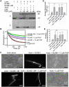
PI3P binds to SCAB1 specifically. A, SCAB1 binds to PI3P in a protein-lipid overlay assay. A hydrophobic membrane spotted with fifteen biologically active phospholipids at 100 pmol was incubated with 1 µg/mL purified SCAB1 and immunoblotted with anti-SCAB1 antibodies. B, The binding affinity of SCAB1 to PI3P. A membrane spotted with eight different phospholipids from 100 to 1.6 pmol was incubated with 1 µg/mL purified SCAB1 and immunoblotted with anti-SCAB1 antibodies. C, CHR domain of SCAB1 binds to PI3P. A membrane spotted with 100 pmol PI3P was incubated with 1 µg/mL purified NTD domain, CHR domain and CTD domain of SCAB1. D, Domain diagram of SCAB1 addressing the beginning and termination of SCAB1 NTD, CHR, and CTD domains. E, Mutation of predicted PI3P binding motifs RXLR-dEER at CHR domain. PI3P binding motifs are shown in blue and dEER in orange at the WT panels. Two predicted motifs (RRIK and KMLH) at positions 228–234 overlap. Dashes indicate identical residues at the mutant panels. F, Triple and quadruple mutants of the PI3P binding motifs of SCAB1 fail to bind to PI3P in a protein-lipid overlay assay. PIP strips were incubated with 1 µg/mL purified wild-type or mutated SCAB1, and immunoblotted with anti-SCAB1 antibodies. G, The binding affinity of SCAB1 to PI3P in MST assay. NT647-labeled GST-SCAB1 or GST-SCAB1-3M recombinant protein is titrated to a varying concentration of PI3P. Error bars represent sd (n = 3). H, Liposome sedimentation assay between the SCAB1 or SCAB1-3M protein and liposomes with PI3P. Liposomes were mixed by a weight ratio of 1:1 DOPC/DOPE and an indicated weight ratio of PI3P. SDS–PAGE and Coomassie Blue staining were performed to detect proteins.
Figure 2
Figure 2
PI3P inhibits SCAB1 to form oligomers in vitro and in vivo. A,The verification of PI3P inhibits the oligomerization of SCAB1 in vitro. Purified SCAB1 (5 mg/mL) was incubated with or without 20-μM PI3P, and then incubated with 50 μM H2O2. B, PI3P on the oligomerization of mutated SCAB1 (SCAB1-3M, SCAB1 110174228M) in vitro. Purified SCAB1-3M (5 mg/mL) or SCAB1 (5 mg/mL) was used. C, The verification of PI3P inhibits the oligomerization of SCAB1 in a concentration-dependent manner in vitro. D, Images of N. benthamiana leaves in split-luciferase complementation assays with varying concentrations of WM. DDM1 was used as a negative control. E, Quantitative analysis of luminescence intensity in (D). Error bars represent sd (n = 30). Statistical significance was determined by One-way ANOVA. The ****P-values compared to 0 μM WM treatment were <0.0001. F, Size exclusion chromatography analysis of Myc-SCAB1 in vivo. Total protein was extracted from 10-day-old Pro35S:Myc-SCAB1 seedlings with or without 10-μM WM treatment and examined by immunoblot with anti-Myc antibody. The fractions are in the collecting order. G, Quantitative analysis of the amount of SCAB1 in the fractions in (F). Error bars represent sd (n = 4). Statistical significance was determined by one-way ANOVA. The **P-value of fraction 4 was <0.01.
Figure 3
Figure 3
PI3P inhibits the F-actin-stabilizing and -bundling activity of SCAB1. A, High-speed cosedimentation assays of SCAB1 and SCAB1-3M with F-actin. Preassembled F-actin was incubated with SCAB1 or SCAB1-3M (1.5 μM) treated with varying concentrations of PI3P. B, Dilution-mediated F-actin depolymerization assay of SCAB1 or SCAB1-3M with or without PI3P. F-actin depolymerization was monitored by tracking the decrease in pyrene fluorescence. C, Epifluorescence images of actin bundles in the presence of SCAB1 or SCAB1-3M with or without PI3P. Actin filaments were visualized by rhodamine–phalloidin staining. Bars = 10 μm. D and E, Statistical analysis of the F-actin bundles in (C). The average filament/cable width (D) and bundling (E, skewness) of actin filamentous structures were measured in ImageJ. Error bars represent sd (n = 50). Statistical significance was determined by one-way ANOVA. The **P-values were <0.01 compared to actin alone.
Figure 4
Figure 4
PI3P inhibits SCAB1-mediated F-actin stabilization in guard cells. A, Confocal images of fABD2 marked F-actin in Col-0, scab1, and OE-SCAB1 guard cells with or without 200 nM LatA and/or 10 μM WM treatments. Bar = 10 μm. B, Statistical analysis of skewness in (A) (n = 50). Statistical significance was determined by one-way ANOVA. The **P-values of Col-0 + WM, scab1, scab1 + WM, and OE-SCAB1 compared to Col-0 <0.01. C, Statistical analysis of occupancy in (A) (n = 50). Statistical significance was determined by one-way ANOVA. The **P-values of Col-0 + WM, scab1, scab1 + WM, and OE-SCAB1 compared to Col-0 < 0.01. D, Confocal images of fABD2 marked F-actin in guard cells of Col-0, scab1, ProSCAB1:SCAB1 in scab1 (COM), and ProSCAB1:SCAB1-3M in scab1 (COM3M) with or without 200-nM LatA treatment. Bar = 10  μm. E, Statistical analysis of skewness in (D) (n = 50). Statistical significance was determined by one-way ANOVA. The **P-values of scab1, COM, and COM3M compared to Col-0 were 0.01. F, Statistical analysis of occupancy in (D) (n = 50). Statistical significance was determined by one-way ANOVA. The **P-values of scab1, COM, and COM3M compared to Col-0 were 0.01.
Figure 5
Figure 5
The F-actin reorganization induced by PI3P during stomatal closure requires SCAB1. A, Confocal images of F-actin in Col-0 guard cells during ABA-induced stomatal closure. F-actin was classified into three groups: Type 1 (radial array), Type 2 (random meshwork), and Type 3 (longitudinal array). Bar = 10 μm. B, Histograms of the reorganization of F-actin in guard cells of Col-0, scab1, scab1 with 10 μM WM, COM, and COM3M in scab1 during stomatal closure induced by 20 μM ABA (n = 50). The experiments were repeated three times and the values represent the average mean ± sem (standard error of mean). Statistical significance was determined by two-way ANOVA. In control, the ****P-values of Col-0 + WM, scab1, scab1 + WM, and COM3M compared to Col-0 were <0.0001 for Types 1 and 2, except for COM, P-values for Type 3 are insignificant. In ABA treatment, the ****P-values of Col-0 + WM, scab1, scab1 + WM, and COM3M compared to Col-0 were <0.0001 for Types 1, 2, and 3, except for COM. C, The corresponding stomatal apertures of Col-0, Col-0 + WM, scab1 with 10 μM WM, scab1, COM, and COM3M (B). Statistical significance was determined by two-way ANOVA. In ABA treatment, the P-values of Col-0 + WM, scab1, scab1 + WM, COM, and COM3M compared to Col-0 were 0.0079 (**), 0.0023 (**), 0.0032 (**), >0.9999, and 0.0032 (**), respectively. D, Real-time qPCR analysis of expression patterns of VPS15 and VPS34 with ABA, mannitol, and water-loss treatments. Seven-day-old seedlings were treated with 20 μM ABA for 3 h, 250 mM mannitol for 3 h, or water loss (seedlings transferred onto dry paper for 2 h) until lost 20% of their fresh weight. RD29A was used as a positive control. Error bars represent sd (n = 3). Statistical significance was determined by two-way ANOVA. For RD29A, the P-values compared to the control were ****<0.0001, ****<0.0001, and ***0.0389, respectively; for VPS15, the P-values compared to the control were ****<0.0001, ****<0.0001, and *0.0101, respectively; for VPS34, the P-values compared to the control were ***0.0004, **0.0013 and 0.4324 (difference is not significant), respectively. E, Size exclusion chromatography analysis of SCAB1 protein under drought stress. Total protein was extracted from 10-day-old seedlings with or without drought treatment (20% of fresh-weight loss), loaded onto a Superdex-200 10/300GL column, and examined by immunoblot with anti-Myc antibody. F, Quantitative analysis of the amount of SCAB1 in fractions of (E). Error bars represent SD (n = 3). Statistical significance was determined by Student’s t test. The p value of fragment 5 was 0.0002.
Figure 6
Figure 6
SCAB1 and PI3P are involved in vacuolar remodeling in guard cells during stomatal closure. A, Time-lapse images of guard cells co-expressing GFP-VAM3 and RFP-SCAB1. Arrows point to the locations at which the vacuolar membrane formed an intravascular structure with SCAB1. Bar = 10 μm. B, The number of vacuole remodeling events with SCAB1 in (A) (n = 17). C, Confocal images of a section of GFP-ΔTip marked the vacuole in Col-0 guard cells during stomatal closure induced by 20 μM ABA. Bar = 10 μm. D, The proportion of different types of vacuoles in guard cells during stomatal closure (n > 20). The experiments were repeated more than three times and the values represent the average mean ± SEM. Statistical significance was determined by two-way ANOVA. In the open group, the P-values of scab1, Col-0 + WM, scab1 + WM, and COM3M compared to Col-0 were 0.0004, <0.0001, <0.0001, and <0.0001 for Type 1, respectively; the P-values of scab1, Col-0 +  WM, scab1 + WM, and COM3M compared to Col-0 were 0.0325, 0.0260, 0.0206, and 0.0593 for Type 2, respectively; and the P values were insignificant for Type 3. In half-close group and close group, the P values of scab1, Col-0 + WM, scab1 + WM, and COM3M compared to Col-0 were insignificant for Types 1, 2, and 3.

Comment in

  • Back to the roots: A focus on plant cell biology.
    Weijers D, Bezanilla M, Jiang L, Roeder AHK, Williams M. Weijers D, et al. Plant Cell. 2022 Jan 20;34(1):1-3. doi: 10.1093/plcell/koab278. Plant Cell. 2022. PMID: 34755878 Free PMC article. No abstract available.

References

    1. Allwood EG, Anthony RG, Smertenko AP, Reichelt S, Drobak BK, Doonan JH, Weeds AG, Hussey PJ (2002) Regulation of the pollen-specific actin-depolymerizing factor LlADF1. Plant Cell 14: 2915–2927 - PMC - PubMed
    1. An CI, Sawada A, Kawaguchi Y, Fukusaki E, Kobayashi A (2005) Transient RNAi induction against endogenous genes in Arabidopsis protoplasts using in vitro-prepared double-stranded RNA. Biosci Biotechnol Biochem 69: 415–418 - PubMed
    1. Assmann SM, Jegla T (2016) Guard cell sensory systems: recent insights on stomatal responses to light, abscisic acid, and CO2. Curr Opin Plant Biol 33: 157–167 - PubMed
    1. Avila EL, Zouhar J, Agee AE, Carter DG, Chary SN, Raikhel NV (2003) Tools to study plant organelle biogenesis. Point mutation lines with disrupted vacuoles and high-speed confocal screening of green fluorescent protein-tagged organelles. Plant Physiol 133: 1673–1676 - PMC - PubMed
    1. Bak G, Lee EJ, Lee Y, Kato M, Segami S, Sze H, Maeshima M, Hwang JU, Lee Y (2013) Rapid structural changes and acidification of guard cell vacuoles during stomatal closure require phosphatidylinositol 3,5-bisphosphate. Plant Cell 25: 2202–2216 - PMC - PubMed

Publication types

MeSH terms

LinkOut - more resources