. 2022 Mar;16(6):1272-1289.
doi: 10.1002/1878-0261.13145. Epub 2021 Dec 18.

FGF7-FGFR2 autocrine signaling increases growth and chemoresistance of fusion-positive rhabdomyosarcomas

Affiliations

FGF7-FGFR2 autocrine signaling increases growth and chemoresistance of fusion-positive rhabdomyosarcomas

Christopher I Milton et al. Mol Oncol. 2022 Mar.

Abstract

Rhabdomyosarcomas are aggressive pediatric soft-tissue sarcomas and include high-risk PAX3-FOXO1 fusion-gene-positive cases. Fibroblast growth factor receptor 4 (FGFR4) is known to contribute to rhabdomyosarcoma progression; here, we sought to investigate the involvement and potential for therapeutic targeting of other FGFRs in this disease. Cell-based screening of FGFR inhibitors with potential for clinical repurposing (NVP-BGJ398, nintedanib, dovitinib, and ponatinib) revealed greater sensitivity of fusion-gene-positive versus fusion-gene-negative rhabdomyosarcoma cell lines and was shown to be correlated with high expression of FGFR2 and its specific ligand, FGF7. Furthermore, patient samples exhibit higher mRNA levels of FGFR2 and FGF7 in fusion-gene-positive versus fusion-gene-negative rhabdomyosarcomas. Sustained intracellular mitogen-activated protein kinase (MAPK) activity and FGF7 secretion into culture media during serum starvation of PAX3-FOXO1 rhabdomyosarcoma cells together with decreased cell viability after genetic silencing of FGFR2 or FGF7 was in keeping with a novel FGF7-FGFR2 autocrine loop. FGFR inhibition with NVP-BGJ398 reduced viability and was synergistic with SN38, the active metabolite of irinotecan. In vivo, NVP-BGJ398 abrogated xenograft growth and warrants further investigation in combination with irinotecan as a therapeutic strategy for fusion-gene-positive rhabdomyosarcomas.

Keywords: FGF7; FGFR2; NVP-BGJ398; autocrine loop; fibroblast growth factor receptor; rhabdomyosarcoma.


Conflict of interest statement

The authors declare no conflict of interest.

Figures

Fig. 1
Fig. 1
Sensitivity of PAX3‐FOXO1 fusion‐positive cells to NVP‐BGJ398 correlates with FGFR2 and FGF7 expression. (A) 2D Rhabdomyosarcoma (RMS) cell line Growth Inhibition at 50% (GI50) values as measured by Methyl Tetrazolium Salt (MTS) after 72 h exposure to NVP‐BGJ398. (B) Scatter plot of FGFR2 (plum) and FGF7 (teal) mRNA expression against the log2 GI50 to NVP‐BGJ398 in RMS cell lines. Each point represents a separate cell line. Spearman r correlation and associated P values with line of best fit (solid) are shown. (C) Representative western blot of FGFR2 protein in fusion‐positive (FP) and fusion‐negative (FN) RMS cells cultured in 10% Fetal Bovine Serum (FBS). (D) RMS cells were incubated with 10% or 0% FBS (serum starved) for 24 h before culture media was collected and subjected to FGF7 enzyme‐linked immunosorbent assay (ELISA). Results are representative of three independent experiments with error bars representing standard deviation, except for (C), which was repeated twice. Significance of differences were assessed using unpaired t‐tests.*P < 0.05, **P < 0.01, ***P < 0.005, ****P < 0.001.
Fig. 2
Fig. 2
FGF7 mRNA and FGFR2 mRNA and protein is highly expressed in FP‐RMS patients. FGFR2 (A) and FGF7 (B) mRNA expression in Rhabdomyosarcoma (RMS) patients from the Innovative Therapies for Children with Cancer/Carte d’Identite’ des Tumeurs (ITCC/CIT) cohort. Sk. Muscle = skeletal muscle, ERMS = embryonal RMS, ARMS = alveolar RMS, NEG = negative, P3F = PAX3‐FOXO1 and P7F = PAX7‐FOXO1. Wilcoxon rank sum test, for P3F FGFR2 is P < 0.001 and FGF7 is P < 0.01 compared to fusion‐negative (FN‐RMS) samples. (n = 101 tumor and 36 normal samples) (C) Representative images of FGFR2 protein expression in a subset of RMS patients on a Tissue Microarray (TMA) by Immunohistochemistry (IHC). Magnification is 10× and 40× with scale bars representing 250 and 50 µm respectively. (n = 12 fusion‐positive and 8 fusion‐negative samples). (D) Quantification of the percentage of FGFR2 positive cells from the TMA IHC was as follows < 10% = 0, 10–25% = 1, > 25% < 50% = 2, ≥ 50% = 3 with scores compared between fusion‐positive (FP‐RMS; blue) and fusion‐negative (FN‐RMS; red) patients using a Mann‐Whitney U test. *P < 0.05; (n = 12 FP‐RMS and 8 FN‐RMS samples).
Fig. 3
Fig. 3
Molecular effects of NVP‐BGJ398 in RMS cell lines in vitro. (A) Representative blot assessing tyrosine phosphorylation on FGFR2 (arrow indicates 120 kDa). RMS01 cells were exposed to 3 h vehicle or drug before 20 min of 25 ng·mL−1 FGF7 and subsequent immunoprecipitation (IP) with a FGFR2 or IgG (Isotype control) antibody. The supernatant of remaining proteins after IP were retained and run as a control to demonstrate depletion in FGFR2 IP lanes compared to IgG control with GAPDH demonstrating equal loading. (B) Representative blot assessing protein phosphorylation in RMS01 cells cultured for 16 h in 10% Fetal Bovine Serum (FBS) or 0% FBS (serum starved) before exposure to vehicle or drug for 3 h and as indicated 25 ng·mL−1 FGF7 for the last 20 min. Quantitation of Fibroblast Growth Factor Receptor Substrate 2α (FRS2α) phosphorylation as measured by electrochemiluminescent assay in RMS01 (C) and RH41 (D) cells subject to the same treatment as in B. Results are representative of two independent experiments, except C and D, which were repeated three times. Error bars represent standard deviation with significance of differences determined by One‐Way ANOVA with Dunnett’s multiple testing correction (n.s. = not significant, **P < 0.01, ***P < 0.005, ****P < 0.001).
Fig. 4
Fig. 4
FGFR2 and FGF7 maintain FP‐RMS cell viability. Effect of 144 h short interfering RNA (siRNA) mediated FGFR2 knockdown on (A) fusion‐positive RMS (FP‐RMS) and (B) fusion‐negative RMS (FN‐RMS) cell viability. (C) Representative western blot of 72 h FGFR2 knockdown in FP‐RMS cells. Effect of 144 h siRNA mediated FGF7 knockdown on (D) FP‐RMS and (E) FN‐RMS cell viability. (F) FGF7 enzyme‐linked immunosorbent assay (ELISA) on media from FP‐RMS lines subjected to FGF7 knockdown. UTC = Untransfected control, NTC = Nontargeting control, Death = positive control siRNA that causes cell death. Results are representative of three independent experiments with error bars representing standard deviation. Significance of difference between siRNAs and NTC were tested by One‐way ANOVA with Dunnett’s multiple testing correction (n.s. = not significant, *P < 0.05, ***P < 0.005, ****P < 0.001).
Fig. 5
Fig. 5
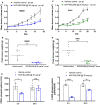
NVP‐BGJ398 inhibits the growth of FP‐RMS xenografts in vivo. Mean volume (A) and weight (B) of RMS01 tumor xenografts after 15 days exposure to vehicle or 30 mg·kg−1 NVP‐BGJ398 quaque die (q.d.). Error bars represent standard error of the mean for control (n = 10) and treated (n = 10) groups. Mean volume (C) and weight (D) of RH41 tumor xenografts after treatment with vehicle or 25 mg·kg−1 NVP‐BGJ398 q.d. for 20 days. Error bars represent standard error of the mean for control (n = 8) and treated (n = 10) groups. (E) Phosphorylation of Fibroblast Growth Factor Receptor Substrate 2α (FRS2α) and the ratio of phospho/total ERK1/2 in RMS01 xenografts at 3 and 24 h post dosing with 30 mg·kg−1 NVP‐BGJ398. Measurements were by electrochemiluminescent assay with error bars representing standard deviation of the mean for control (n = 3) and treated (n = 3) groups. Unpaired t‐tests with Welch’s correction were used to assess significance of differences. (n.s. = not significant, *P < 0.05, **P < 0.01, ****P < 0.001).
Fig. 6
Fig. 6
Combining NVP‐BGJ398 with SN38 is efficacious against FP‐RMS cells in vitro. (A) Representative images of wells from clonogenic assays in which fusion‐positive rhabdomyosarcoma (FP‐RMS) cell lines RMS01 and RH41 were exposed to the indicated drugs for 5 days before 7 days growth in media alone. Quantification of RMS01 (B) and RH41 (C) cell growth from clonogenic assays as described above. Results are representative of four independent experiments with significance of differences measured by One‐way ANOVA with Dunnett’s multiple testing correction. ****P < 0.001, Bliss scores, > 0 ≤ 1 = synergistic. (D) Representative western blots assessing protein levels in RMS01 cells in vitro after exposure to the indicated drugs for 8 or 120 h. Results are representative of two independent experiments. Cell viability of RMS01 (E) and RH41 (F) cells after 144 h FGFR2 or FGF7 knockdown in combination with either DMSO vehicle (gray) or SN38 (blue/green), relative to nontargeting control (NTC). Results are representative of three independent experiments with error bars representing standard deviation from the mean. Significance of differences were assessed using unpaired t‐tests with Welch’s correction. ***P < 0.005, ****P < 0.001.

References

    1. Zhang X, Ibrahimi OA, Olsen SK, Umemori H, Mohammadi M, Ornitz DM. Receptor specificity of the fibroblast growth factor family. The complete mammalian FGF family. J Biol Chem. 2006;23:15694–700. - PMC - PubMed
    1. Turner N, Grose R. Fibroblast growth factor signalling: from development to cancer. Nat Rev Cancer. 2010;2:116–29. - PubMed
    1. Itoh N, Ornitz DM. Fibroblast growth factors: from molecular evolution to roles in development, metabolism and disease. J Biochem. 2011;2:121–30. - PMC - PubMed
    1. Oladipupo SS, Smith C, Santeford A, Park C, Sene A, Wiley LA, et al. Endothelial cell FGF signaling is required for injury response but not for vascular homeostasis. Proc Natl Acad Sci USA. 2014;111:13379–84. - PMC - PubMed
    1. Dell'Era P, Ronca R, Coco L, Nicoli S, Metra M, Presta M. Fibroblast growth factor receptor‐1 is essential for in vitro cardiomyocyte development. Circ Res. 2003;5:414–20. - PubMed

Publication types