. 2021 Dec 1;131(23):e127171.
doi: 10.1172/JCI127171.

MIF but not MIF-2 recruits inflammatory macrophages in an experimental polymicrobial sepsis model

Affiliations

MIF but not MIF-2 recruits inflammatory macrophages in an experimental polymicrobial sepsis model

Pathricia Veronica Tilstam et al. J Clin Invest. .

Abstract

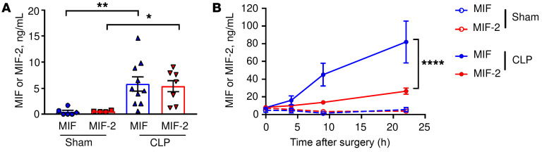
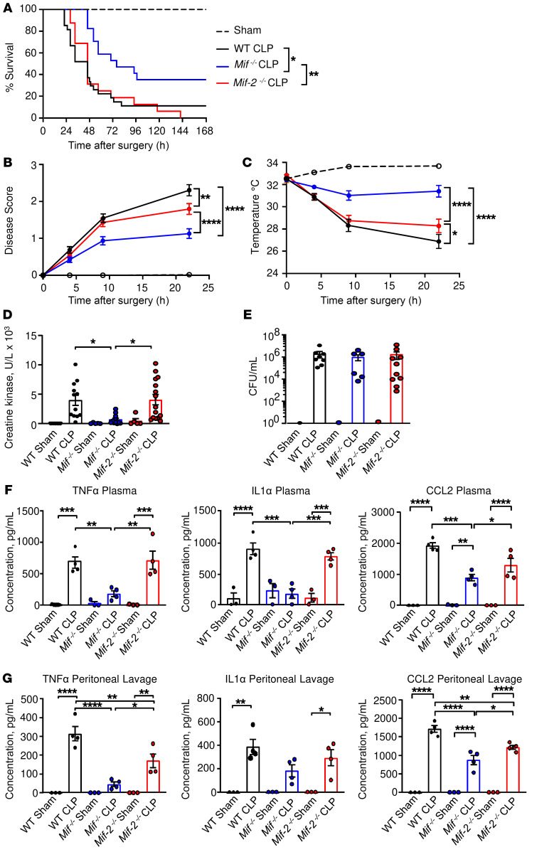
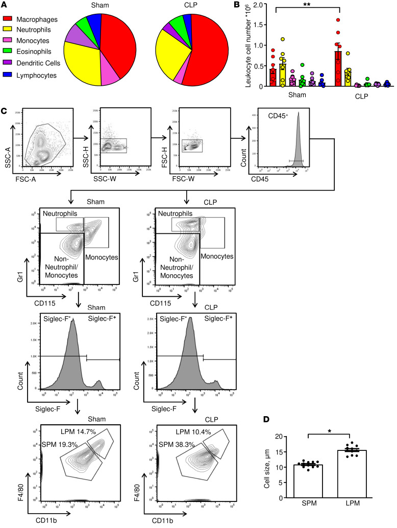
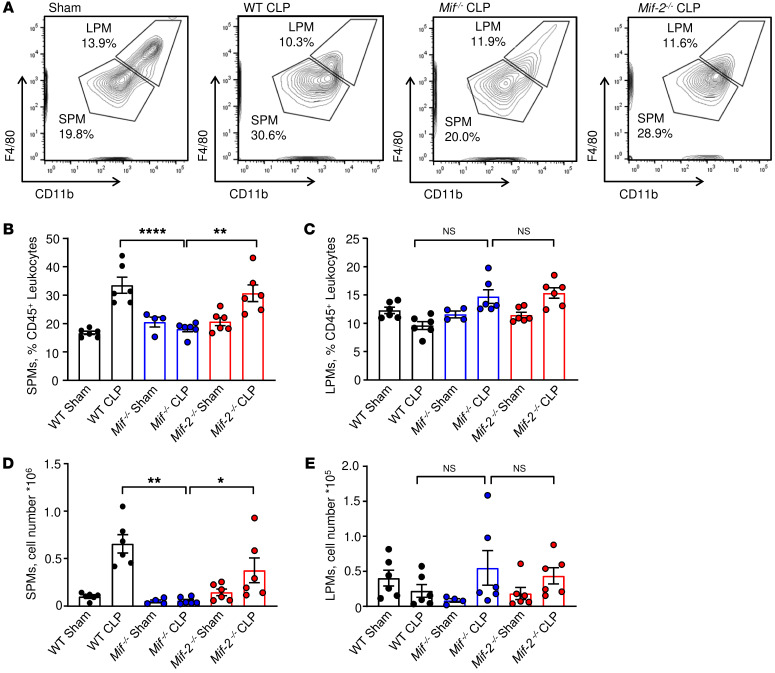
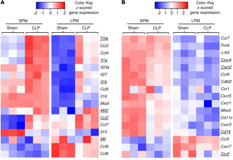
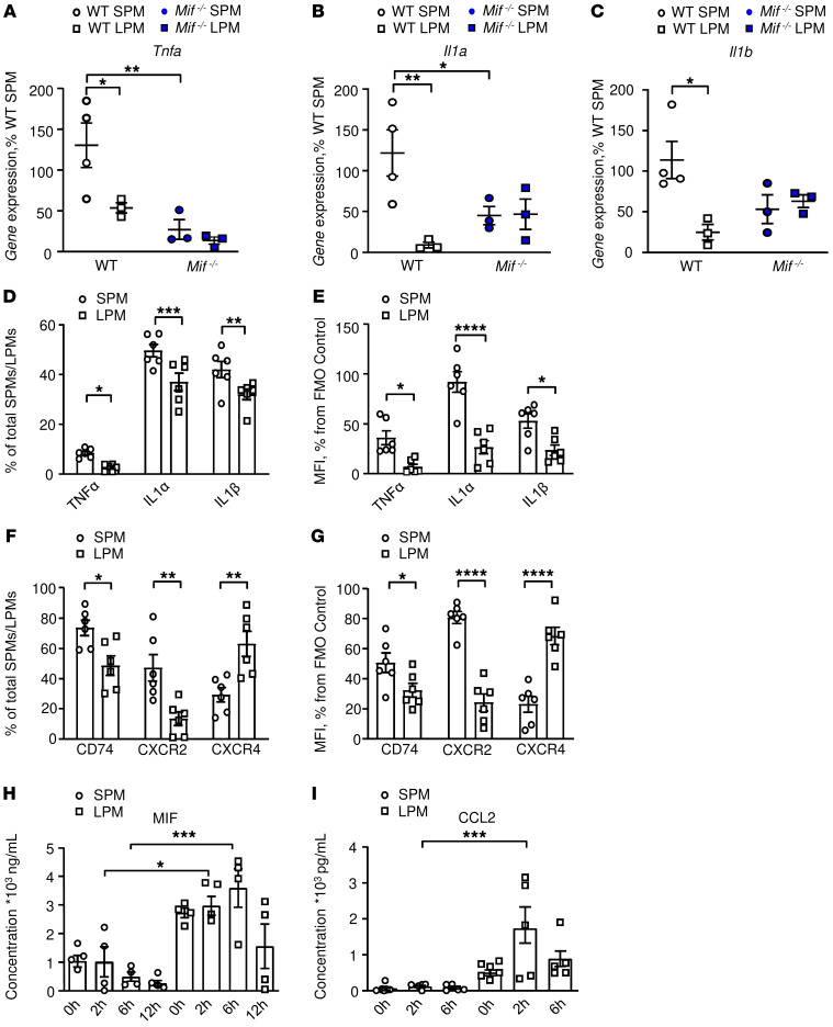
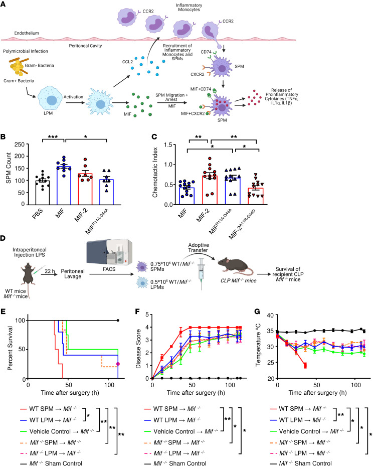
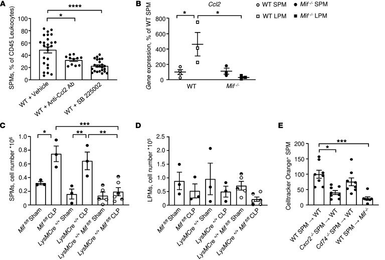
Excessive inflammation drives the progression from sepsis to septic shock. Macrophage migration inhibitory factor (MIF) is of interest because MIF promoter polymorphisms predict mortality in different infections, and anti-MIF antibody improves survival in experimental models when administered 8 hours after infectious insult. The recent description of a second MIF superfamily member, D-dopachrome tautomerase (D-DT/MIF-2), prompted closer investigation of MIF-dependent responses. We subjected Mif-/- and Mif-2-/- mice to polymicrobial sepsis and observed a survival benefit with Mif but not Mif-2 deficiency. Survival was associated with reduced numbers of small peritoneal macrophages (SPMs) that, in contrast to large peritoneal macrophages (LPMs), were recruited into the peritoneal cavity. LPMs produced higher quantities of MIF than SPMs, but SPMs expressed higher levels of inflammatory cytokines and the MIF receptors CD74 and CXCR2. Adoptive transfer of WT SPMs into Mif-/- hosts reduced the protective effect of Mif deficiency in polymicrobial sepsis. Notably, MIF-2 lacks the pseudo-(E)LR motif present in MIF that mediates CXCR2 engagement and SPM migration, supporting a specific role for MIF in the recruitment and accumulation of inflammatory SPMs.

Keywords: Cytokines; Immunology; Inflammation; Macrophages; Mouse models.


Figures

Figure 1
Figure 1. MIF and MIF-2 levels increase in sepsis.
WT mice underwent CLP or sham surgery and (A) peritoneal lavage at 22 hours and (B) peripheral blood at 0, 4, 9, and 22 hours were analyzed for MIF and MIF-2 by ELISA. (A) n = 4 to 10 animals, 1-way ANOVA with Tukey’s multiple-comparison test. (B) n = 6 to 22 animals, 2-way ANOVA with Bonferroni’s multiple-comparison test. *P < 0.05; **P < 0.01; ****P < 0.0001.
Figure 2
Figure 2. Delayed onset of septic shock in Mif–/– but not Mif-2–/– mice.
(A) WT, Mif–/–, or Mif-2–/– mice were subjected to CLP (n = 16–19 animals) or sham surgery (n = 13 animals), and survival was monitored for at least 7 days. Log-rank (Mantel-Cox) test was performed to determine significance. (B) Disease score (see Methods) and (C) surface body temperature were recorded in the immediate postoperative period. Two-way ANOVA with Bonferroni’s multiple-comparison test. (D) Creatine kinase (CK) levels and (E) bacterial CFUs determined at 22 hours after CLP or sham surgery in plasma or heparinized whole blood, respectively. Data points are from 5 or more independent experiments and reflect n = 14 to 15 animals per group. For CFUs, each symbol represents 1 animal, n = 6–17 (Mann-Whitney test). (F and G) TNF-α, IL-1α, and CCL2 plasma (F) and peritoneal fluid (G) concentrations in WT, Mif–/–, and Mif-2–/– mice. Cytokine/chemokine levels were measured by flow cytometry using the LEGENDplex Multi Analyte Flow Assay Kit. n = 3 to 4 mice per group, 1-way ANOVA with Tukey’s multiple-comparison test. *P < 0.05; **P < 0.01; ***P < 0.001; ****P < 0.0001.
Figure 3
Figure 3. Macrophages are the predominant cell type in CLP and can be classified into SPM and LPM subpopulations.
(A) Peritoneal cells were stained for flow cytometry and relative numbers of CD11b+F4/80+ macrophages, CD115Gr1hi neutrophils, CD115+Gr1hi monocytes, Siglec F+ eosinophils, CD11c+ DCs, and CD3+/CD19+ lymphocytes; (A) was determined as percentage of CD45+ cells and (B) measured as absolute cell number. Plot of n = 7 mice from 2 separate experiments. Two-way ANOVA with Sidak’s multiple-comparison test. (C) Representative gating strategy for the identification of small (SPM) and large (LPM) peritoneal macrophages in the peritoneal lavage of WT mice 22 hours after CLP or sham surgery. (D) Cytospins of SPMs and LPMs isolated from peritoneal lavage of septic mice and stained with Wright-Giemsa stain. Macrophage size was determined by ImageJ software from n = 11 animals. SPMs measured 10.9 ± 0.3 μm; the size of LPMs was 15.6 ± 0.5 μm (unpaired, 2-tailed Student’s t test). *P < 0.05; **P < 0.01.
Figure 4
Figure 4. MIF deficiency is associated with reduced i.p. SPM numbers.
(A) Representative gating strategy for SPM and LPM analysis in WT, Mif–/–, and Mif-2–/– mice after CLP or sham surgery. Relative cell numbers and absolute cell count analyses of SPMs (B and D) and LPMs (C and E) in the peritoneal lavage of WT, Mif–/–, and Mif-2–/– mice after CLP or sham surgery. Flow cytometric analyses were performed on peritoneal cells collected 22 hours after CLP or sham surgery and included CountBright absolute counting beads in n = 4 to 6 animals/genotype. *P < 0.05; **P < 0.01; ****P < 0.0001 by 1-way ANOVA with Tukey’s multiple-comparison test (B) or Kruskal-Wallis with Dunn’s multiple-comparison test (CE).
Figure 5
Figure 5. SPMs exhibit a more proinflammatory phenotype than LPMs.
Visualization of differentially expressed genes in SPM-sham, LPM-sham, SPM-CLP, and LPM-CLP isolated from WT mice 22 hours after surgery showing differentially expressed cytokines and chemokines (A) and receptors (B) as a heatmap with z-scored FPKM values. Underlined genes are discussed in Results.
Figure 6
Figure 6. LPMs express higher levels of MIF and CCL2 than SPMs.
(AC) Confirmation of MIF-dependent regulation of Tnfa, Il1a, and Il1b expression by SPMs in contrast to LPMs. Quantitative expression by qPCR of (A) Tnfa, (B) Il1a, and (C) Il1b in SPMs and LPMs isolated from WT and Mif–/– animals 22 hours after CLP. n = 4 animals per group with replication (2-way ANOVA with Sidak’s multiple-comparison test). (DG) We confirmed cellular protein expression in SPMs and LPMs by flow cytometry. Intracellular cytokine expression assessed by flow cytometry in post-CLP SPMs and LPMs showing (D) cytokine-expressing macrophages as percentage of total SPMs/LPMs and (E) MFI as percentage of fluorescence-minus-one (FMO) control. (F and G) Flow cytometry analysis of the MIF receptors CD74, CXCR2, and CXCR4 expressed by SPMs and LPMs after CLP, (F) expressed as percentage of total SPMs/LPMs, and as (G) MFI percentage of FMO control. Results are from 3 independent experiments, n = 6 animals per group, 2-way ANOVA with Sidak’s multiple-comparison test. (H and I) In vitro confirmation of induced production of MIF and CCL2 by LPMs versus SPMs. (H) MIF and (I) CCL2 production from WT SPMs and LPMs stimulated with LPS (100 ng/mL, 1 × 10⁵ cells/well). Cytokine concentrations in supernatants were measured by ELISA. The results are from 3 independent experiments, and data are expressed as mean ± SEM, n = 5 to 11 samples per time point. Significance determined by 1-way ANOVA with Tukey’s multiple-comparison test (H) or Kruskal-Wallis test with Dunn’s multiple-comparison test (I). *P < 0.05; **P < 0.01; ***P < 0.001; ****P < 0.0001.
Figure 7
Figure 7. Adoptive transfer of WT SPMs induces premature mortality in Mif–/– CLP mice.
(A) Hypothesis for the role of LPM-expressed MIF and CCL2 in the recruitment and retention of SPMs into peritonea followed by SPM cytokine production. (B) SPM migration into LPS-conditioned peritonea of WT mice measured 12 hours after the i.p. injection of 1 mg/kg recombinant MIF, MIF-2, or MIF lacking the pseudo-(E)LR motif (MIFR11A–D44A). Results are normalized against control group. Data from 3 independent experiments, n = 6 to 12 mice per group (1-way ANOVA with Tukey’s multiple-comparison test). (C) Directed migration in response to CCL2 of SPMs stimulated with recombinant MIF, MIF-2, MIFR11A–D44A, or MIF-2 with recombinant amino acid insertion of the pseudo-(E)LR motif (MIF-2A11R–G44D). Shown is the relative reduction in the CCL2-triggered chemotactic index. Data from 3 independent experiments, n = 12 samples per group (1-way ANOVA with Tukey’s multiple-comparison test). (D) Experimental scheme for the adoptive transfer of WT SPMs/LPMs and Mif–/– SPMs/LPMs into Mif–/– recipient mice. SPMs/LPMs were isolated by FACS from WT and Mif–/– donor mice, 0.75 × 10⁶ SPMs/mouse and 0.5 × 10⁵ LPMs/mouse transferred by i.p. injection into Mif–/– mice after CLP surgery. (E) Kaplan-Meier survival plots; (F) disease score; (G) surface body temperature of Mif–/– CLP mice after adoptive transfer of WT SPM (red), WT LPM (blue), vehicle control (green), Mif–/– SPM (dotted orange), and Mif–/– LPM (dotted magenta). Sham surgery control (black). n = 4 to 6 mice per group from 4 independent experiments. P values determined by log-rank (Mantel-Cox) test (E), Kruskal-Wallis test with Dunn’s multiple-comparison test (F), or 1-way ANOVA with Tukey’s multiple-comparison test (G). Significance for F and G was determined at 36 hours after injection. Images (A and D) created with Biorender.com. *P < 0.05; **P < 0.01; ***P < 0.001.
Figure 8
Figure 8. CCL2 and MIF regulation of LPMs and SPMs during i.p. sepsis.
(A) Impact of CCL2 antagonism (R&D Systems, AF-479-NA, 25 μg/kg, i.p.) and CXCR2 antagonism (SB225002, 10 mg/kg, i.p.) on SPM migration into LPS-conditioned peritonea (LPS 12.5 mg/kg, i.p., 2 hours previously). Data are from n = 11 to 19 animals (1-way ANOVA with Tukey’s multiple-comparison test). (B) Quantitative expression of Ccl2 by SPMs and LPMs from WT and Mif–/– mice 22 hours after CLP induction (n = 4 mice per group, 2-way ANOVA with Sidak’s multiple-comparison test). (C and D) Myeloid-derived MIF mediates i.p. SPM accumulation. Absolute cell count analyses of SPMs (C) and LPMs (D) in the peritoneal lavage of LysM-Cre+/+ Miffl/fl, LysM-Cre+/+, or Miffl/fl mice after sham or CLP surgery. LysM-Cre+/– Miffl/fl (visualized in the figure as half-filled circles) demonstrated similar SPM and LPM cell numbers as homozygous LysM-Cre+/+ Miffl/fl mice and were combined as 1 group. Flow cytometric analyses were performed on peritoneal cells collected 22 hours after CLP or sham surgery and included CountBright absolute counting beads in n = 3 animals/genotype. One-way ANOVA with Tukey’s multiple-comparison test. (E) Impact of Cxcr2 or Cd74 deficiency on SPM migration into LPS-conditioned peritonea. SPMs were isolated from WT, Cxcr2–/–, or Cd74–/– mice (after LPS injection, 12.5 mg/kg); labeled with CellTracker Orange and 1 × 10⁶ cells transferred (i.v.) into WT mice; and followed 2 hours later by LPS challenge (12.5 mg/kg, i.p.). Labeled CellTracker Orange+ SPMs were measured in the peritoneal lavage by flow cytometry 18 hours later (Mif–/– recipients also shown as controls). Data are from n = 4 to 8 animals per group and representative of 2 replicate experiments, 1-way ANOVA with Tukey’s multiple-comparison test. *P < 0.05; **P < 0.01; ***P < 0.001; ****P < 0.0001.

References

    1. Vincent JL, et al. Assessment of the worldwide burden of critical illness: the intensive care over nations (ICON) audit. Lancet Respir Med. 2014;2(5):380–386. doi: 10.1016/S2213-2600(14)70061-X. - DOI - PubMed
    1. Hotchkiss RS, et al. Sepsis-induced immunosuppression: from cellular dysfunctions to immunotherapy. Nat Rev Immunol. 2013;13(12):862–874. doi: 10.1038/nri3552. - DOI - PMC - PubMed
    1. Volbeda M, et al. Glucocorticosteroids for sepsis: systematic review with meta-analysis and trial sequential analysis. Intensive Care Med. 2015;41(7):1220–1234. doi: 10.1007/s00134-015-3899-6. - DOI - PMC - PubMed
    1. Brown KA, et al. Targeting cytokines as a treatment for patients with sepsis: a lost cause or a strategy still worthy of pursuit? Int Immunopharmacol. 2016;36:291–299. doi: 10.1016/j.intimp.2016.04.041. - DOI - PubMed
    1. Marti-Carvajal AJ, et al. Human recombinant protein C for severe sepsis and septic shock in adult and paediatric patients. Cochrane Database Syst Rev. 2012;12:CD004388. - PMC - PubMed

Publication types

MeSH terms