. 2022 Feb 9;96(3):e0110321.
doi: 10.1128/JVI.01103-21. Epub 2021 Dec 1.

ATF6-Mediated Unfolded Protein Response Facilitates Adeno-associated Virus 2 (AAV2) Transduction by Releasing the Suppression of the AAV Receptor on Endoplasmic Reticulum Stress

Affiliations

ATF6-Mediated Unfolded Protein Response Facilitates Adeno-associated Virus 2 (AAV2) Transduction by Releasing the Suppression of the AAV Receptor on Endoplasmic Reticulum Stress

Mengtian Cui et al. J Virol. .

Abstract

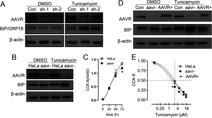
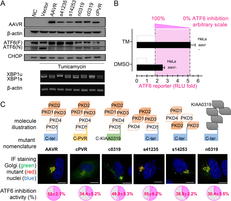
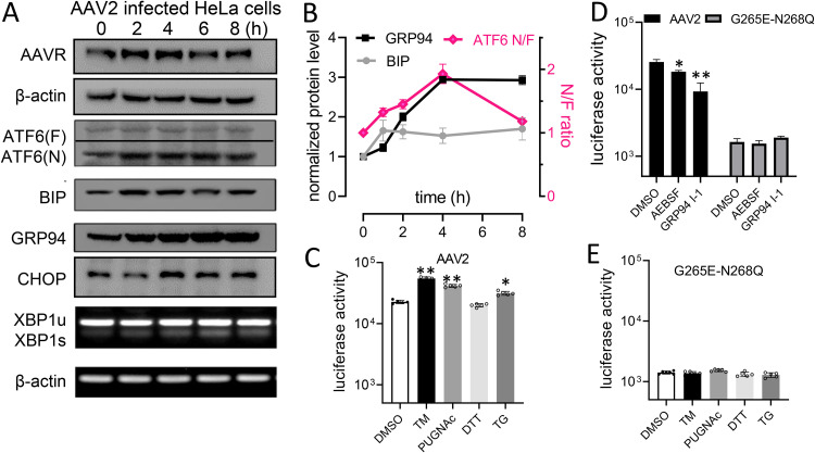
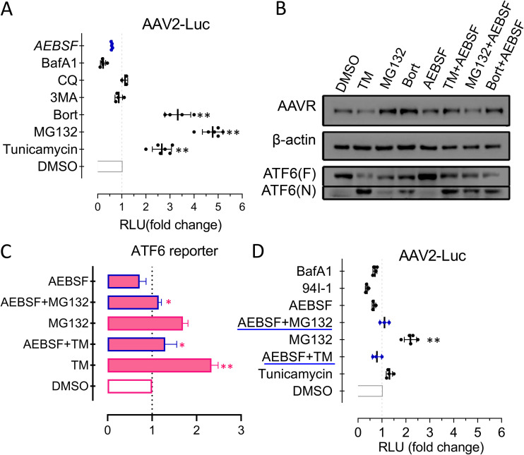
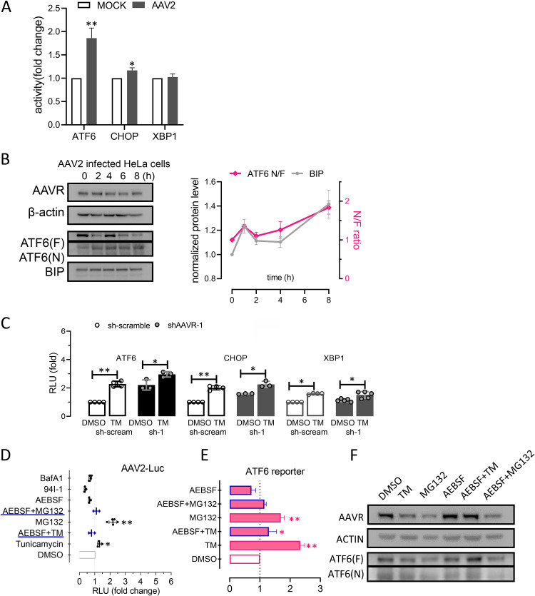
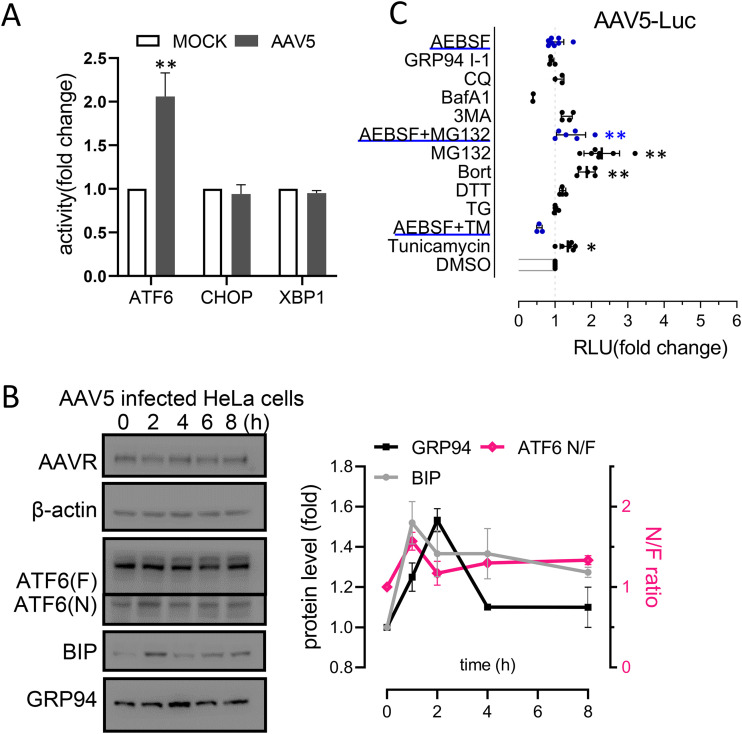
Adeno-associated virus (AAV) is extensively used as a viral vector to deliver therapeutic genes during human gene therapy. A high-affinity cellular receptor (AAVR) for most serotypes was recently identified; however, its biological function as a gene product remains unclear. In this study, we used AAVR knockdown cell models to show that AAVR depletion significantly attenuated cells to activate unfolded protein response (UPR) pathways when exposed to the endoplasmic reticulum (ER) stress inducer, tunicamycin. By analyzing three major UPR pathways, we found that ATF6 signaling was most affected in an AAVR-dependent fashion, distinct from CHOP and XBP1 branches. AAVR capacity in UPR regulation required the full native AAVR protein, and AAV2 capsid binding to the receptor altered ATF6 dynamics. Conversely, the transduction efficiency of AAV2 was associated with changes in ATF6 signaling in host cells following treatment with different small molecules. Thus, AAVR served as an inhibitory molecule to repress UPR responses via a specificity for ATF6 signaling, and the AAV2 infection route involved the release from AAVR-mediated ATF6 repression, thereby facilitating viral intracellular trafficking and transduction. IMPORTANCE The native function of the AAVR as an ER-Golgi localized protein is largely unknown. We showed that AAVR acted as a functional molecule to regulate UPR signaling under induced ER stress. AAVR inhibited the activation of the transcription factor, ATF6, whereas receptor binding to AAV2 released the suppression effects. This finding has expanded our understanding of AAV infection biology in terms of the physiological properties of AAVR in host cells. Importantly, our research provides a possible strategy which may improve the efficiency of AAV-mediated gene delivery during gene therapy.

Keywords: AAV receptor; AAV transduction; ATF6 signal; UPR.


Conflict of interest statement

The authors declare no conflict of interest.

Figures

FIG 1
FIG 1
AAVR depletion enhances tunicamycin (TM)-induced endoplasmic reticulum (ER) stress responses in HeLa cells. (A) HeLa cells were transfected with short hairpin RNAs (shRNAs) sh-1 and sh-2 targeting AAVR for 24 h. Western blotting for BIP/GRP78 was performed with or without 2.5 μmol/L TM for 6 h. (B) An AAVR knockdown cell line (aavr–) was prepared from shRNA lentivirus infection and compared with parental HeLa cells on TM treatment in BIP expression. (C) A CCK-8 time course assay comparing aavr– and HeLa cell growth. (D) Transfection of an AAVR-expressing plasmid into aavr– cells (AAVR+) and Western blotting for BIP with or without TM. (E) 48-h dose response curves of TM treatments by CCK-8 assay. Data are expressed as the mean ± standard deviation (n = 3). Statistical significance was determined using Student’s t test. *, P < 0.05; **, P < 0.01.
FIG 2
FIG 2
AAVR knockdown enhances ATF6 signaling in tunicamycin (TM)-induced stress on the unfolded protein response (UPR) in HeLa cells. (A) Western blots for ATF6 activation on both expression of full-length ATF(F) and cleaved ATF6(N) induced by TM for 6 h in aavr– and parental HeLa cells. Levels of BIP and GRP94 upstream and downstream of ATF6 signaling were detected. (B) CHOP expression in aav– and HeLa cells with or without TM stress. (C) XBP1s and XBP1u mRNA levels during IRE1 pathway activation by semiquantitative PCR. (D) Luciferase reporter assays showing UPR pathway activation. HeLa and aavr– cells were transfected with 0.2 μg firefly luciferase reporter and 0.04 μg pTK-Renilla plasmid for 24 h with/without 2.5 μM TM for 6 h. Data are presented as the mean ± standard deviation (n ≥ 3). Statistical significance was determined using the unpaired Student’s t test. *, P < 0.05; **, P < 0.01.
FIG 3
FIG 3
The effect of AAVR and mutants on inhibition of ATF6-mediated unfolded protein response (UPR) following tunicamycin stress. (A) Transfection of various AAVR mutants into aavr– cells and UPR assessment via ATF6, CHOP, and XBP1 activation (Western blots). (B) Illustration of defining a quantitative measurement on AAVR inhibition of ATF6 signaling by luciferase assays. (C) Intracellular localization of transfected AAVR mutants and their impact on inhibiting ATF6 activation. Scale bar = 10 μm. Data are presented as the mean ± standard deviation (n ≥ 3). Statistical significance was determined using the unpaired Student’s t test. *, P < 0.05; **, P < 0.01.
FIG 4
FIG 4
AAV2 infection alters ATF6-mediated unfolded protein response (UPR) signaling and activates transduction in HeLa cells. (A) Western blots showing ATF6, CHOP, and XBP1 expression during AAV2 infection time courses. (B) Quantification of data from panel A by densitometry (n = 3). (C) Comparison of AAV2 with a nonreceptor binding mutant, G265D-N268Q, for transduction in HeLa cells following treatments of various reagents of UPR modulation. (D) Comparison of infection of AAV2 and the G265D-N268Q mutant following inhibition of ATF6 or downstream GRP94. Data are presented as the mean ± standard deviation (n ≥ 3). Statistical significance was determined using the unpaired Student’s t test. *, P < 0.05; **, P < 0.01.
FIG 5
FIG 5
The effects of ATF6-mediated unfolded protein response following treatment with different reagents to modulate AAV2 transduction. (A) AAV2 infection of a luciferase reporter in HeLa cells treated with reagents to assess AAV transduction. (B) Western blots showing ATF6 activation comparing AEBSF treatments (with/without) to inhibit ATF6 cleavage. (C) Reporter assays for ATF6 activation. (D) AAV2 transduction efficiency in HeLa cells. Data are presented as the mean ± standard deviation (n ≥ 3). Statistical significance was determined using the unpaired Student’s t test. *, P < 0.05; **, P < 0.01.
FIG 6
FIG 6
Activation of ATF6-mediated unfolded protein response and AAV2 transduction in monkey primary hepatocytes. (A) Reporter assays for ATF6, CHOP, and XBP1 activation in monkey primary hepatocytes following AAV2 infection for 6 h. (B) Western blot and densitometric quantification of ATF6, BIP, and GRP94 during AAV2 infection time courses. (C) Luciferase reporter assays showing ATF6, CHOP, and XBP1 activation. Monkey primary hepatocytes were transfected with AAVR-targeted short hairpin RNA (shRNA) or a scrambled control(sh-scream) for 24 h. (D) Reporter assays showing ATF6 activation in monkey primary hepatocytes treated with reagents. (E) AAV2 infection of a luciferase reporter in monkey primary hepatocytes. Data are presented as the mean ± standard deviation (n ≥ 3). Statistical significance was determined using the unpaired Student’s t test. *, P < 0.05; **, P < 0.01.
FIG 7
FIG 7
Activation of ATF6-mediated unfolded protein response facilitates AAV5 transduction in HeLa cells. (A) Reporter assays showing ATF6, CHOP, and XBP1 activation in HeLa cells at 6 h post-AAV5 infection. (B) Western blot and densitometric quantification of ATF6, BIP, GRP94, and CHOP during AAV5 infection time courses. (C) AAV5 transduction in HeLa cells treated with various reagents that modulate AAV transduction. Data are presented as the mean ± standard deviation (n ≥ 3). Statistical significance was determined using the unpaired Student’s t test. *, P < 0.05; **, P < 0.01.

Similar articles

Cited by

References

    1. Gao G, Vandenberghe LH, Alvira MR, Lu Y, Calcedo R, Zhou X, Wilson JM. 2004. Clades of adeno-associated viruses are widely disseminated in human tissues. J Virol 78:6381–6388. 10.1128/JVI.78.12.6381-6388.2004. - DOI - PMC - PubMed
    1. Gao GP, Alvira MR, Wang L, Calcedo R, Johnston J, Wilson JM. 2002. Novel adeno-associated viruses from rhesus monkeys as vectors for human gene therapy. Proc Natl Acad Sci USA 99:11854–11859. 10.1073/pnas.182412299. - DOI - PMC - PubMed
    1. Mendell JR, Al-Zaidy SA, Rodino-Klapac LR, Goodspeed K, Gray SJ, Kay CN, Boye SL, Boye SE, George LA, Salabarria S, Corti M, Byrne BJ, Tremblay JP. 2021. Current clinical applications of in vivo gene therapy with AAVs. Mol Ther 29:464–488. 10.1016/j.ymthe.2020.12.007. - DOI - PMC - PubMed
    1. Hacker UT, Bentler M, Kaniowska D, Morgan M, Büning H. 2020. Towards clinical implementation of adeno-associated virus (AAV) vectors for cancer gene therapy: current status and future perspectives. Cancers 12:1889. 10.3390/cancers12071889. - DOI - PMC - PubMed
    1. Gruntman AM, Flotte TR. 2018. The rapidly evolving state of gene therapy. FASEB J 32:1733–1740. 10.1096/fj.201700982R. - DOI - PubMed

Publication types

MeSH terms

Supplementary concepts