. 2022 Feb 23;96(4):e0165521.
doi: 10.1128/JVI.01655-21. Epub 2021 Dec 1.

Hepatitis B Virus X Protein Expression Is Tightly Regulated by N6-Methyladenosine Modification of Its mRNA

Affiliations

Hepatitis B Virus X Protein Expression Is Tightly Regulated by N6-Methyladenosine Modification of Its mRNA

Geon-Woo Kim et al. J Virol. .

Abstract

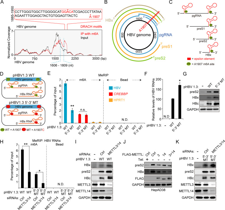
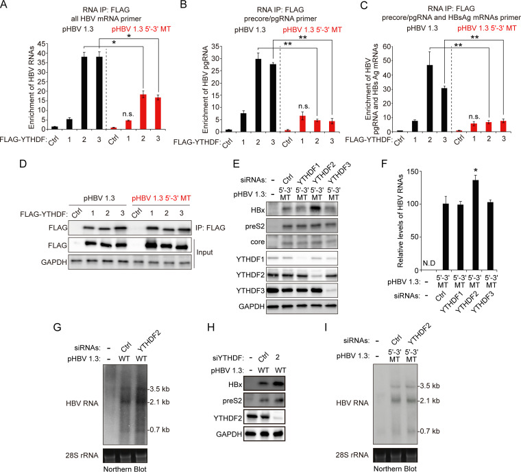
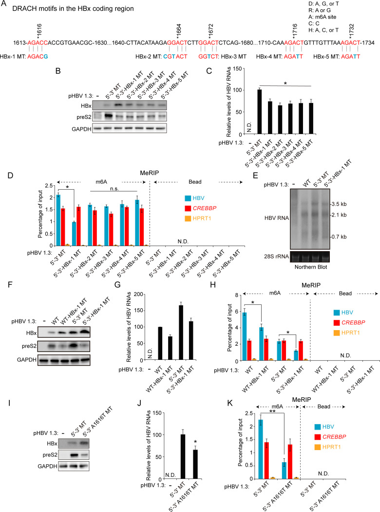
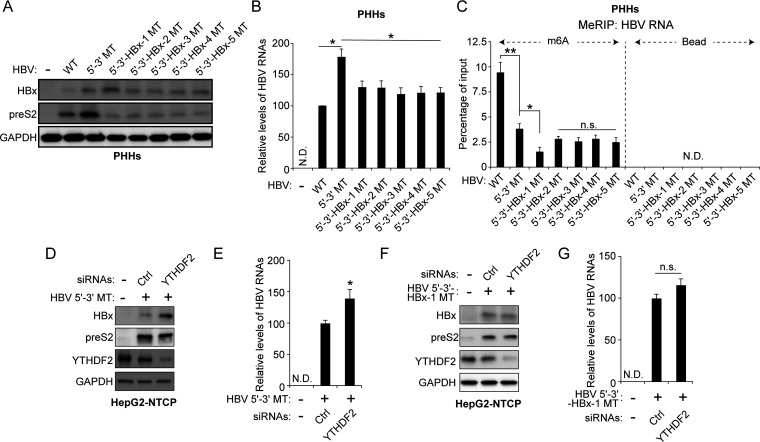
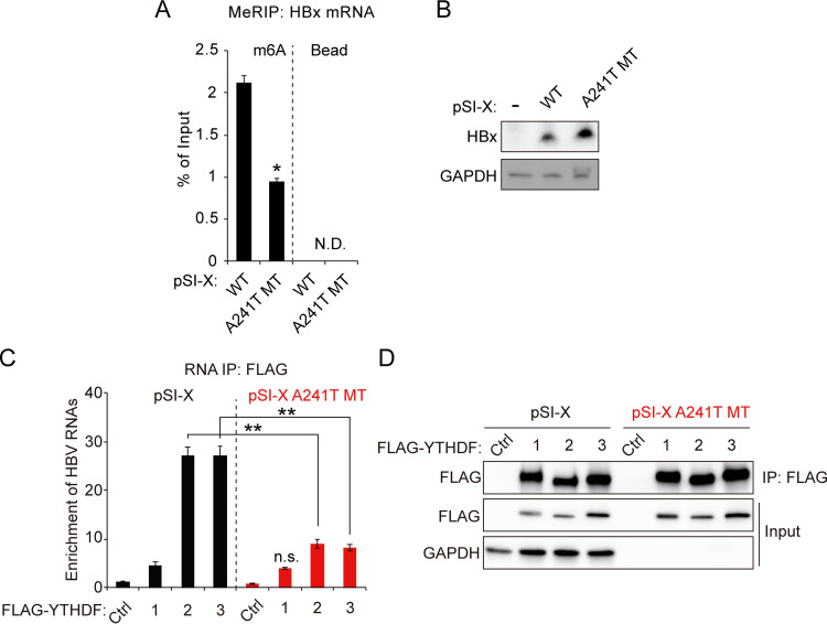
Hepatitis B virus (HBV) encodes a regulatory protein, termed HBx, that has been intensely studied in the past and shown to play a key role(s) in viral transcription and replication. In addition, a huge body of work exists in the literature related to signal transduction and possible mechanism(s) leading to hepatocarcinogenesis associated with infection. We have previously reported that HBV transcripts are modified by N6-methyladenosine (m6A) at the single consensus DRACH motif at nucleotides (nt) 1905 to 1909 in the epsilon structural element, and this m6A modification affects the HBV life cycle. In this study, we present evidence that additional variants of m6A (DRACH) motifs located within nt 1606 to 1809 correspond to the coding region of HBx mRNA and 3' untranslated region (UTR) of other viral mRNAs. Using the mutants of additional m6A sites in nt 1606 to 1809 and a depletion strategy of m6A methyltransferases (METTL3/14) and reader proteins (YTHDFs), we show that m6A modification at nt 1616, located in the HBx coding region, regulates HBx protein expression. The HBx RNA and protein expression levels were notably increased by the silencing of m6A reader YTHDF2 and methyltransferases as well as the mutation of m6A sites in the HBx coding region. However, other viral protein expression levels were not affected by the m6A modification at nt 1616. Thus, m6A modifications in the HBx open reading frame (ORF) downregulate HBx protein expression, commonly seen during HBV transfections, transgenic mice, and natural infections of human hepatocytes. These studies identify the functional role of m6A modification in the subtle regulation of HBx protein expression consistent with its possible role in establishing chronic hepatitis. IMPORTANCE N6-methyladenosien (m6A) modifications recently have been implicated in the HBV life cycle. Previously, we observed that m6A modification occurs in the adenosine at nt 1907 of the HBV genome, and this modification regulates the viral life cycle. Here, we identified an additional m6A site located in nt 1616 of the HBV genome. This modification negatively affects HBx RNA and protein expression. In the absence of m6A methyltransferases (METTL3/14) and reader protein (YTHDF2), the HBx RNA and protein expression were increased. Using HBV mutants that lack m6A in the HBx coding region, we present the unique positional effects of m6A in the regulation of HBx protein expression.

Keywords: HBV life cycle; HBx protein; N6-methyladenosine; hepatitis B virus.


Conflict of interest statement

The authors declare no conflict of interest.

Figures

FIG 1
FIG 1
Regulation of HBx protein expression by m6A modification sites other than the nt 1907 m6A site. (A) The m6A peaks are identified in nt 1606 to 1809 and nt 1885 to 1950 of the HBV genome using methylated RNA immunoprecipitation (IP) sequencing. The major peak in 1885 to 1950 includes a single m6A consensus motif at nt 1907, located on the lower stem-loop of the epsilon element. The minor peak corresponds to nt 1606 to 1809. (B) The location of the 1907 m6A site in the pictorial representation of transcripts within the HBV genome is indicated by a red line across the HBV DNA/transcripts. (C) Schematic showing the position of the 1907 m6A site, indicated by the green circle in all of the HBV RNAs. (D) Schematics indicate the 5′ and 3′ m6A sites in the epsilon elements of HBV pgRNA. Green circles indicate the m6A site, and red circles indicate the A1907C mutation in HBV pgRNA. pHBV 1.3 5′–3′ MT contains A1907C mutation at the 5′ and 3′ ends. (E to G) The indicated plasmids were transfected in Huh7 cells for 72 h. m6A methylated RNAs were immunoprecipitated from total RNA using an anti-m6A antibody. (E) m6A-methylated HBV RNAs, CREBBP, and HPRT1 RNAs were analyzed by input RNA levels by RT-qPCR. CREBBP and HPRT1 serve as positive and negative controls (Ctrl), respectively. (F) The HBV RNA levels were analyzed by RT-qPCR. (G) The indicated proteins were analyzed by immunoblotting. (H) Huh7 cells expressing the HBV WT or 5′–3′ MT were transfected with METTL3/14 siRNAs. Total RNA was immunoprecipitated using an anti-m6A antibody. m6A-methylated RNAs were analyzed by RT-qPCR. (I) Huh7 cells expressing the HBV WT were transfected with METTL3/14 siRNAs. After 48 h, cellular lysates were isolated and the indicated proteins were analyzed by Western blotting. (J) The indicated plasmids were transfected into HepAD38 cells grown in the absence or presence of tetracycline for 48 h. Cellular lysates were prepared from these cells and analyzed by Western blotting. (K) Huh7 cells expressing the HBV 5′–3′ MT were transfected with METTL3/14 siRNAs. After 48 h, cellular lysates were isolated and the indicated proteins were analyzed by Western blotting. In panels E, F, and H, the error bars represent the SDs from three independent experiments. The P values were calculated via an unpaired Student's t test. *, P < 0.05; **, P < 0.01. WT, wild type; MT, mutant; ND, nondetect.
FIG 2
FIG 2
Silencing of m6A reader YTHDF2 increases HBx mRNA and protein expression via interaction with m6A sites of the HBx coding region. (A to D) Huh7 cells expressing the HBV WT or 5′–3′ MT were transfected with the FLAG-YTHDF expression plasmids. After 48 h, total RNA and cell lysates were isolated from these cells. Total RNA was immunoprecipitated using an anti-FLAG antibody. (A to C) The immunoprecipitated HBV RNA levels were analyzed by the indicated primers. (D) The indicated proteins were analyzed by Western blotting. (E and F) Huh7 cells were transfected with pHBV 1.3 5′–3′ MT plasmid and then treated with YTHDF2 siRNAs for 48 h. Cell lysates and total RNA were isolated and analyzed by immunoblotting (E) and RT-qPCR (F). (G and H) Huh7 cells expressing HBV WT genome were transfected with the YTHDF2 siRNAs. After 48 h, total RNA and cellular lysates were extracted from these cells for Northern blotting (G) and Western blotting (H). (I) Huh7 cells expressing HBV 5′–3′ MT genome were transfected with the YTHDF2 siRNAs. After 48 h, total RNA and cellular lysates were extracted from these cells for Northern blotting. In panels A to C and F, the error bars represent the SDs from three independent experiments. The P values are calculated via an unpaired Student's t test. *, P < 0.05; **, P < 0.01. WT, wild type; MT, mutant; IP, immunoprecipitation; n.s., nonsignificant; ND, not detected.
FIG 3
FIG 3
Of the additional variant m6A sites in the HBx coding region, m6A modification at nt 1616 of the HBV genome regulates HBx protein expression. (A) The location of m6A consensus DRACH motifs within the minor m6A peak (1613 to 1734 nt) of the HBV genome is shown in red. Blue characters indicate mutations to disrupt the DRACH motifs in the HBx coding region. HBx-1, -2, -3, -4, or -5 MT contains the mutation of the DRACH motif at the position indicated in blue. (B to D) The indicated HBV expression plasmids were transfected into Huh7 cells for 72 h. Cell lysates and total RNA were extracted from these cells. (B) The indicated proteins were analyzed by immunoblotting. (C) HBV RNAs were analyzed by RT-qPCR. (D) m6A-methylated RNAs were immunoprecipitated from total RNA using an anti-m6A antibody, and m6A-methylated HBV RNA levels were normalized to input RNA levels by RT-qPCR. (E) Huh7 cells were transfected with pHBV 1.3 WT, 5′–3′ MT, or 5′–3′-HBx-1 MT plasmid for 72 h. Cellular HBV RNAs were analyzed by Northern blotting. (F to H) Huh7 cells were transfected with the indicated plasmids for 72 h. Cell lysates and total RNA were extracted from these cells. (F) The indicated protein expression levels were analyzed by Western blotting. (G) HBV RNA levels were analyzed by RT-qPCR. (H) m6A-methylated RNAs were immunoprecipitated from total RNA using an anti-m6A antibody and m6A-methylated HBV RNAs were normalized by input RNA levels by RT-qPCR. (I to K) Huh7 cells were transfected with pHBV 1.3 5′–3′ MT or pHBV 1.3 5′–3′-A1616T MT plasmid for 72 h. (I) The indicated proteins were analyzed by Western blotting. (J) HBV RNA levels were analyzed by RT-qPCR. (K) m6A methylated HBV RNAs were immunoprecipitated from total RNA using an anti-m6A antibody and normalized by input RNA levels by RT-qPCR. In panels C, D, G, H, J, and K, the error bars represent the SDs from three independent experiments. The P values were calculated via an unpaired Student's t test. *, P < 0.05; **, P < 0.01. n.s., nonsignificant; ND, not detected.
FIG 4
FIG 4
Regulation of HBx protein by m6A modification in the HBV-infected PHHs and HepG2-NTCP cells. (A to C) HBV particles prepared from the indicated HBV genome-transfected cells were used to infect primary human hepatocytes (PHHs) for 10 days. Total RNA and cellular lysates were extracted from these cells. (A) The indicated proteins were analyzed by Western blotting. (B) HBV RNA levels were analyzed by RT-qPCR. (C) Total RNAs extracted from the cells shown in panel A were immunoprecipitated using an anti-m6A antibody to identify m6A methylated HBV RNAs. These were normalized by input RNA levels by RT-qPCR. (D to G) HepG2-NTCP cells were infected with infectious virus particles prepared from HBV 5′–3′ MT- or 5′–3′-HBx-1 MT-expressing cells for 8 days. HepG2-NTCP cells were also transfected with control and/or YTHDF2 siRNAs for 48 h. Total RNA and cellular lysates were isolated from these cells. (D and F) The indicated proteins were analyzed by Western blotting. (E and G) HBV RNA levels were assayed by RT-qPCR. In panels B, C, E, and G, the error bars represent the SDs from three independent experiments. The P values were calculated via an unpaired Student's t test. *, P < 0.05; **, P < 0.01; n.s., nonsignificant; ND, not detected.
FIG 5
FIG 5
m6A modification in HBx coding region affects HBx expression by interaction with YTHDF2. (A and B) Huh7 cells were transfected with pSI-X WT or A241T MT plasmid for 48 h. (A) Total RNA and cell lysates were extracted. m6A methylated RNAs were enriched by an anti-m6A antibody and normalized by input RNA levels using RT-qPCR. (B) The indicated proteins were assayed by immunoblotting. (C and D) Huh7 cells expressing pSI-X WT or pSI-X A241T MT were transfected with FLAG-YTHDFs expression plasmids for 48 h. The RNA-protein complexes were immunoprecipitated using an anti-FLAG antibody. (C) Immunoprecipitated HBx mRNA levels were normalized by input HBx mRNA using RT-qPCR. (D) The indicated proteins were analyzed by Western blotting. In panels A and C the error bars represent the SDs from three independent experiments. The P values were calculated via an unpaired Student's t test. *, P < 0.05; **, P < 0.01. IP, immunoprecipitation; n.s., nonsignificant; N.D., not detected.

Similar articles

Cited by

References

    1. Hu J, Protzer U, Siddiqui A. 2019. Revisiting hepatitis B virus: challenges of curative therapies. J Virol 93:e01032-19. 10.1128/JVI.01032-19. - DOI - PMC - PubMed
    1. Seeger C, Mason WS. 2015. Molecular biology of hepatitis B virus infection. Virology 479-480:672–686. 10.1016/j.virol.2015.02.031. - DOI - PMC - PubMed
    1. Yan H, Zhong GC, Xu GW, He WH, Jing ZY, Gao ZC, Huang Y, Qi YH, Peng B, Wang HM, Fu LR, Song M, Chen P, Gao WQ, Ren BJ, Sun YY, Cai T, Feng XF, Sui JH, Li WH. 2012. Sodium taurocholate cotransporting polypeptide is a functional receptor for human hepatitis B and D virus. Elife 1:e00049. 10.7554/eLife.00049. - DOI - PMC - PubMed
    1. Bouchard MJ, Schneider RJ. 2004. The enigmatic X gene of hepatitis B virus. J Virol 78:12725–12734. 10.1128/JVI.78.23.12725-12734.2004. - DOI - PMC - PubMed
    1. Slagle BL, Andrisani OM, Bouchard MJ, Lee CG, Ou JH, Siddiqui A. 2015. Technical standards for hepatitis B virus X protein (HBx) research. Hepatology 61:1416–1424. 10.1002/hep.27360. - DOI - PMC - PubMed

Publication types

MeSH terms