

Biomaterials direct functional B cell response in a material-specific manner
- PMID: 34851674
- PMCID: PMC8635437
- DOI: 10.1126/sciadv.abj5830
Biomaterials direct functional B cell response in a material-specific manner
Abstract
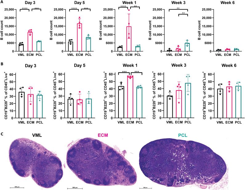
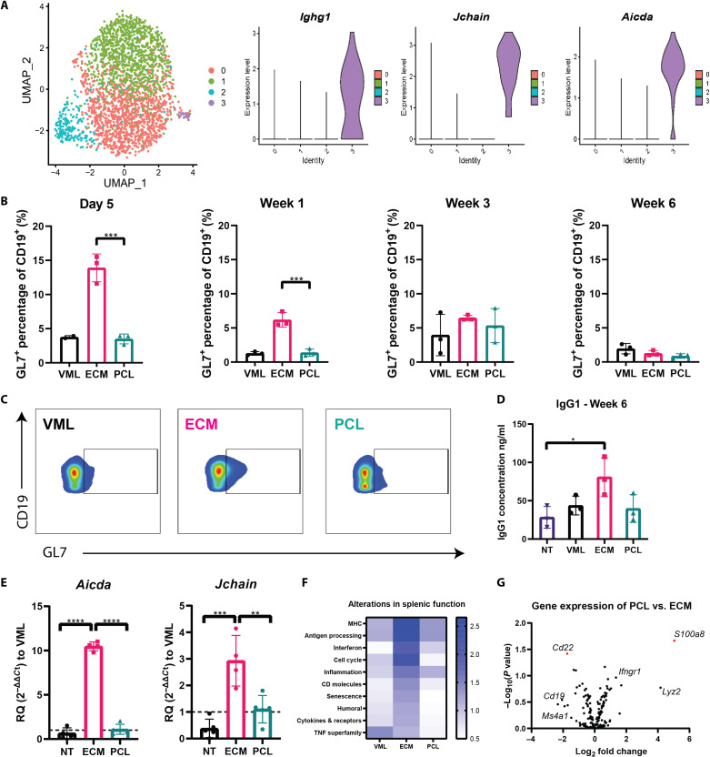
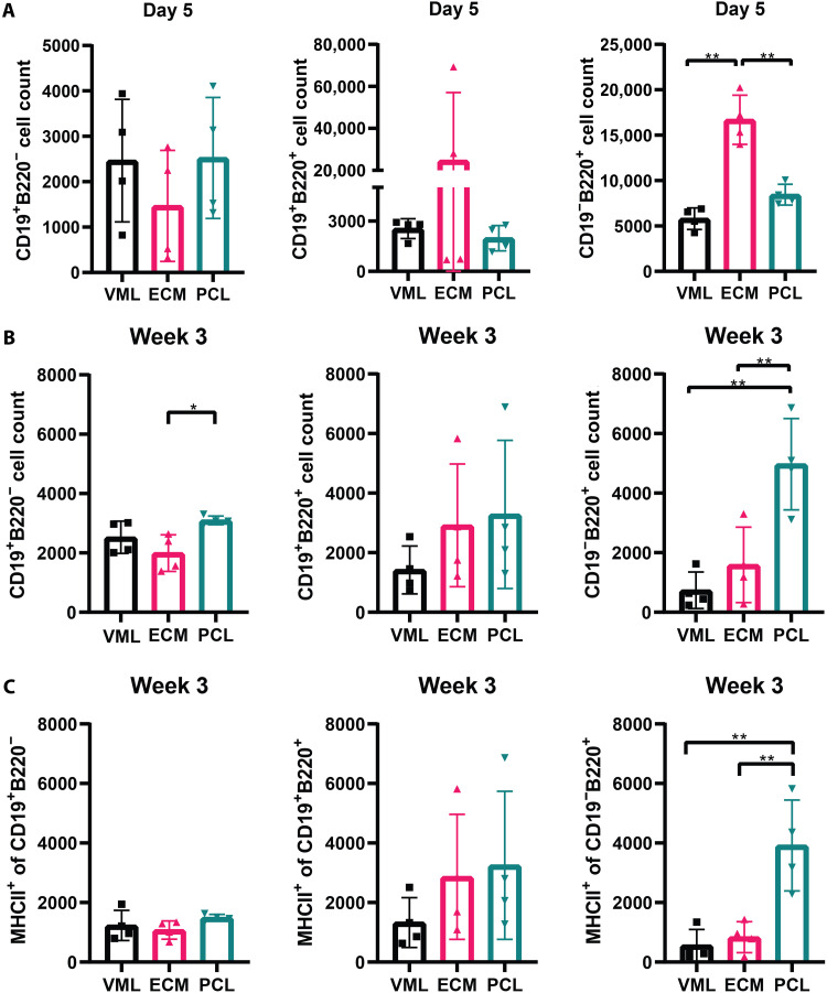
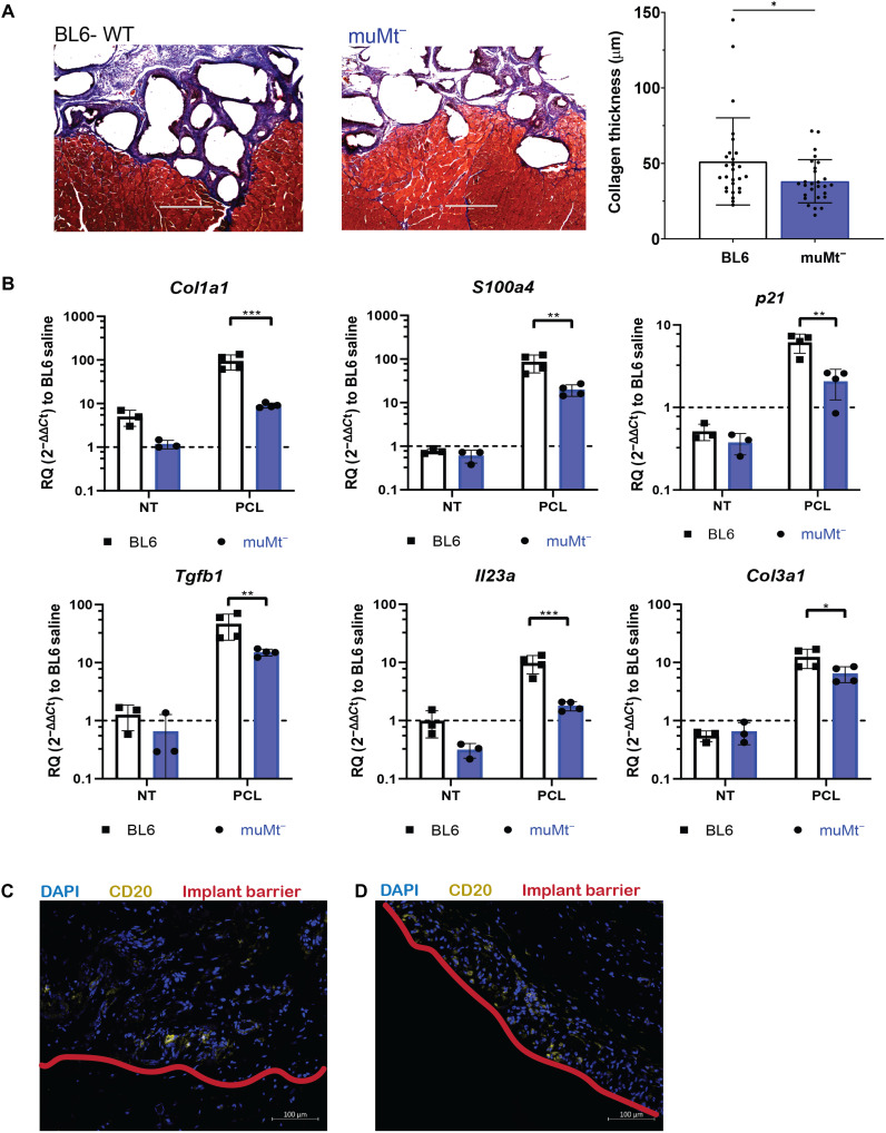
B cells are an adaptive immune target of biomaterials development in vaccine research but, despite their role in wound healing, have not been extensively studied in regenerative medicine. To probe the role of B cells in biomaterial scaffold response, we evaluated the B cell response to biomaterial materials implanted in a muscle wound using a biological extracellular matrix (ECM), as a reference for a naturally derived material, and synthetic polyester polycaprolactone (PCL), as a reference for a synthetic material. In the local muscle tissue, small numbers of B cells are present in response to tissue injury and biomaterial implantation. The ECM materials induced mature B cells in lymph nodes and antigen presentation in the spleen. The synthetic PCL implants resulted in prolonged B cell presence in the wound and induced an antigen-presenting phenotype. In summary, the adaptive B cell immune response to biomaterial induces local, regional, and systemic immunological changes.
Figures
Similar articles
-
T lymphocytes as critical mediators in tissue regeneration, fibrosis, and the foreign body response.Acta Biomater. 2021 Oct 1;133:17-33. doi: 10.1016/j.actbio.2021.04.023. Epub 2021 Apr 24. Acta Biomater. 2021. PMID: 33905946 Review.
-
Extracellular matrix-based biomaterials as adipose-derived stem cell delivery vehicles in wound healing: a comparative study between a collagen scaffold and two xenografts.Stem Cell Res Ther. 2020 Nov 27;11(1):510. doi: 10.1186/s13287-020-02021-x. Stem Cell Res Ther. 2020. PMID: 33246508 Free PMC article.
-
The role of biomaterials and scaffolds in immune responses in regenerative medicine: macrophage phenotype modulation by biomaterial properties and scaffold architectures.Biomater Sci. 2021 Dec 7;9(24):8090-8110. doi: 10.1039/d1bm00840d. Biomater Sci. 2021. PMID: 34762077 Review.
-
The Scaffold Immune Microenvironment: Biomaterial-Mediated Immune Polarization in Traumatic and Nontraumatic Applications<sup/>Tissue Eng Part A. 2017 Oct;23(19-20):1044-1053. doi: 10.1089/ten.TEA.2016.0304. Epub 2016 Nov 9. Tissue Eng Part A. 2017. PMID: 27736323 Free PMC article.
-
Key players in the immune response to biomaterial scaffolds for regenerative medicine.Adv Drug Deliv Rev. 2017 May 15;114:184-192. doi: 10.1016/j.addr.2017.07.006. Epub 2017 Jul 13. Adv Drug Deliv Rev. 2017. PMID: 28712923 Review.
Cited by
-
Immunomodulatory Biomaterials and Emerging Analytical Techniques for Probing the Immune Micro-Environment.Tissue Eng Regen Med. 2023 Feb;20(1):11-24. doi: 10.1007/s13770-022-00491-z. Epub 2022 Oct 14. Tissue Eng Regen Med. 2023. PMID: 36241939 Free PMC article. Review.
-
Linguistic Analysis Identifies Emergent Biomaterial Fabrication Trends for Orthopaedic Applications.Adv Healthc Mater. 2023 Apr;12(10):e2202591. doi: 10.1002/adhm.202202591. Epub 2023 Feb 5. Adv Healthc Mater. 2023. PMID: 36657736 Free PMC article. Review.
-
Biomaterial-Based Regenerative Strategies for Volumetric Muscle Loss: Challenges and Solutions.Adv Wound Care (New Rochelle). 2025 Mar;14(3):159-175. doi: 10.1089/wound.2024.0079. Epub 2024 Jul 10. Adv Wound Care (New Rochelle). 2025. PMID: 38775429 Review.
-
Murine gut microbiota dysbiosis via enteric infection modulates the foreign body response to a distal biomaterial implant.Proc Natl Acad Sci U S A. 2025 May 20;122(20):e2422169122. doi: 10.1073/pnas.2422169122. Epub 2025 May 12. Proc Natl Acad Sci U S A. 2025. PMID: 40354538
-
Immunoengineering Biomaterials for Musculoskeletal Tissue Repair across Lifespan.Adv Mater. 2024 Jul;36(28):e2311646. doi: 10.1002/adma.202311646. Epub 2024 May 7. Adv Mater. 2024. PMID: 38416061 Free PMC article. Review.
References
-
- Couzin-Frankel J., Cancer immunotherapy. Science 342, 1432–1433 (2013). - PubMed
-
- Steinman L., Immune therapy for autoimmune diseases. Science 305, 212–216 (2004). - PubMed
-
- Chung L., Maestas D. R., Housseau F., Elisseeff J. H., Key players in the immune response to biomaterial scaffolds for regenerative medicine. Adv. Drug Deliv. Rev. 114, 184–192 (2017). - PubMed
-
- Wolfram D., Rabensteiner E., Grundtman C., Böck G., Mayerl C., Parson W., Almanzar G., Hasenöhrl C., Piza-Katzer H., Wick G., T regulatory cells and TH17 cells in peri–silicone implant capsular fibrosis. Plast. Reconstr. Surg. 129, 327e–337e (2012). - PubMed
-
- Singh A., Biomaterials innovation for next generation ex vivo immune tissue engineering. Biomaterials 130, 104–110 (2017). - PubMed
Grants and funding
LinkOut - more resources
Full Text Sources
Molecular Biology Databases
