. 2021 Dec 3;7(49):eabj9805.
doi: 10.1126/sciadv.abj9805. Epub 2021 Dec 1.

Emergence and global spread of Listeria monocytogenes main clinical clonal complex

Affiliations

Emergence and global spread of Listeria monocytogenes main clinical clonal complex

Alexandra Moura et al. Sci Adv. .

Abstract

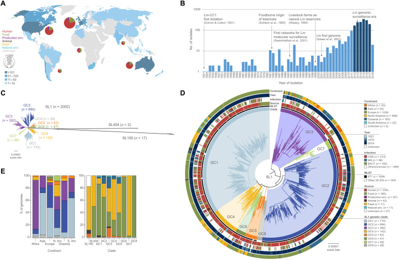
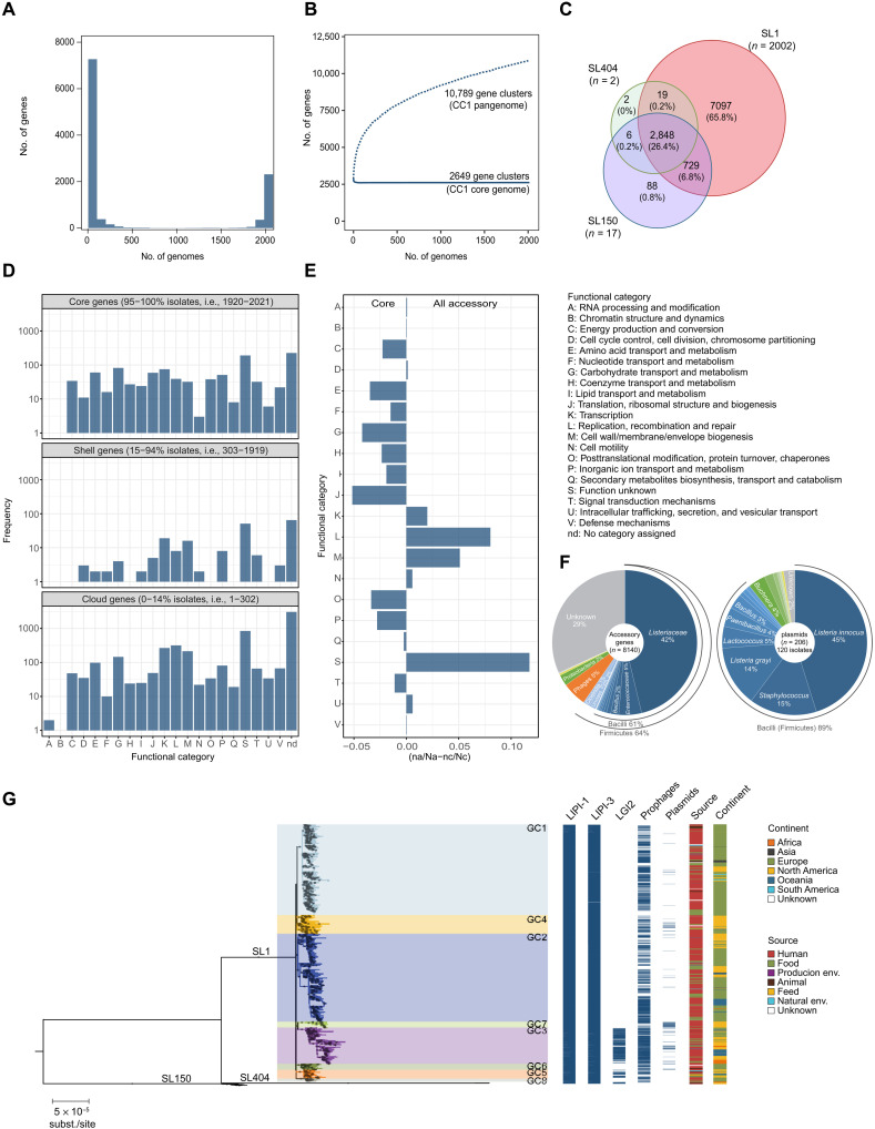
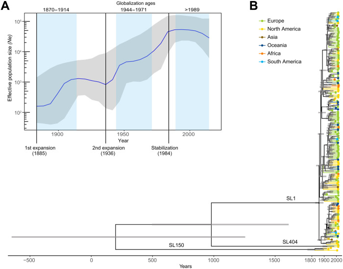
The bacterial foodborne pathogen Listeria monocytogenes clonal complex 1 (Lm-CC1) is the most prevalent clonal group associated with human listeriosis and is strongly associated with cattle and dairy products. Here, we analyze 2021 isolates collected from 40 countries, covering Lm-CC1 first isolation to present days, to define its evolutionary history and population dynamics. We show that Lm-CC1 spread worldwide from North America following the Industrial Revolution through two waves of expansion, coinciding with the transatlantic livestock trade in the second half of the 19th century and the rapid growth of cattle farming and food industrialization in the 20th century. In sharp contrast to its global spread over the past century, transmission chains are now mostly local, with limited inter- and intra-country spread. This study provides an unprecedented insight into L. monocytogenes phylogeography and population dynamics and highlights the importance of genome analyses for a better control of pathogen transmission.


Figures

Fig. 1.
Fig. 1.. Geographical and temporal distribution of the isolates used in this study (N = 2021) and phylogenetic analyses.
(A) Geographical distribution and source distribution. Sampled countries are colored in blue, with hue gradient according to the number of isolates. Pie charts are proportional to the number of isolates sampled in each continent and represent the repartition of sample source types, using the source color key indicated in (D). Eight isolates had unknown sampling location and are not shown in the map. (B) Temporal distribution of isolates collected in this study. Darker blue bars indicate the period for which exhaustive clinical sampling was obtained for seven countries spanning three continents (2012–2017; US, FR, UK, DK, NL, AU, and NZ). (C) Unrooted maximum likelihood phylogenetic tree of 2021 Lm-CC1 genomes. The tree was generated from analysis (GTR+F+G4 model, 1000 ultrafast bootstraps) of a 1.29-Mb recombination-purged core genome alignment. (D) Midpoint-rooted maximum likelihood phylogenetic tree of 2002 SL1 genomes based on a recombination-purged core genome alignment of 1.29 Mb. The four external rings indicate the world region, year, type of infection, and source type, respectively. The two inner rings indicate ST1 isolates and the eight SL1 GCs identified in this study, respectively. (E) Percentage of genomes by world region (left) and clade (right). Partitions are colored by world regions and clade, using the same color code as in (D).
Fig. 2.
Fig. 2.. Lm-CC1 pangenome analysis.
(A) Frequency of sampled gene families. (B) Pan- and core-gene families sampled. (C) Venn diagram showing the number of gene families present in at least one sublineage member. (D) Distribution of the functional categories of the clusters of orthologous genes across the Lm-CC1 pangenome. (E) Differential proportion of each assigned COG (cluster of orthologous group) category in core versus accessory genome, calculated as the difference between the ratio of each category (n) and the total number of hits (N) among each gene pool set, as in (naccessory/Naccessory-ncore/Ncore). (F) Taxonomic classification of accessory genes and plasmids, based on eggNOG-mapper v.2 and MOB-suite v.2, respectively. (G) Distribution of Listeria genomic islands, prophages, and plasmids across Lm-CC1 phylogeny. The midpoint-rooted maximum likelihood phylogenetic tree (GTR+F+G4 model, 1000 ultra-fast bootstraps) was inferred from the 1.29-Mb recombination-purged core genome alignment of 2021 Lm-CC1 genomes.
Fig. 3.
Fig. 3.. Bayesian temporal and demographic analyses on a representative 200 isolate dataset of Lm-CC1.
(A) Bayesian skyline plot (BSP) with the estimation of Lm-CC1 effective population size (Ne). The y axis refers to the predicted number of individuals (log scale), and the x axis refers to the time scale (in years). The median population size is marked in blue, with its 95% high posterior density (HPD) in gray. Blue vertical panels delimitate the three globalization ages (1870–1914, 1944–1971, and 1989–present). (B) Bayesian time-calibrated tree. Nodes represent the estimated mean divergence times, and gray bars represent the 95% HPD CIs of node age. Scale indicates time (in years). Terminal branches and tips are colored by continents, as indicated in the key panel.
Fig. 4.
Fig. 4.. Phylogeography of sublineage SL1.
(A) Time-calibrated phylogeny based on the 1956 SL1 genomes (full view of Lm-CC1 in fig. S10). Pies at the nodes represent the probability of ancestral geographical locations, estimated using PastML using the MPPA method with an F81-like model. (B) Inferred spread of SL1 populations across continents. The first introductions of each phylogroup are represented by arrows from their estimated world region origin. Gray labels and arrows denote clades with uncertain ancestral origin among datasets. (C) Proportion of intercontinental transitions per 10-year bins, normalized by the total number of phylogenetic branches per bin.
Fig. 5.
Fig. 5.. Transmission dynamics of sublineage SL1: Evidence of local expansion after global spread.
(A) Each point summarizes the relative risk that a pair of isolates has an MRCA within a defined time frame and between different spatial scales (within the same country, within the same continent, or within different continents), relative to the risk that a pair of isolates from countries separated by >1000 km has an MRCA in the same range (set as the reference value, “ref”). Error bars represent the 95% CIs, based on 100 bootstrap time-calibrated trees. (B) Proportion of pairs of isolates within the same country (France) sharing an MRCA of 5 or less years in function of the spatial distance within and between administrative departments (shown in the map). The green line indicates the mean proportion of genetically close strains regardless of the geographical location. (C) Relative risk for a pair of isolates to share an MRCA of 5 or less years when (left) both are coming from Paris compared to when coming from another department (P = 0.43) and (right) when coming from the same department in France, except Paris, compared to when coming from different departments (P < 0.001, see Materials and Methods for details).

Similar articles

Cited by

References

    1. Swaminathan B., Gerner-Smidt P., The epidemiology of human listeriosis. Microbes Infect. 9, 1236–1243 (2007). - PubMed
    1. Charlier C., Perrodeau É., Leclercq A., Cazenave B., Pilmis B., Henry B., Lopes A., Maury M. M., Moura A., Goffinet F., Dieye H. B., Thouvenot P., Ungeheuer M.-N. N., Tourdjman M., Goulet V., de Valk H., Lortholary O., Ravaud P., Lecuit M.; MONALISA Study Group , Clinical features and prognostic factors of listeriosis: The MONALISA national prospective cohort study. Lancet Infect. Dis. 17, 510–519 (2017). - PubMed
    1. Orsi R. H., den Bakker H. C., Wiedmann M., Listeria monocytogenes lineages: Genomics, evolution, ecology, and phenotypic characteristics. Int. J. Med. Microbiol. 301, 79–96 (2011). - PubMed
    1. Ragon M., Wirth T., Hollandt F., Lavenir R., Lecuit M., Le Monnier A., Brisse S., A new perspective on Listeria monocytogenes evolution. PLOS Pathog. 4, e1000146 (2008). - PMC - PubMed
    1. Cantinelli T., Chenal-Francisque V., Diancourt L., Frezal L., Leclercq A., Wirth T., Lecuit M., Brisse S., “Epidemic clones” of Listeria monocytogenes are widespread and ancient clonal groups. J. Clin. Microbiol. 51, 3770–3779 (2013). - PMC - PubMed