. 2022 Mar 24;139(12):1807-1819.
doi: 10.1182/blood.2021013523.

Disease progression and clinical outcomes in telomere biology disorders

Affiliations

Disease progression and clinical outcomes in telomere biology disorders

Marena R Niewisch et al. Blood. .

Abstract

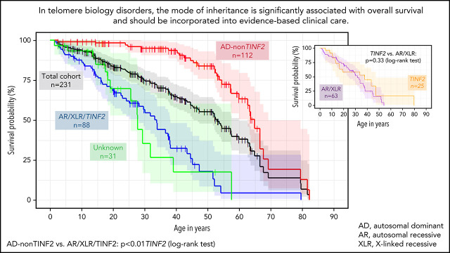
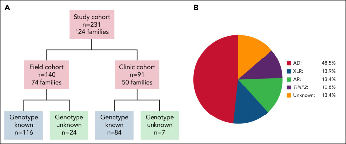
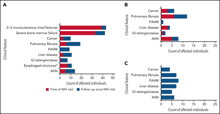
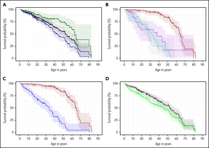
Dyskeratosis congenita related telomere biology disorders (DC/TBDs) are characterized by very short telomeres caused by germline pathogenic variants in telomere biology genes. Clinical presentations can affect all organs, and inheritance patterns include autosomal dominant (AD), autosomal recessive (AR), X-linked (XLR), or de novo. This study examined the associations between mode of inheritance with phenotypes and long-term clinical outcomes. Two hundred thirty-one individuals with DC/TBDs (144 male, 86.6% known genotype, median age at diagnosis 19.4 years [range 0 to 71.6]), enrolled in the National Cancer Institute's Inherited Bone Marrow Failure Syndrome Study, underwent detailed clinical assessments and longitudinal follow-up (median follow-up 5.2 years [range 0 to 36.7]). Patients were grouped by inheritance pattern, considering AD-nonTINF2, AR/XLR, and TINF2 variants separately. Severe bone marrow failure (BMF), severe liver disease, and gastrointestinal telangiectasias were more prevalent in AR/XLR or TINF2 disease, whereas pulmonary fibrosis developed predominantly in adults with AD disease. After adjusting for age at DC/TBD diagnosis, we observed the highest cancer risk in AR/XLR individuals. At last follow-up, 42% of patients were deceased with a median overall survival (OS) of 52.8 years (95% confidence interval [CI] 45.5-57.6), and the hematopoietic cell or solid organ transplant-free median survival was 45.3 years (95% CI 37.4-52.1). Significantly better OS was present in AD vs AR/XLR/TINF2 disease (P < .01), while patients with AR/XLR and TINF2 disease had similar survival probabilities. This long-term study of the clinical manifestations of DC/TBDs creates a foundation for incorporating the mode of inheritance into evidence-based clinical care guidelines and risk stratification in patients with DC/TBDs. This trial was registered at www.clinicaltrials.gov as #NCT00027274.


Figures

None
Graphical abstract
Figure 1.
Figure 1.
Overview of study cohort and distribution of inheritance pattern subgroups. (A) Cohort overview. Field cohort: participants completed questionnaires and the study team reviewed their medical records. 32 field cohort participants were also members of clinic cohort families. Clinic cohort: participants evaluated by specialists at the National Institutes of Health Warren G. Magnuson Clinical Center. (B) Schematic depiction of the distribution of inheritance pattern in the 231 study subjects. AD, autosomal dominant inheritance; AR, autosomal recessive inheritance; XLR, X-linked recessive inheritance; TINF2, TINF2 heterozygotes.
Figure 2.
Figure 2.
Clinical complications in the clinic cohort. (A) Development of clinical complications over time in 91 patients (clinic cohort). Red shaded areas show number of participants with clinical features (indicated on the y-axis) at time of evaluation at the National Institutes of Health. Blue shaded areas indicate the additional number of patients who developed these complications during follow-up. Mucocutaneous triad features include nail dysplasia, lacy reticular skin pigmentation, oral leukoplakia. Severe BMF: ANC <500/mm3, platelets <20 000/mm3, and/or Hb <8.0 g/dL OR cytopenia requiring treatment (HCT, regular platelet or red blood cell transfusions, or androgen treatment) and/or diagnosis of myelodysplastic syndrome or leukemia. (B) Clinical complications occurring with no prior hematopoietic cell transplantation. (C) Clinical complications occurring following HCT. AVN, avascular osteonecrosis of hip(s), knees, and/or shoulder(s); GI, gastrointestinal; PAVM, pulmonary arteriovenous malformation. *Esophageal strictures in 2 cases were diagnosed following HCT without history of gut GvHD.
Figure 3.
Figure 3.
Kaplan Meier estimates of the combined field and clinic cohorts. (A) OS of combined cohorts: OS of complete cohort (field and clinic cohort, n = 231, black line) and divided by sex: 87 females (green line) and 144 males (blue line), P < .01. (B) OS according to inheritance pattern. Red line shows autosomal dominant/nonTINF2 (AD, n = 112); turquoise line shows autosomal recessive/X-linked recessive (AR/XLR, n = 63). Purple line indicates disease due to TINF2 (n = 25), and gray shows 31 patients with unknown genotype (AD vs AR/XLR P < .01, AD vs TINF2 P < .01, AR/XLR vs TINF2 P = .33). (C) OS of AD-nonTINF2 vs all other inheritance pattern groups: nonTINF2 AD disease (dark red, n = 112) vs all other inheritance pattern groups (AR/XLR/TINF2, blue, n = 88 P < .01). (D) OS compared with transplant-free survival. OS of 231 patients with telomere biology disorders (black line), with 77 of 231 receiving either HCT, liver transplant, and/or lung transplant. Transplant-free survival is indicated by the green line. One patient receiving a kidney transplant was not included in the calculations. Observed survival (stair-step lines) are shown; shaded areas show 95% confidence intervals. AD, autosomal dominant; AR, autosomal recessive; XLR, X-linked recessive.

Comment in

References

    1. de Lange T, Shiue L, Myers RM, et al. . Structure and variability of human chromosome ends. Mol Cell Biol. 1990;10(2):518-527. - PMC - PubMed
    1. Zinsser F. Atrophia cutis reticularis cum pigmentatione dystrophia unguium et leukoplakia oris; poikiloderma atrophicans vascularis Jacobi. Inkonographia Dermatologia. 1906;5:219-223.
    1. Heiss NS, Knight SW, Vulliamy TJ, et al. . X-linked dyskeratosis congenita is caused by mutations in a highly conserved gene with putative nucleolar functions. Nat Genet. 1998;19(1):32-38. - PubMed
    1. Mitchell JR, Wood E, Collins K. A telomerase component is defective in the human disease dyskeratosis congenita. Nature. 1999;402(6761):551-555. - PubMed
    1. Ward SC, Savage SA, Giri N, et al. . Beyond the triad: inheritance, mucocutaneous phenotype, and mortality in a cohort of patients with dyskeratosis congenita. J Am Acad Dermatol. 2018;78(4):804-806. - PMC - PubMed

Publication types

Associated data