. 2021 Dec 14;54(12):2877-2892.e7.
doi: 10.1016/j.immuni.2021.11.001. Epub 2021 Nov 4.

Lipid nanoparticles enhance the efficacy of mRNA and protein subunit vaccines by inducing robust T follicular helper cell and humoral responses

Affiliations

Lipid nanoparticles enhance the efficacy of mRNA and protein subunit vaccines by inducing robust T follicular helper cell and humoral responses

Mohamad-Gabriel Alameh et al. Immunity. .

Erratum in

Abstract

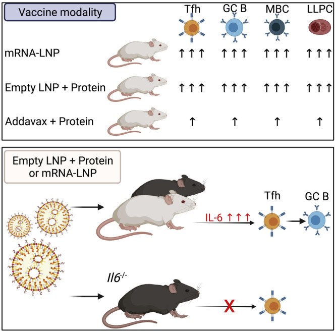
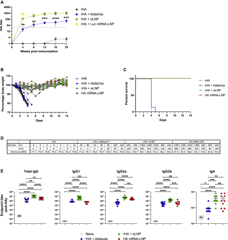
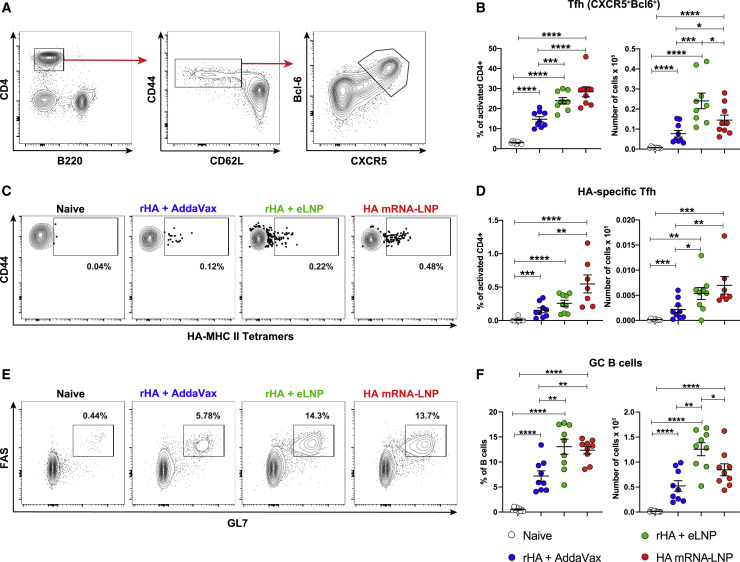
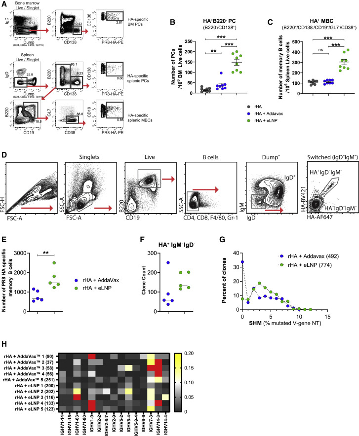
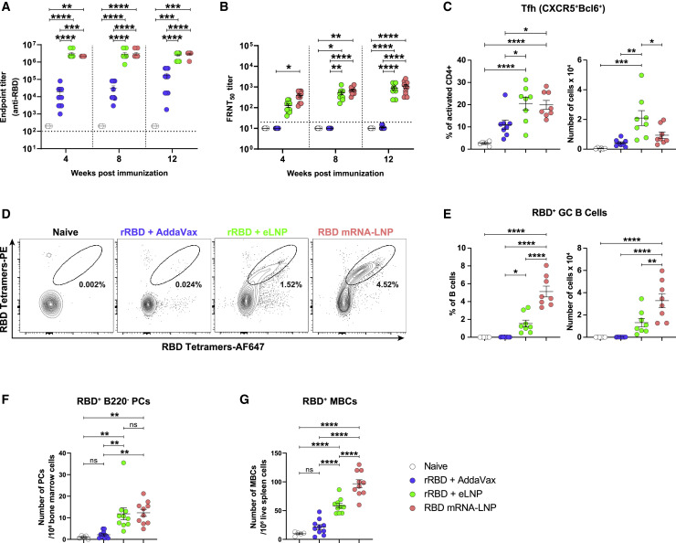
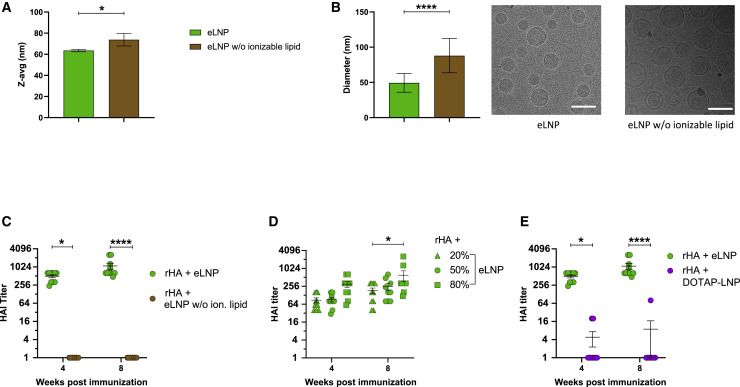
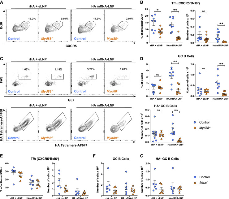
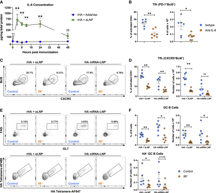
Adjuvants are critical for improving the quality and magnitude of adaptive immune responses to vaccination. Lipid nanoparticle (LNP)-encapsulated nucleoside-modified mRNA vaccines have shown great efficacy against severe acute respiratory syndrome coronavirus 2 (SARS-CoV-2), but the mechanism of action of this vaccine platform is not well-characterized. Using influenza virus and SARS-CoV-2 mRNA and protein subunit vaccines, we demonstrated that our LNP formulation has intrinsic adjuvant activity that promotes induction of strong T follicular helper cell, germinal center B cell, long-lived plasma cell, and memory B cell responses that are associated with durable and protective antibodies in mice. Comparative experiments demonstrated that this LNP formulation outperformed a widely used MF59-like adjuvant, AddaVax. The adjuvant activity of the LNP relies on the ionizable lipid component and on IL-6 cytokine induction but not on MyD88- or MAVS-dependent sensing of LNPs. Our study identified LNPs as a versatile adjuvant that enhances the efficacy of traditional and next-generation vaccine platforms.

Keywords: IL-6; SARS-CoV-2; Tfh cell; adjuvant; germinal centers; influenza virus; lipid nanoparticle; vaccine.


Conflict of interest statement

Declaration of interests In accordance with the University of Pennsylvania policies and procedures and our ethical obligations as researchers, we report that D.W. and N.P. are named on a patent describing the use of nucleoside-modified mRNA in lipid nanoparticles as a vaccine platform. We have disclosed those interests fully to the University of Pennsylvania, and we have in place an approved plan for managing any potential conflicts arising from licensing of our patents. K.K. is an employee of BioNTech. P.J.C.L., B.L.M., and Y.K.T. are employees of Acuitas Therapeutics, a company involved in the development of mRNA-LNP therapeutics. Y.K.T., D.W., and M.G.A. are named on patents that describe lipid nanoparticles for delivery of nucleic acid therapeutics, including mRNA and the use of modified mRNA in lipid nanoparticles as a vaccine platform. The Icahn School of Medicine at Mount Sinai has filed patent applications regarding SARS-CoV-2 and influenza virus vaccines that name F.K. as co-inventor.

Figures

None
Graphical abstract
Figure 1
Figure 1
LNP-adjuvanted HA mRNA and protein subunit vaccines induce durable, protective humoral responses (A) BALB/c mice received a single IM immunization with 10 μg of rHA mixed with eLNP (an equal amount of LNP as in 30 μg mRNA-LNP), 30 μg Luc mRNA-LNP, or AddaVax. PR8 HAI titers were followed for 20 weeks. Data are pooled from two independent experiments. n = 10 mice/group. The horizontal dotted line represents the protective HAI value (1:40). Data are shown as mean + SEM. (B–D) BALB/c mice were immunized ID with 10 μg of rHA, rHA + Addavax, or rHA + eLNP or 10 μg HA mRNA-LNP. Animals were terminally bled 9 months after injection, and immune sera were transferred into naive mice. Mice were challenged with PR8 virus, and survival and weight loss were followed daily. n = 7 mice/group in two independent experiments. (B) Weight of serum-transferred mice as percentage of starting weight. (C) Survival as percentage of total number of live animals per group. (D) HAI titers of pre-transfer immune sera and sera of injected mice at the time of infection (2 h after transfer). The lowest body weight percentage reached during 14 days after infection is shown. (E) Titers of HA-binding IgG, IgG1, IgG2a, IgG2b, and IgA were determined by endpoint dilution ELISA using sera of immunized mice collected 4 weeks after IM immunization. n = 10 animals per treatment group, n = 6 naive animals in two independent experiments. Statistical analysis: In (A), two-way ANOVA with Bonferroni’s multiple comparisons test; comparisons of AddaVax and eLNP groups are shown for each time point; ∗∗∗p ≤ 0.001, ∗∗∗∗p ≤ 0.0001. In (E), one-way ANOVA with Bonferroni’s multiple comparisons test was performed on log transformed values; p ≤ 0.05, ∗∗p ≤ 0.01, ∗∗∗p ≤ 0.001, ∗∗∗∗p ≤ 0.0001. See also Figure S1.
Figure 2
Figure 2
LNP-adjuvanted HA mRNA and protein subunit vaccines induce robust antigen-specific Tfh cell and GC B cell responses BALB/c mice received a single IM immunization with PR8 HA mRNA-LNP or rHA mixed with eLNP or AddaVax as described in Figure 1. Tfh and GC B cell responses were examined at day 12 in dLNs by flow cytometry. (A and B) Tfh cell (CD4+B220CD44hiCD62LBcl6+CXCR5+) representative gating strategy (A) and percentages and absolute numbers (B). (C and D) HA-specific Tfh cell (HA-MHC class II tetramer+ Tfh cells) representative flow cytometry (C) and percentages and absolute numbers (D). (E and F) GC B cell (CD4/CD8CD19+FAS+GL7+) representative flow cytometry (E) and percentages and absolute numbers (F). n = 7–9 mice/group. Data were combined from three independent experiments. Symbols represent individual animals. Data shown are mean + SEM. Statistical analysis: unpaired two-tailed Mann-Whitney U test was conducted. p ≤ 0.05, ∗∗p ≤ 0.01, ∗∗∗p ≤ 0.001, ∗∗∗∗p ≤ 0.0001. See also Figures S2 and S3.
Figure 3
Figure 3
The LNP-adjuvanted protein vaccine induces strong antigen-specific LLPC and class-switched, somatically mutated MBC responses (A–C) BALB/c mice received a single IM immunization with rHA or rHA mixed with eLNP or AddaVax as described in Figure 1. PR8 HA-specific LLPC and MBC responses were examined in bone marrow and spleen, respectively, 20 weeks after immunization. n = 8 mice/group. Symbols represent individual animals. Data shown are mean + SEM and are pooled from two independent experiments. (A) Gating strategy for measuring HA-specific LLPC and MBC responses. (B and C) PR8 HA-specific LLPC (IgDDumpB220CD138+PR8 HA+, B) and MBC (IgDDumpCD138B220+CD19+CD38+GL7PR8 HA+, C) responses. (D–H) Immune repertoire profiling of PR8 HA-binding MBCs. Mice received a single ID immunization with rHA formulated with eLNP or AddaVax. PR8 HA-specific MBCs were sorted from spleens 80 days after immunization. (D) Sorting strategy. (E) Number of PR8 HA-specific MBCs recovered from the sort. (F) Clone counts of IgMIgD HA-binding cells. Symbols represent individual animals. (G) HA-binding clones binned by their somatic hypermutation (SHM) percentage in the IGHV gene (compared with the nearest germline gene). Each clone counts only once. (H) IGHV gene usage in sorted HA+ MBCs (the heatmap indicates the fraction of clones). Red cells denote values that are between 0.21–0.59. In (G) and (H), IGHV genes present in at least 6 mice were included. Values in parentheses denote clone numbers. In (D)–(H), data were obtained from one experiment; n = 5 per group. Statistical analysis: in (B), (C), (E), and (F), unpaired two-tailed Mann-Whitney U test was conducted. ∗∗p ≤ 0.01, ∗∗∗p ≤ 0.001. See also Figure S4.
Figure 4
Figure 4
LNP-adjuvanted SARS-CoV-2 mRNA and protein subunit vaccines induce robust cellular and humoral immune responses BALB/c mice received a single IM immunization with 10 μg of RBD mRNA-LNP or recombinant SARS-CoV-2 RBD (rRBD) formulated with eLNP or AddaVax. (A) RBD-specific IgG titers were determined by endpoint dilution ELISA. Horizontal dotted line represents the limit of detection. (B) Neutralizing Ab titers were measured using a VSV-based pseudovirus neutralization assay. Horizontal dotted line represents the limit of detection. Focus reduction neutralization titer 50% (FRNT50) titers below the limit of detection are reported as half of the limit of detection. (C–E) Tfh and GC B cell responses were determined in dLNs 12 days after immunization. n = 8 mice/group. (C) Percentages and absolute numbers of Tfh cells (CD4+B220CD44hiCD62LBcl6+CXCR5+). (D) Representative frequencies of RBD-specific GC B cells (CD4/CD8CD19+FAS+GL7+RBD+). (E) Frequencies and absolute numbers of RBD-specific GC B cells (defined as in D). (F and G) Antigen-specific (F) LLPCs (IgDDumpB220CD138+RBD+) in bone marrow and (G) MBCs (IgDDumpCD138B220+CD19+CD38+GL7RBD+) in the spleen 12 weeks after immunization. In (A) (B), (F), and (G), n = 10 mice/group. In (A)–(C) and (E)–(G), symbols represent individual animals. Data are pooled from two independent experiments. Data shown are mean + SEM. Statistical analysis: in (A) and (B), two-way ANOVA with Bonferroni’s multiple comparisons test; differences are only shown within time points. In (C) and (E)–(G), unpaired two-tailed Mann-Whitney U test; p ≤ 0.05, ∗∗p ≤ 0.01, ∗∗∗p ≤ 0.001, ∗∗∗∗p ≤ 0.0001. See also Figures S5 and S6.
Figure 5
Figure 5
The ionizable lipid component is critical for the adjuvant activity of LNPs (A and B) Physicochemical characterization of the eLNPs with or without ionizable lipids determined by (A) DLS or (B) cryo-EM. The scale bars represent 100 nm. (C) BALB/c mice received a single IM immunization with rHA mixed with eLNPs with or without the ionizable lipid, and PR8 HAI titers were determined 4 and 8 weeks after vaccine administration. (D) BALB/c mice received a single IM immunization with 10 μg of rHA mixed with eLNPs with various amounts of ionizable lipid, and PR8 HAI titers were determined 4 and 8 weeks after vaccine administration. (E) BALB/c mice received a single IM immunization with 10 μg of rHA mixed with eLNP or DOTAP-LNP, and PR8 HAI titers were determined 4 and 8 weeks after vaccine administration. In (C)–(E), n = 10 mice/group. Symbols represent individual animals. Data are pooled from two independent experiments. In (A)–(E), data are shown as mean + SEM. Statistical analysis: in (A) and (B), unpaired two-tailed Mann-Whitney U test. In (C)–(E), two-way ANOVA with Bonferroni’s multiple comparisons test; only significant differences within the same time point are shown. p ≤ 0.05, ∗∗p ≤ 0.01, ∗∗∗∗p ≤ 0.0001.
Figure 6
Figure 6
Sensing through MyD88 enhances the potency of mRNA-LNP vaccines but is not required for LNP adjuvanticity Myd88−/−, Mavs−/− and control mice received a single IM immunization with 30 μg PR8 HA mRNA-LNP or 30 μg rHA + eLNP. Tfh and GC B cell responses were examined 12 days after immunization in dLNs by flow cytometry. (A and B) Tfh cell (CD4+B220CD44hiCD62LBcl6+CXCR5+) representative flow cytometry (A) and percentages and absolute numbers (B) in Myd88−/− and control mice. (C and D) GC B cell (CD3CD19+FAS+GL7+) and HA-specific GC B cell (RBDHA+) representative flow cytometry (C) and percentages and absolute numbers (D). (E and F) Percentages and absolute numbers of Tfh cells (E) and absolute numbers of GC B cells and HA-specific GC B cells (F) in Mavs−/− and control mice. In (A)–(D), n = 7–8 mice/group. In (E) and (F), n = 6 mice/group. Data were combined from three independent experiments in (A)–(D) and two independent experiments in (E) and (F). In (B) and (D)–(F), symbols represent individual animals. Data shown are mean + SEM. Statistical analysis: unpaired two-tailed Mann-Whitney U test was conducted. p ≤ 0.05, ∗∗p ≤ 0.01, ∗∗∗p ≤ 0.001, ∗∗∗∗p ≤ 0.0001.
Figure 7
Figure 7
IL-6 is crucial for LNP-induced GC reactions (A) BALB/c mice received a single IM immunization with 10 μg rHA adjuvanted with eLNP or AddaVax. IL-6 levels were determined by ELISA in dLN lysates at various times after immunization. (B) BALB/c mice received anti-IL-6 or isotype mAb 1 day prior to and every other day after a single IM immunization with 10 μg rHA + eLNP. Tfh cell (CD4+B220CD44hiCD62LPD-1+CXCR5+) percentages and absolute numbers were analyzed in dLNs 5 days after immunization. (C–F) Il6−/− and control mice received a single IM immunization with 30 μg PR8 HA mRNA-LNP or 30 μg rHA + eLNP. Twelve days later, Tfh and GC B cell responses were examined in dLNs by flow cytometry. (C and D) Tfh cell (CD4+B220CD44hiCD62LBcl6+CXCR5+) representative flow cytometry (C) and percentages and absolute numbers (D). (E and F) GC B cell (CD3CD19+FAS+GL7+, top panel) and antigen-specific GC B cell (RBDHA+, bottom panel) representative flow cytometry (E) and percentages and absolute numbers (F). In (A)–(F), n = 6–7 mice/group. Data were combined from two independent experiments. In (B), (D), and (F), symbols represent individual animals. Data are shown as mean + SEM. Statistical analysis: unpaired two-tailed Mann-Whitney U test was conducted. p ≤ 0.05, ∗∗p ≤ 0.01, ∗∗∗p ≤ 0.001, ∗∗∗∗p ≤ 0.0001. See also Figure S7.

Comment in

  • Fatballs foster fabulous follicles.
    Turley JL, Lavelle EC. Turley JL, et al. Immunity. 2021 Dec 14;54(12):2695-2697. doi: 10.1016/j.immuni.2021.11.009. Immunity. 2021. PMID: 34910938 Free PMC article.

Similar articles

Cited by

References

    1. Akira S., Takeda K. Toll-like receptor signalling. Nat. Rev. Immunol. 2004;4:499–511. - PubMed
    1. Alameh M.G., Weissman D., Pardi N. Messenger RNA-Based Vaccines Against Infectious Diseases. Curr. Top. Microbiol. Immunol. 2020 doi: 10.1007/82_2020_202. Published online April 17, 2020. - DOI - PubMed
    1. Amanat F., Stadlbauer D., Strohmeier S., Nguyen T.H.O., Chromikova V., McMahon M., Jiang K., Arunkumar G.A., Jurczyszak D., Polanco J., et al. A serological assay to detect SARS-CoV-2 seroconversion in humans. Nat. Med. 2020;26:1033–1036. - PMC - PubMed
    1. Arunachalam P.S., Scott M.K.D., Hagan T., Li C., Feng Y., Wimmers F., Grigoryan L., Trisal M., Edara V.V., Lai L., et al. Systems vaccinology of the BNT162b2 mRNA vaccine in humans. Nature. 2021;596:410–416. - PMC - PubMed
    1. Awasthi S., Hook L.M., Pardi N., Wang F., Myles A., Cancro M.P., Cohen G.H., Weissman D., Friedman H.M. Nucleoside-modified mRNA encoding HSV-2 glycoproteins C, D, and E prevents clinical and subclinical genital herpes. Sci. Immunol. 2019;4:eaaw7083. - PMC - PubMed

Publication types

MeSH terms