Aortic Stiffness, Pulse Pressure, and Cerebral Pulsatility Progress Despite Best Medical Management: The OXVASC Cohort
- PMID: 34852644
- PMCID: PMC7612543
- DOI: 10.1161/STROKEAHA.121.035560
Aortic Stiffness, Pulse Pressure, and Cerebral Pulsatility Progress Despite Best Medical Management: The OXVASC Cohort
Abstract
Background: Increased cerebral arterial pulsatility is associated with cerebral small vessel disease, recurrent stroke, and dementia despite the best medical treatment. However, no study has identified the rates and determinants of progression of arterial stiffness and pulsatility.
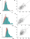
Methods: In consecutive patients within 6 weeks of transient ischemic attack or nondisabling stroke (OXVASC [Oxford Vascular Study]), arterial stiffness (pulse wave velocity [PWV]) and aortic systolic, aortic diastolic, and aortic pulse pressures (aoPP) were measured by applanation tonometry (Sphygmocor), while middle cerebral artery (MCA) peak (MCA-PSV) and trough (MCA-EDV) flow velocity and Gosling pulsatility index (PI; MCA-PI) were measured by transcranial ultrasound (transcranial Doppler, DWL Doppler Box). Repeat assessments were performed at the 5-year follow-up visit after intensive medical treatment and agreement determined by intraclass correlation coefficients. Rates of progression and their determinants, stratified by age and sex, were determined by mixed-effects linear models, adjusted for age, sex, and cardiovascular risk factors.
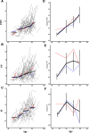
Results: In 188 surviving, eligible patients with repeat assessments after a median of 5.8 years. PWV, aoPP, and MCA-PI were highly reproducible (intraclass correlation coefficients, 0.71, 0.59, and 0.65, respectively), with progression of PWV (2.4%; P<0.0001) and aoPP (3.5%; P<0.0001) but not significantly for MCA-PI overall (0.93; P=0.22). However, PWV increased at a faster rate with increasing age (0.009 m/s per y/y; P<0.0001), while aoPP and MCA-PI increased significantly above the age of 55 years (aoPP, P<0.0001; MCA-PI, P=0.009). Higher aortic systolic blood pressure and diastolic blood pressure predicted a greater rate of progression of PWV and aoPP, but not MCA-PI, although current MCA-PI was particularly strongly associated with concurrent aoPP (P<0.001).
Conclusions: Arterial pulsatility and aortic stiffness progressed significantly after 55 years of age despite the best medical treatment. Progression of stiffness and aoPP was determined by high blood pressure, but MCA-PI predominantly reflected current aoPP. Treatments targetting cerebral pulsatility may need to principally target aortic stiffness and pulse pressure to have the potential to prevent cerebral small vessel disease.
Keywords: arterial pressure; follow-up studies; leukoaraiosis; linear models; vascular stiffness.
Conflict of interest statement
None.
Figures


Similar articles
-
Increased cerebral arterial pulsatility in patients with leukoaraiosis: arterial stiffness enhances transmission of aortic pulsatility.Stroke. 2012 Oct;43(10):2631-6. doi: 10.1161/STROKEAHA.112.655837. Epub 2012 Aug 23. Stroke. 2012. PMID: 22923446
-
Blood flow pattern in the middle cerebral artery in relation to indices of arterial stiffness in the systemic circulation.Am J Hypertens. 2012 Mar;25(3):319-24. doi: 10.1038/ajh.2011.223. Epub 2011 Nov 24. Am J Hypertens. 2012. PMID: 22113170
-
Low Heart Rate Is Associated with Cerebral Pulsatility after TIA or Minor Stroke.Ann Neurol. 2022 Dec;92(6):909-920. doi: 10.1002/ana.26480. Epub 2022 Aug 29. Ann Neurol. 2022. PMID: 36054225 Free PMC article.
-
Arterial stiffness and cerebral hemodynamic pulsatility during cognitive engagement in younger and older adults.Exp Gerontol. 2018 Jan;101:54-62. doi: 10.1016/j.exger.2017.11.004. Epub 2017 Nov 10. Exp Gerontol. 2018. PMID: 29129735
-
New Insights Into Cerebrovascular Pathophysiology and Hypertension.Stroke. 2022 Apr;53(4):1054-1064. doi: 10.1161/STROKEAHA.121.035850. Epub 2022 Mar 8. Stroke. 2022. PMID: 35255709 Free PMC article. Review.
Cited by
-
Stiffness and Elasticity of Aorta Assessed Using Computed Tomography Angiography as a Marker of Cardiovascular Health-A Cross-Sectional Study.J Clin Med. 2024 Jan 10;13(2):384. doi: 10.3390/jcm13020384. J Clin Med. 2024. PMID: 38256515 Free PMC article.
-
Association between blood-brain barrier permeability and changes in pulse wave velocity following a recent small subcortical infarct.Hypertens Res. 2024 Sep;47(9):2495-2502. doi: 10.1038/s41440-024-01764-x. Epub 2024 Jun 28. Hypertens Res. 2024. PMID: 38942814
-
Reduced neurovascular coupling is associated with increased cardiovascular risk without established cerebrovascular disease: A cross-sectional analysis in UK Biobank.J Cereb Blood Flow Metab. 2025 May;45(5):897-907. doi: 10.1177/0271678X241302172. Epub 2024 Nov 22. J Cereb Blood Flow Metab. 2025. PMID: 39576882 Free PMC article.
-
The Carotid Siphon as a Pulsatility Modulator for Brain Protection: Role of Arterial Calcification Formation.J Pers Med. 2025 Aug 4;15(8):356. doi: 10.3390/jpm15080356. J Pers Med. 2025. PMID: 40863418 Free PMC article. Review.
-
Associations of Cerebrovascular Regulation and Arterial Stiffness With Cerebral Small Vessel Disease: A Systematic Review and Meta-Analysis.J Am Heart Assoc. 2023 Dec 5;12(23):e032616. doi: 10.1161/JAHA.123.032616. Epub 2023 Nov 28. J Am Heart Assoc. 2023. PMID: 37930079 Free PMC article.
References
-
- Smith EE, Egorova S, Blacker D, Killiany RJ, Muzikansky A, Dickerson BC, Tanzi RE, Albert MS, Greenberg SM, Guttmann CR. Magnetic resonance imaging white matter hyperintensities and brain volume in the prediction of mild cognitive impairment and dementia. Arch Neurol. 2008; 65:94–100. doi: 10.1001/archneurol.2007.23 - PubMed
-
- Teodorczuk A, Firbank MJ, Pantoni L, Poggesi A, Erkinjuntti T, Wallin A, Wahlund LO, Scheltens P, Waldemar G, Schrotter G, et al. ; LADIS Group. Relationship between baseline white-matter changes and development of late-life depressive symptoms: 3-year results from the LADIS study. Psychol Med. 2010; 40:603–610. doi: 10.1017/S0033291709990857 - PubMed
-
- Inzitari D, Pracucci G, Poggesi A, Carlucci G, Barkhof F, Chabriat H, Erkinjuntti T, Fazekas F, Ferro JM, Hennerici M, et al. ; LADIS Study Group. Changes in white matter as determinant of global functional decline in older independent outpatients: three year follow-up of LADIS (leukoaraiosis and disability) study cohort. BMJ. 2009; 339:b2477. doi: 10.1136/bmj.b2477 - PMC - PubMed
Publication types
MeSH terms
Grants and funding
LinkOut - more resources
Full Text Sources
Research Materials
Miscellaneous

