

Zinc finger E-Box binding homeobox 2 (ZEB2)-induced astrogliosis protected neuron from pyroptosis in cerebral ischemia and reperfusion injury
- PMID: 34852714
- PMCID: PMC8809936
- DOI: 10.1080/21655979.2021.2012551
Zinc finger E-Box binding homeobox 2 (ZEB2)-induced astrogliosis protected neuron from pyroptosis in cerebral ischemia and reperfusion injury
Abstract
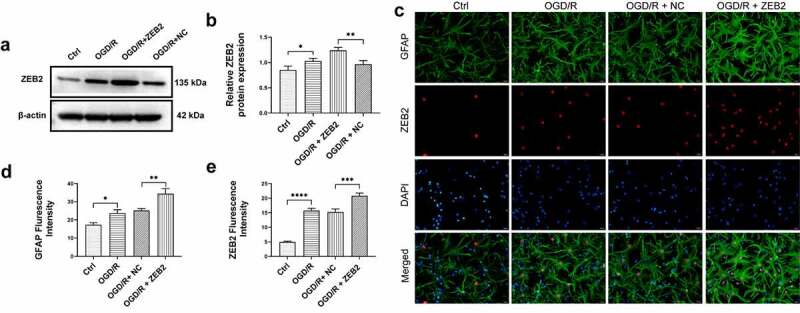
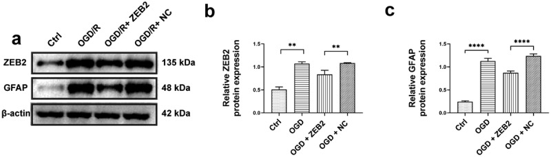
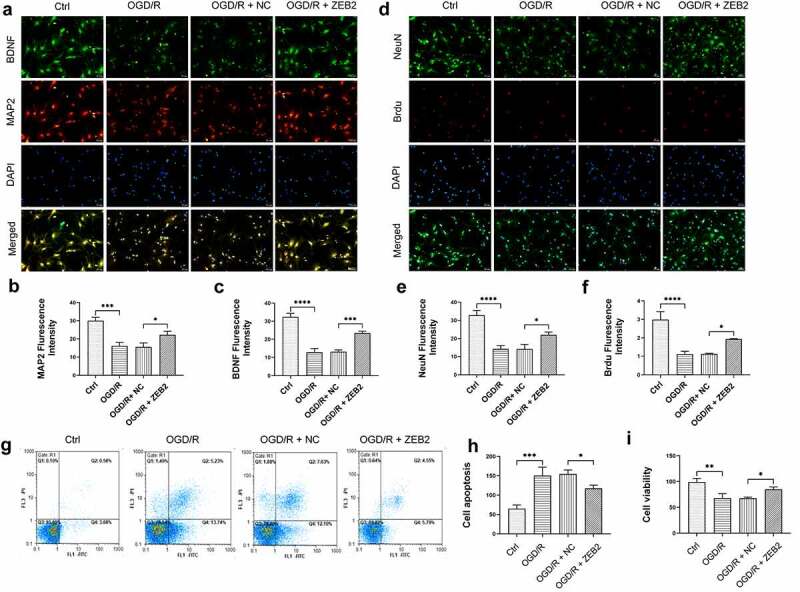
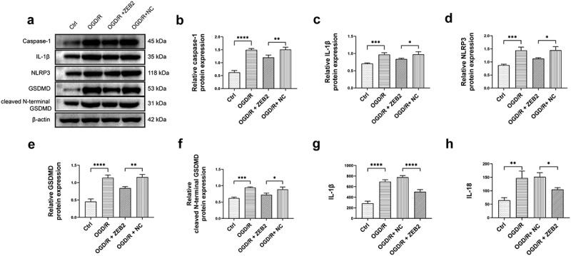
Ischemia injury can cause cell death or impairment of neuron and astrocytes, and thus induce loss of nerve function. central nervous systems injury induces an aberrant activation of astrocytes called astrogliosis. Pyroptosis, which is a kind of programmed cell death, was proved play an important role in ischemia injury. Zinc Finger E-Box Binding Homeobox 2 (ZEB2) promoted neuron astrogliosis, which play a protected role in neuron regeneration. However, its precise mechanism remains unclear. This study investigated the mechanism of ZEB2 on astrogliosis and neuron regeneration after cerebral ischemia reperfusion condition. To confirm our hypothesis, Neurons and astrocytes were isolated from fetal Sprague Dawley rats, in vivo Middle Cerebral Artery Occlusion/reperfusion (MCAO/R) rat model and in vitro oxygen-glucose deprivation/reperfusion (OGD/R)-treated astrocytes and neurocytes model were constructed. Our results showed that ZEB2 was expressed in nucleus of astrocyte and upregulated after OGD/R induction, ZEB2 promoted astrogliosis. Further upregulation of ZEB2 promoted the astrogliosis, which promoted neuron proliferation and regeneration by decreased pyroptosis. Moreover, this finding was further confirmed in vivo MCAO/R rat model. Overexpression of ZEB2 promoted astrogliosis, which decreased infarct volume and accumulated recovery of neurological function by alleviated pyroptosis. This finding revealed that ZEB2 was a regulator of the astrogliosis after ischemia/reperfusion injury, and then astrogliosis promoted neuron regeneration via decreased neuron pyroptosis. Therefore, ZEB2 may be a potential therapeutic target for ischemia/reperfusion injury.
Keywords: Mcao/R; ZEB2; astrocyte; neural regeneration; pyroptosis.
Conflict of interest statement
No potential conflict of interest was reported by the author(s).
Figures
Similar articles
-
Blocking epidermal growth factor receptor attenuates reactive astrogliosis through inhibiting cell cycle progression and protects against ischemic brain injury in rats.J Neurochem. 2011 Nov;119(3):644-53. doi: 10.1111/j.1471-4159.2011.07446.x. Epub 2011 Sep 28. J Neurochem. 2011. PMID: 21883215
-
TLR3 ligand Poly IC Attenuates Reactive Astrogliosis and Improves Recovery of Rats after Focal Cerebral Ischemia.CNS Neurosci Ther. 2015 Nov;21(11):905-13. doi: 10.1111/cns.12469. CNS Neurosci Ther. 2015. PMID: 26494128 Free PMC article.
-
Lipocalin-2-mediated astrocyte pyroptosis promotes neuroinflammatory injury via NLRP3 inflammasome activation in cerebral ischemia/reperfusion injury.J Neuroinflammation. 2023 Jun 23;20(1):148. doi: 10.1186/s12974-023-02819-5. J Neuroinflammation. 2023. PMID: 37353794 Free PMC article.
-
Astrogliosis and glial scar in ischemic stroke - focused on mechanism and treatment.Exp Neurol. 2025 Mar;385:115131. doi: 10.1016/j.expneurol.2024.115131. Epub 2024 Dec 27. Exp Neurol. 2025. PMID: 39733853 Review.
-
Progress in Research on Regulated Cell Death in Cerebral Ischaemic Injury After Cardiac Arrest.J Cell Mol Med. 2025 Feb;29(3):e70404. doi: 10.1111/jcmm.70404. J Cell Mol Med. 2025. PMID: 39936900 Free PMC article. Review.
Cited by
-
Comprehensive analysis of genetic associations and single-cell expression profiles reveals potential links between migraine and multiple diseases: a phenome-wide association study.Front Neurol. 2024 Feb 7;15:1301208. doi: 10.3389/fneur.2024.1301208. eCollection 2024. Front Neurol. 2024. PMID: 38385040 Free PMC article.
-
LncRNA THRIL Functions as a Marker for Carotid Artery Stenosis and Affects the Biological Function of Human Aortic Endothelial Cell.J Inflamm Res. 2023 Jun 8;16:2437-2446. doi: 10.2147/JIR.S409679. eCollection 2023. J Inflamm Res. 2023. PMID: 37313306 Free PMC article.
-
Crosstalk Between miRNA and Protein Expression Profiles in Nitrate-Exposed Brain Cells.Mol Neurobiol. 2023 Jul;60(7):3855-3872. doi: 10.1007/s12035-023-03316-9. Epub 2023 Mar 27. Mol Neurobiol. 2023. PMID: 36971918
-
The interplay of transition metals in ferroptosis and pyroptosis.Cell Div. 2024 Aug 3;19(1):24. doi: 10.1186/s13008-024-00127-9. Cell Div. 2024. PMID: 39097717 Free PMC article. Review.
-
The opposite effect of ELP4 and ZEB2 on TCF7L2-mediated microglia polarization in ischemic stroke.J Cell Commun Signal. 2025 Jan 16;19(1):e12061. doi: 10.1002/ccs3.12061. eCollection 2025 Mar. J Cell Commun Signal. 2025. PMID: 39822731 Free PMC article.
References
-
- Kerendi F, Kin H, Halkos ME, et al. Remote postconditioning. brief renal ischemia and reperfusion applied before coronary artery reperfusion reduces myocardial infarct size via endogenous activation of adenosine receptors. Basic Res Cardiol. 2005;100(5):404–412. - PubMed
-
- Suzuki K, Matsumaru, Y, Takeuchi, M, et al. Effect of mechanical thrombectomy without vs with intravenous thrombolysis on functional outcome among patients with acute ischemic stroke: the SKIP randomized clinical trial. JAMA. 2021 Jan 19;325(3):244–253. doi:10.1001/jama.2020.23522. - DOI - PMC - PubMed
MeSH terms
Substances
LinkOut - more resources
Full Text Sources
Research Materials