. 2021 Dec 1;28(1):82.
doi: 10.1186/s12929-021-00780-0.

A nuanced role of the small loop of hepatitis B virus small envelope protein in virion morphogenesis and secretion

Affiliations

A nuanced role of the small loop of hepatitis B virus small envelope protein in virion morphogenesis and secretion

Chih-Hsu Chang et al. J Biomed Sci. .

Abstract

Background: The virion secretion mechanism of human hepatitis B virus (HBV) remains to be investigated. In our current study, we characterized a reverse transcriptase mutant, which changed from the YMDD motif to YMHA. We noted that this mutant YMHA secreted no virions in the medium. Because of the overlapping open reading frame (ORF) between the polymerase and the envelope genes, the lack of virion secretion is likely due to corresponding concurrent mutations in a small loop of the envelope protein (HBsAg, HBV surface antigen). In literature, small loop mutations are thought to affect virion secretion of hepatitis delta virus (HDV), but not HBV.

Methods: Here, we revisited the relationship between the small loop and virion secretion by site-directed mutagenesis and native agarose gel electrophoresis.

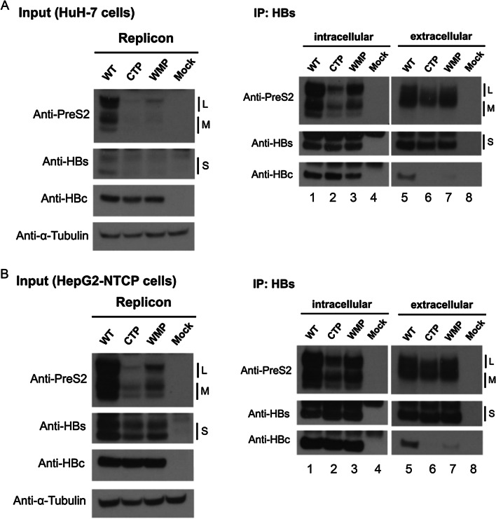
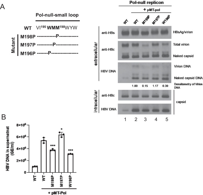
Results: A proline substitution at residue 196 or 198 in the small loop blocked both HBV genome-containing and genome-free virion secretion, but not the secretion of 22-nm HBsAg subviral particles. Surprisingly, a leucine substitution at residue 196 enhanced genome-containing virion secretion. It is also intriguing that a proline-197, sandwiched by residue 196 and 198, exhibited no apparent defect in secreted virions, with or without containing an HBV genome. By complementation assay, we demonstrated that the wild type small envelope protein alone is sufficient to rescue the virion secretion defect of a small loop mutant M198P.

Conclusions: The effect of the small loop mutation of HBV small envelope protein on virion secretion is position-dependent. It warrants further investigation how the small loop of HBsAg plays a subtle role in HBV morphogenesis and secretion of virions with or without containing an HBV genome.

Keywords: Envelope protein; HBV; Hepatitis B virus; Proline substitution; Small loop; Virion secretion.


Conflict of interest statement

We declare no competing financial or non-financial interests. The funders had no role in the design of the study; in the collection, analyses, or interpretation of data; in the writing of the manuscript, or in the decision to publish the results.

Figures

Fig. 1
Fig. 1
HBV virion secretion is blocked by mutations within a cytosolic small loop of the envelope protein (HBsAg). A The predicted topology of the folded HBV envelope protein on the ER membrane contains a cytosolic large loop CYL-I and a small loop CYL-II. A conceptual model for HBV virion assembly and secretion postulates a matrix role for these two cytosolic loops in their interactions with nucleocapsids. B A motif YMDD in the wild type reverse transcriptase (RT) domain has been engineered into a YMHA motif. Plasmid DNAs of wild type and mutant HBV DNAs were transfected into a human hepatoma cell line HuH-7. This YMHA mutant secreted no virions in the medium as detected by native agarose gel electrophoresis and Western blot analysis using an anti-HBc antibody. The polymerase gene overlaps with the envelope gene. C The YMHA polymerase mutation caused simultaneous changes of amino acid sequences from WMM to CTP at position 196–198 within the small loop of the envelope protein. The CTP triple mutations were dissociated by analyzing individual mutations 196C, 197T, and 198P, respectively. Right panel: Lack of virion secretion was detected only in the triple mutant CTP and a single proline substitution mutant 198P. Red asterisk * highlights the strongly reduced levels of HBc core protein signal of total virions in mutants CTP and M198P
Fig. 2
Fig. 2
No apparent difference in the subcellular distribution of HBV envelope HBs and core protein HBc between wild type HBV and mutants containing a proline substitution at the small loop of HBs. The intracellular distributions of HBs and HBc were examined by confocal microscopy using anti-surface antibody (green) and anti-core antibody (red)
Fig. 3
Fig. 3
Similar core-envelope interactions were detected between wild type and small loop mutants in HuH7 cells and HepG2-NTCP cells by immunoprecipitation assay and Western blot analysis. A, B Bead-associated anti-HBs antibody was used to immunoprecipitate intracellular lysate, followed by Western blot analyses using anti-preS2, anti-HBs and anti-core antibodies. Intracellular input. B While intracellular core protein signals were similar between WT and mutants (lanes 1–3), no core protein signal and virion secretion were observed in the extracellular compartment (lanes 6–7). L, M, and S: large, middle and small envelope protein
Fig. 4
Fig. 4
Secretion of genome-containing virions is also affected by proline substitution in the small loop at amino acid 196–198 in a position-dependent manner. A HuH-7 cells was cotransfected with a polymerase-deficient replicon plasmid and a wild type polymerase expression vector (pMT-pol). Surprisingly, unlike mutants 196P and 198P, a proline substitution at amino acid 197 resulted in no virion secretion deficiency. Secreted viral particles in the media were enriched by PEG before native agarose gel, followed by Western and Southern blot analyses. Lane 1: A very faint and weak HBV DNA signal can still be visualized from the Pol-null replicon in both intracellular and extracellular panels. Under the strong CMV promoter in the context of pCHT-9/3091 (Materials and Methods), this Pol-null replicon is a bit leaky in polymerase expression and viral DNA synthesis. The virion-associated DNAs were quantified by densitometry and Image J software. B Virion-associated HBV DNA genome in the supernatant of HuH-7 cells was analyzed by qPCR. HBV virions were immunoprecipitated with a rabbit anti-HBs antibody and treated with nuclease digestion before viral DNA extraction. HBV DNA signals of small loop mutants cotransfected with pMT-Pol were compared individually to the WT-HBV control. GE: genomic equivalent. Bar graph statistics by one-way ANOVA tests. ***P < 0.001; *P < 0.05
Fig. 5
Fig. 5
Two lamivudine-resistant polymerase mutants are normal in virion secretion. A Polymerase mutants YIDD created corresponding changes in the overlapping envelope ORF—W196L and W196S. B No apparent defect in virion secretion was detected in two polymerase YIDD mutants containing envelope mutations W196L and W196S, respectively. Mutant W196L exhibited increased secretion of genome-containing virions. Virion-associated DNAs were quantified by densitometry and Image J software. C Virion-associated HBV DNA was analyzed by qPCR as described in Fig. 4B. HBV DNA signals of small loop mutants cotransfected with pMT-Pol were compared individually to the WT-HBV control. GE: genomic equivalent. Bar graph statistics by one-way ANOVA tests. ***P < 0.001; **P < 0.01
Fig. 6
Fig. 6
Only the small envelope protein can rescue the virion secretion defect of the proline substitution mutant M198P. A A cartoon illustrates that the envelope protein ORF can encode a total of 6 different protein products, including large, middle and small envelope proteins. A proline substitution mutation (red asterisk*) is supposed to be present in all 6 envelope proteins. B Western blot analysis detected respective protein products from large, middle and small envelope protein expression vectors. This expression vector is a core-null replicon, which contains no core protein, and is expressing a wild type polymerase and various mutant envelope proteins. Left panel outlines the respective mutations at the initiation codons of large, middle, and small envelope proteins. C Cotransfection with the core-null replicon containing large or middle envelope protein did not restore virion secretion. Only the small envelope protein can successfully rescue the virion secretion defect of the small loop mutant M198P. Red asterisk * highlights the HBc core protein signal and HBV DNA signal by complementation with the small S envelope protein. Virion-associated DNAs were quantified by densitometry and Image J software. D Virion-associated HBV DNA was analyzed by qPCR as described in Fig. 4B. HBV DNA signals of the complementation experiments were compared individually to the mutant M198P. GE: genomic equivalent. Bar graph statistics by one-way ANOVA tests. ***P < 0.001

Similar articles

Cited by

References

    1. Blumberg BS. Hepatitis B virus, the vaccine, and the control of primary cancer of the liver. Proc Natl Acad Sci U S A. 1997;94:7121–7125. - PMC - PubMed
    1. Shih C, Yang CC, Choijilsuren G, Chang CH, Liou AT. Hepatitis B virus. Trends Microbiol. 2018;26(4):386–387. - PubMed
    1. Shih C, Chou SF, Yang CC, Huang JY, Choijilsuren G, Jhou RS. Control and eradication strategies of hepatitis B virus. Trends Microbiol. 2016;24(9):739–749. - PubMed
    1. Sureau C, Negro F. The hepatitis delta virus: replication and pathogenesis. J Hepatol. 2016;64:S102–S116. - PubMed
    1. Gerin JL, Ford EC, Purcell RH. Biochemical characterization of Australia antigen. Evidence for defective particles of hepatitis B virus. Am J Pathol. 1975;81(3):651–668. - PMC - PubMed

MeSH terms

Substances