

Cancer associated-fibroblast-derived exosomes in cancer progression
- PMID: 34852849
- PMCID: PMC8638446
- DOI: 10.1186/s12943-021-01463-y
Cancer associated-fibroblast-derived exosomes in cancer progression
Abstract
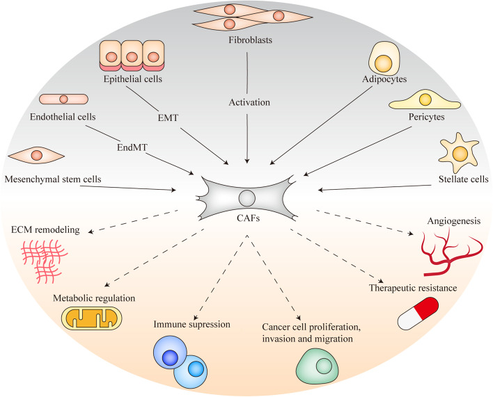
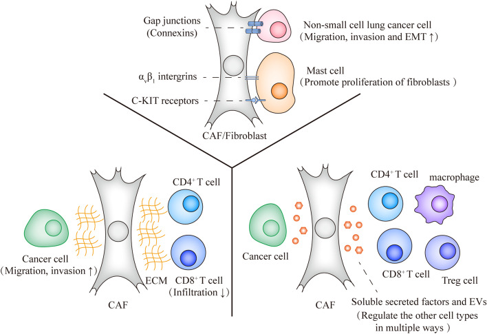
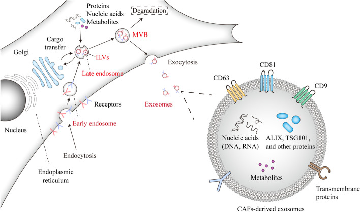
To identify novel cancer therapies, the tumor microenvironment (TME) has received a lot of attention in recent years in particular with the advent of clinical successes achieved by targeting immune checkpoint inhibitors (ICIs). The TME consists of multiple cell types that are embedded in the extracellular matrix (ECM), including immune cells, endothelial cells and cancer associated fibroblasts (CAFs), which communicate with cancer cells and each other during tumor progression. CAFs are a dominant and heterogeneous cell type within the TME with a pivotal role in controlling cancer cell invasion and metastasis, immune evasion, angiogenesis and chemotherapy resistance. CAFs mediate their effects in part by remodeling the ECM and by secreting soluble factors and extracellular vesicles. Exosomes are a subtype of extracellular vesicles (EVs), which contain various biomolecules such as nucleic acids, lipids, and proteins. The biomolecules in exosomes can be transmitted from one to another cell, and thereby affect the behavior of the receiving cell. As exosomes are also present in circulation, their contents can also be explored as biomarkers for the diagnosis and prognosis of cancer patients. In this review, we concentrate on the role of CAFs-derived exosomes in the communication between CAFs and cancer cells and other cells of the TME. First, we introduce the multiple roles of CAFs in tumorigenesis. Thereafter, we discuss the ways CAFs communicate with cancer cells and interplay with other cells of the TME, and focus in particular on the role of exosomes. Then, we elaborate on the mechanisms by which CAFs-derived exosomes contribute to cancer progression, as well as and the clinical impact of exosomes. We conclude by discussing aspects of exosomes that deserve further investigation, including emerging insights into making treatment with immune checkpoint inhibitor blockade more efficient.
Keywords: CAFs; TME; biomarkers; cancer cells; exosomes; immune cells.
© 2021. The Author(s).
Conflict of interest statement
The authors declare that they have no competing interests.
Figures
References
-
- Hanahan D, Weinberg RA. Hallmarks of cancer: the next generation. Cell. 2011;144(5):646–674. - PubMed
-
- Herschman HR. Primary response genes induced by growth factors and tumor promoters. Annu Rev Biochem. 1991;60:281–319. - PubMed
-
- Dvorak HF. Tumors: Wounds That Do Not Heal-A Historical Perspective with a Focus on the Fundamental Roles of Increased Vascular Permeability and Clotting. Semin Thromb Hemost. 2019;45(6):576–592. - PubMed
-
- Dvorak HF. Tumors: wounds that do not heal. Similarities between tumor stroma generation and wound healing. N Engl J Med. 1986;315(26):1650–1659. - PubMed
-
- Sundaram GM, Quah S, Sampath P. Cancer: the dark side of wound healing. FEBS J. 2018;285(24):4516–4534. - PubMed
Publication types
MeSH terms
Substances
Grants and funding
LinkOut - more resources
Full Text Sources
Medical
Research Materials
