

Digoxin targets low density lipoprotein receptor-related protein 4 and protects against osteoarthritis
- PMID: 34853001
- PMCID: PMC9082564
- DOI: 10.1136/annrheumdis-2021-221380
Digoxin targets low density lipoprotein receptor-related protein 4 and protects against osteoarthritis
Abstract
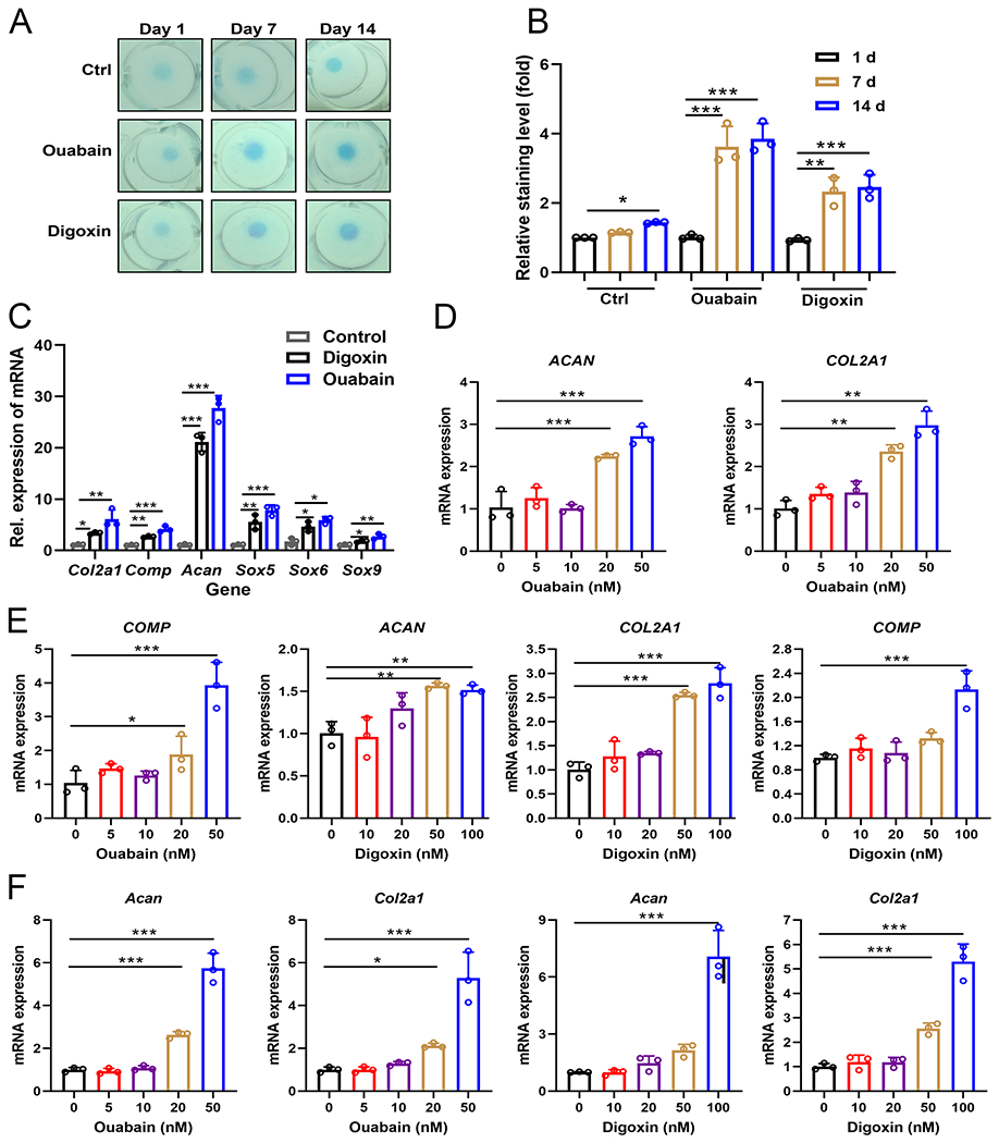
Objectives: Dysregulated chondrocyte metabolism is closely associated with the pathogenesis of osteoarthritis (OA). Suppressing chondrocyte catabolism to restore cartilage homeostasis has been extensively explored, whereas far less effort has been invested toward enhancing chondrocyte anabolism. This study aimed to repurpose clinically approved drugs as potential stimulators of chondrocyte anabolism in treating OA.
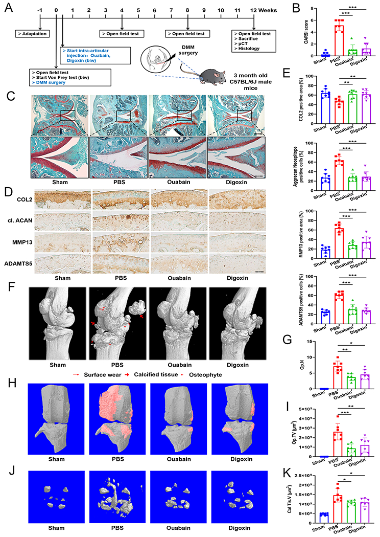
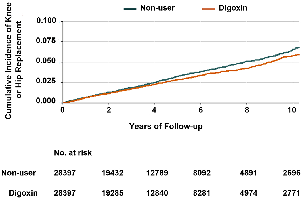
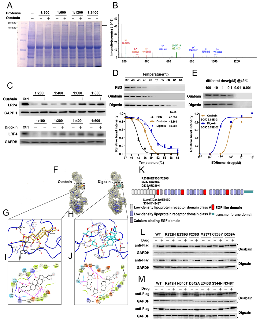
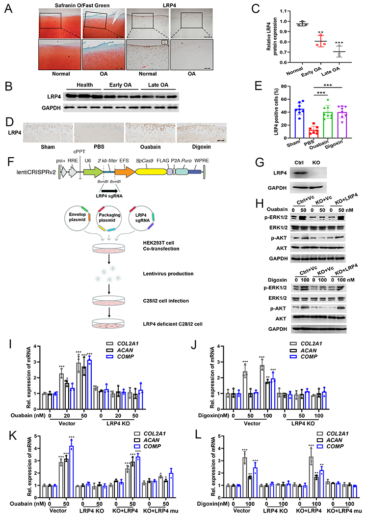
Methods: Screening of a Food and Drug Administration-approved drug library; Assays for examining the chondroprotective effects of digoxin in vitro; Assays for defining the therapeutic effects of digoxin using a surgically-induced OA model; A propensity-score matched cohort study using The Health Improvement Network to examine the relationship between digoxin use and the risk of joint OA-associated replacement among patients with atrial fibrillation; identification and characterisation of the binding of digoxin to low-density lipoprotein receptor-related protein 4 (LRP4); various assays, including use of CRISPR-Cas9 genome editing to delete LRP4 in human chondrocytes, for examining the dependence on LRP4 of digoxin regulation of chondrocytes.
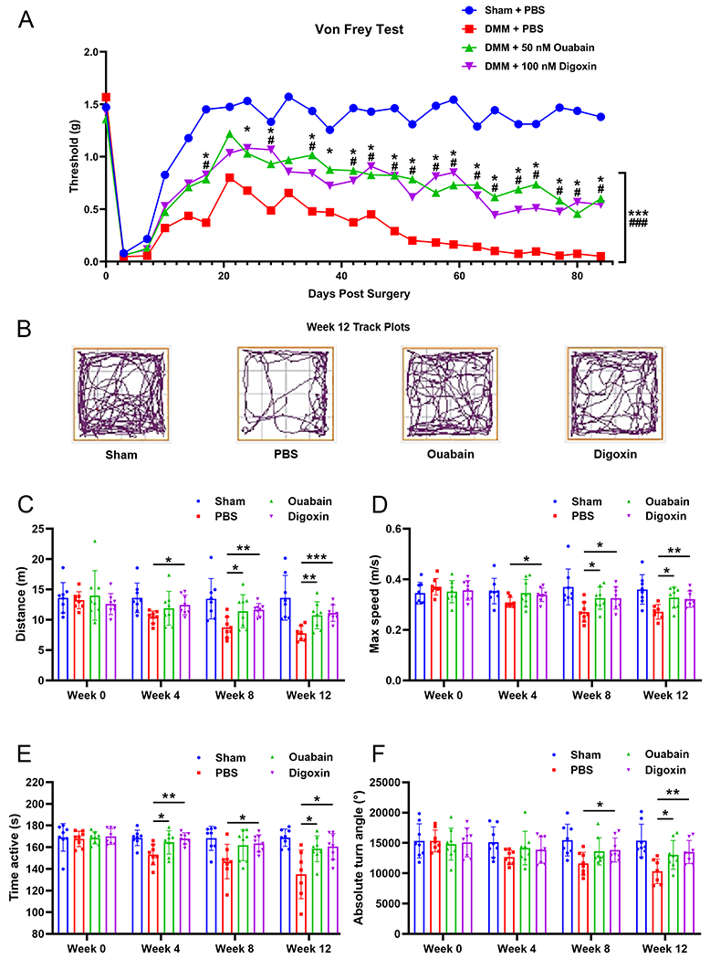
Results: Serial screenings led to the identification of ouabain and digoxin as stimulators of chondrocyte differentiation and anabolism. Ouabain and digoxin protected against OA and relieved OA-associated pain. The cohort study of 56 794 patients revealed that digoxin use was associated with reduced risk of OA-associated joint replacement. LRP4 was isolated as a novel target of digoxin, and deletion of LRP4 abolished digoxin's regulations of chondrocytes.
Conclusions: These findings not only provide new insights into the understanding of digoxin's chondroprotective action and underlying mechanisms, but also present new evidence for repurposing digoxin for OA.
Keywords: chondrocytes; osteoarthritis; therapeutics.
© Author(s) (or their employer(s)) 2022. No commercial re-use. See rights and permissions. Published by BMJ.
Conflict of interest statement
Competing interests: None declared.
Figures
Similar articles
-
Digoxin protects against intervertebral disc degeneration via TNF/NF-κB and LRP4 signaling.Front Immunol. 2023 Sep 18;14:1251517. doi: 10.3389/fimmu.2023.1251517. eCollection 2023. Front Immunol. 2023. PMID: 37790932 Free PMC article.
-
Agrin mediates chondrocyte homeostasis and requires both LRP4 and α-dystroglycan to enhance cartilage formation in vitro and in vivo.Ann Rheum Dis. 2016 Jun;75(6):1228-35. doi: 10.1136/annrheumdis-2015-207316. Epub 2015 Aug 19. Ann Rheum Dis. 2016. PMID: 26290588 Free PMC article.
-
Agkistrodon ameliorates pain response and prevents cartilage degradation in monosodium iodoacetate-induced osteoarthritic rats by inhibiting chondrocyte hypertrophy and apoptosis.J Ethnopharmacol. 2019 Mar 1;231:545-554. doi: 10.1016/j.jep.2018.12.004. Epub 2018 Dec 5. J Ethnopharmacol. 2019. PMID: 30529425
-
Molecular regulation of articular chondrocyte function and its significance in osteoarthritis.Histol Histopathol. 2011 Mar;26(3):377-94. doi: 10.14670/HH-26.377. Histol Histopathol. 2011. PMID: 21210351 Review.
-
The metabolic characteristics and changes of chondrocytes in vivo and in vitro in osteoarthritis.Front Endocrinol (Lausanne). 2024 May 24;15:1393550. doi: 10.3389/fendo.2024.1393550. eCollection 2024. Front Endocrinol (Lausanne). 2024. PMID: 38854686 Free PMC article. Review.
Cited by
-
WTAP-mediated m6A modification of FRZB triggers the inflammatory response via the Wnt signaling pathway in osteoarthritis.Exp Mol Med. 2024 Feb;56(1):156-167. doi: 10.1038/s12276-023-01135-5. Epub 2024 Jan 4. Exp Mol Med. 2024. PMID: 38172596 Free PMC article.
-
Near Infrared Responsive Gold Nanorods Attenuate Osteoarthritis Progression by Targeting TRPV1.Adv Sci (Weinh). 2024 Apr;11(16):e2307683. doi: 10.1002/advs.202307683. Epub 2024 Feb 15. Adv Sci (Weinh). 2024. PMID: 38358041 Free PMC article.
-
LRP4 and Agrin Are Modulated by Cartilage Degeneration and Involved in β-Catenin Signaling in Human Articular Chondrocytes.Int J Mol Sci. 2025 Jan 24;26(3):1007. doi: 10.3390/ijms26031007. Int J Mol Sci. 2025. PMID: 39940775 Free PMC article.
-
Exploring the anticancer mechanism of cardiac glycosides using proteome integral solubility alteration approach.Cancer Med. 2024 Sep;13(18):e70252. doi: 10.1002/cam4.70252. Cancer Med. 2024. PMID: 39350574 Free PMC article.
-
Digoxin protects against intervertebral disc degeneration via TNF/NF-κB and LRP4 signaling.Front Immunol. 2023 Sep 18;14:1251517. doi: 10.3389/fimmu.2023.1251517. eCollection 2023. Front Immunol. 2023. PMID: 37790932 Free PMC article.
References
-
- Hunter DJ, Bierma-Zeinstra S. Osteoarthritis. The Lancet 2019;393:1745–59. - PubMed
-
- Hunter DJ, Schofield D, Callander E. The individual and socioeconomic impact of osteoarthritis. Nat Rev Rheumatol 2014;10:437–41. - PubMed
-
- Martel-Pelletier J, Barr AJ, Cicuttini FM, et al. Osteoarthritis. Nat Rev Dis Primers 2016;2:16072. - PubMed
Publication types
MeSH terms
Substances
Grants and funding
LinkOut - more resources
Full Text Sources
Medical
Miscellaneous
