. 2022 Jan 12;42(2):255-263.
doi: 10.1523/JNEUROSCI.1557-21.2021. Epub 2021 Dec 1.

Hypocretin/Orexin Interactions with Norepinephrine Contribute to the Opiate Withdrawal Syndrome

Affiliations

Hypocretin/Orexin Interactions with Norepinephrine Contribute to the Opiate Withdrawal Syndrome

Ronald McGregor et al. J Neurosci. .

Abstract

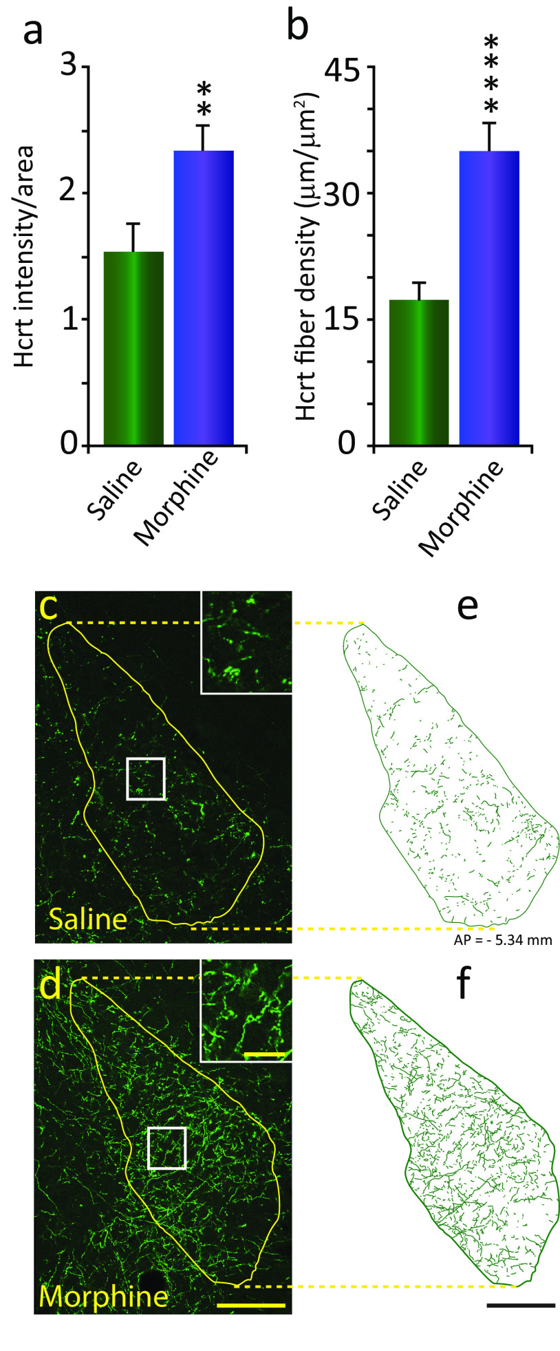
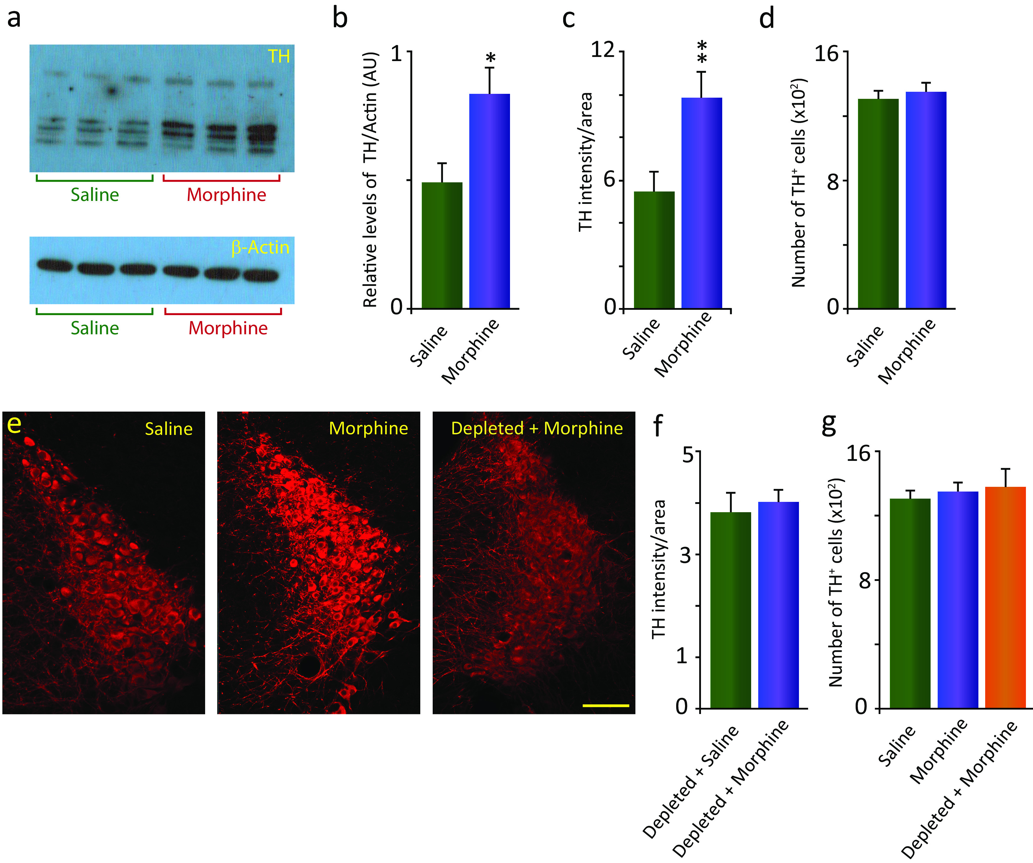
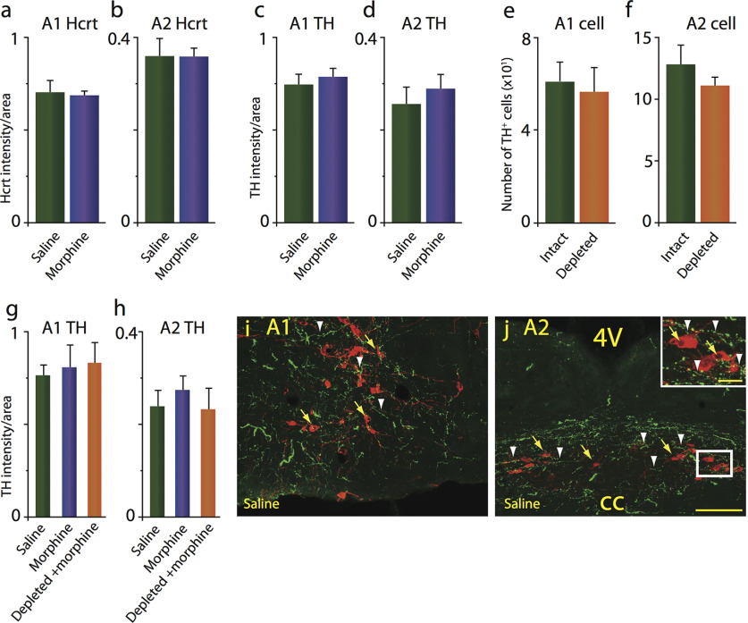
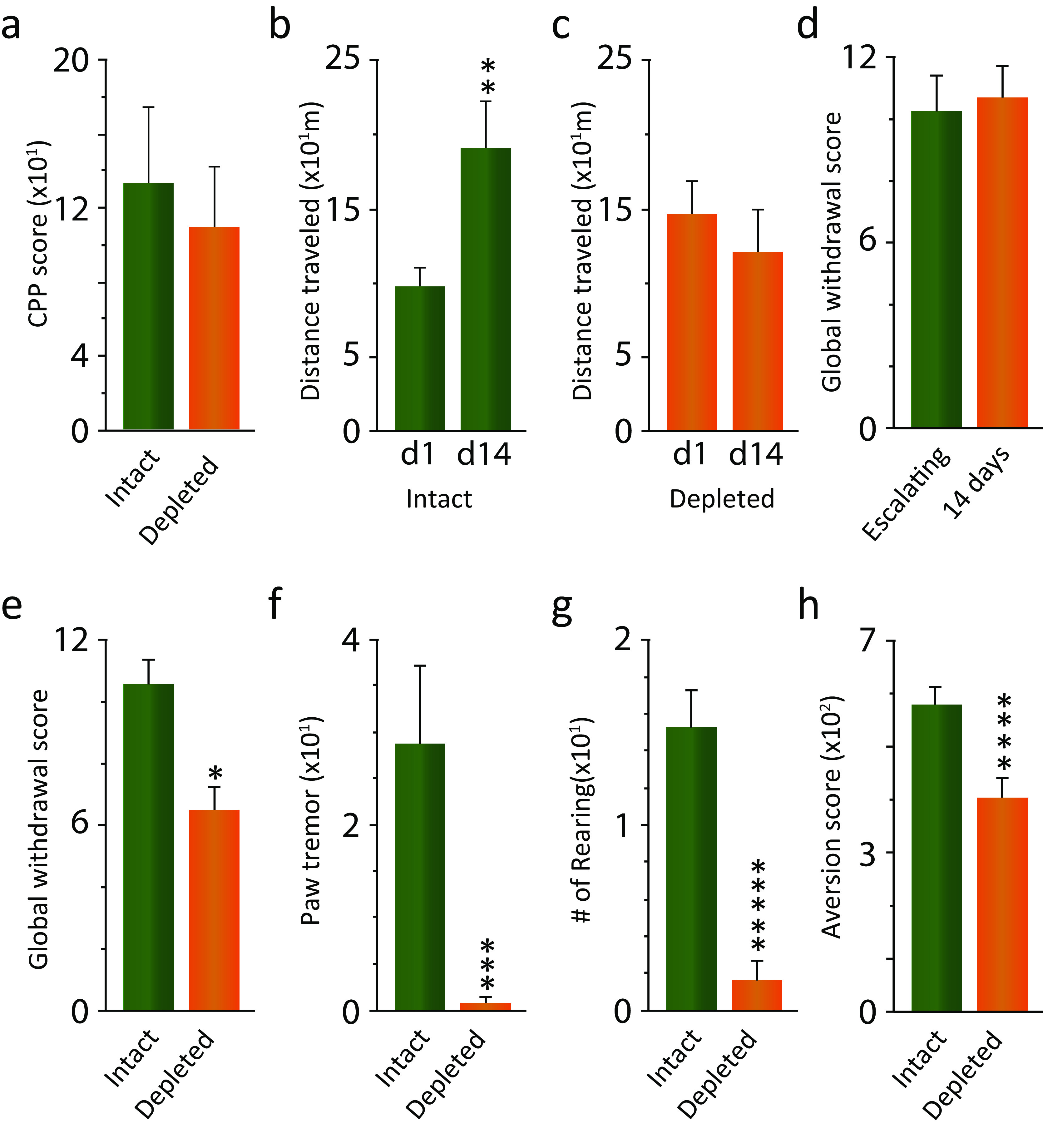
We previously found that human heroin addicts and mice chronically exposed to morphine exhibit a significant increase in the number of detected hypocretin/orexin (Hcrt)-producing neurons. However, it remains unknown how this increase affects target areas of the hypocretin system involved in opioid withdrawal, including norepinephrine containing structures locus coeruleus (LC) and A1/A2 medullary regions. Using a combination of immunohistochemical, biochemical, imaging, and behavioral techniques, we now show that the increase in detected hypocretin cell number translates into a significant increase in hypocretin innervation and tyrosine hydroxylase (TH) levels in the LC without affecting norepinephrine-containing neuronal cell number. We show that the increase in TH is completely dependent on Hcrt innervation. The A1/A2 regions were unaffected by morphine treatment. Manipulation of the Hcrt system may affect opioid addiction and withdrawal.SIGNIFICANCE STATEMENT Previously, we have shown that the hypothalamic hypocretin system undergoes profound anatomic changes in human heroin addicts and in mice exposed to morphine, suggesting a role of this system in the development of addictive behaviors. The locus coeruleus plays a key role in opioid addiction. Here we report that the hypothalamic hypocretin innervation of the locus coeruleus increases dramatically with morphine administration to mice. This increase is correlated with a massive increase in tyrosine hydroxylase expression in locus coeruleus. Elimination of hypocretin neurons prevents the tyrosine hydroxylase increase in locus coeruleus and dampens the somatic and affective components of opioid withdrawal.

Keywords: addiction; anatomy; hypocretin; locus coeruleus; opioids; withdrawal.


Figures

Figure 1.
Figure 1.
Morphine treatment increases Hcrt axonal labeling in the locus coeruleus. a, Morphine treatment (once a day, 14 d, 50 mg/kg, s.c.) resulted in a significant increase of Hcrt immunofluorescence intensity per unit of area (52.3%) compared with saline treatment in the LC of intact-DTA-Hcrt animals. All error bars in the figures are the SEM. b, This was the result of a significant increase (201.4%) in the density of Hcrt axons. c, d, Confocal images of representative sections of Hcrt axons in the LC of intact-DTA-Hcrt animals after saline (c) or morphine (d) treatment. The difference in Hcrt innervation is visually apparent. Inserts in c and d show higher magnifications of the square area, shown in white. e, f, Hcrt fiber tracings of the LC in a saline-treated (e) or morphine-treated (f) intact-DTA-Hcrt animal. The confocal images of the LC were obtained from the same section double stained for Hcrt and TH (Fig 2e, saline and morphine). Scale bars: 100 µm; insert, 20 µm. ****p = 0.004, **p = 0.022. n = 4/condition.
Figure 2.
Figure 2.
Morphine treatment increases TH immunofluorescence intensity in the LC without increasing the number of labeled cells. This effect is dependent on Hcrt innervation. a, Top, Western blot assay showing a significant increase (69.9%) in the levels of TH in intact-DTA-Hcrt animals treated with morphine (14 d, 50 mg/kg, s.c.) compared with saline animals (left, saline; right, morphine; n = 6/condition). Bottom, β-Actin control. b, Relative density of bands expressed as the ratio of TH and β-actin in animal groups. c, Elevation of TH levels was reflected in a significant increase in the TH immunofluorescence intensity per unit of area (80.1%) compared with saline in the LC (n = 4/condition). d, No significant difference in the number of TH-expressing cells in the LC in animals treated with morphine compared with saline was observed (n = 4/condition). e, Confocal images of representative sections of the LC of intact-DTA-Hcrt animals after saline (left) or morphine (middle) treatment. The difference in TH immunofluorescence is visually recognizable. When Hcrt neurons were depleted (right), there was no difference in TH immunofluorescence between animals injected with morphine and animals injected with saline (n = 4/condition). f, Group data showing no significant difference in TH labeling in depleted-DTA-Hcrt mice given saline and depleted-DTA-Hcrt mice given 14 d of 50 mg/kg morphine. g, There is no significant difference in the number of TH-expressing cells in the LC of mice lacking Hcrt neurons and treated with morphine compared with intact animals treated with saline or morphine (n = 4/condition). Scale bar, 100 µm. **p = 0.029, *p = 0.035.
Figure 3.
Figure 3.
Morphine treatment does not alter Hcrt axon density, TH levels, or TH cell numbers in the A1/A2 medullary norepinephrine-containing regions. a, b, Intact-DTA-Hcrt animals treated with morphine (14 d, 50 mg/kg) did not show a significant change in Hcrt axonal labeling in the A1 (a) and the A2 (b) regions. c, d, TH levels remained comparable to baseline levels after morphine treatment in the A1 (c) and A2 (d) regions. e, f, TH+ cell counts were unaffected by the elimination of Hcrt neurons in depleted-DTA-Hcrt animals in the A1 (e) or the A2 (f) regions. g, h, Deletion of Hcrt neurons did not affect TH levels in the A1 (g) and the A2 (h) regions after morphine treatment compared with intact-DTA-Hcrt animals treated with saline or morphine. i, j, Confocal images of representative sections of the A1 (i) and A2 (j) of an intact-DTA-Hcrt animal after saline treatment, illustrating the distribution of Hcrt fibers in green (white arrowheads) and TH+ neurons in red (yellow arrows). Insert in j shows a higher magnification of white square. Scale bars: 100 µm; insert, 20 µm. cc, Central canal; 4V, fourth ventricle. n = 4/condition.
Figure 4.
Figure 4.
The rewarding properties of morphine are not significantly affected by the lack of Hcrt neurons. But Hcrt depletion prevents locomotor sensitization and reduces both the somatic signs of naloxone-precipitated withdrawal and naloxone conditioned place aversion in morphine-dependent animals. a, Preference for the chamber paired with morphine did not significantly differ between intact-DTA-Hcrt and depleted-DTA-Hcrt animals (n = 12/condition). b, Intact-DTA-Hcrt animals developed sensitization to repeated doses of morphine (n = 6). c, Depletion of Hcrt neurons completely eliminated this addiction-linked behavior (n = 6). d, Five days of escalating morphine and 14 d daily 50 mg/kg morphine doses resulted in comparable global symptom withdrawal scores (n = 6/condition). e, Elimination of Hcrt neurons in depleted-DTA-Hcrt animals resulted in a significant reduction in the global withdrawal score compared with intact-DTA-Hcrt animals. f, g, Paw tremor (f) and rearing (g) showed the largest reduction of the somatic withdrawal signs (n = 6/condition). h, Lack of Hcrt neurons significantly reduced the aversion to the chamber paired with naloxone (n = 10/condition). *****p = 0.0001, ****p = 0.002, ***p = 0.007, **p = 0.04, *p = 0.043.

Similar articles

Cited by

References

    1. Akbarian S, Rios M, Liu RJ, Gold SJ, Fong HF, Zeiler S, Coppola V, Tessarollo L, Jones KR, Nestler EJ, Aghajanian GK, Jaenisch R (2002) Brain-derived neurotrophic factor is essential for opiate-induced plasticity of noradrenergic neurons. J Neurosci 22:4153–4162. - PMC - PubMed
    1. Aston-Jones G, Smith RJ, Sartor GC, Moorman DE, Massi L, Tahsili-Fahadan P, Richardson KA (2010) Lateral hypothalamic orexin/hypocretin neurons: a role in reward-seeking and addiction. Brain Res 1314:74–90. 10.1016/j.brainres.2009.09.106 - DOI - PMC - PubMed
    1. Baimel C, Bartlett SE, Chiou LC, Lawrence AJ, Muschamp JW, Patkar O, Tung LW, Borgland SL (2015) Orexin/hypocretin role in reward: implications for opioid and other addictions. Br J Pharmacol 172:334–348. 10.1111/bph.12639 - DOI - PMC - PubMed
    1. Barateau L, Jaussent I, Lopez R, Boutrel B, Leu-Semenescu S, Arnulf I, Dauvilliers Y (2016) Smoking, alcohol, drug use, abuse and dependence in narcolepsy and idiopathic hypersomnia: a case-control study. Sleep 39:573–580. 10.5665/sleep.5530 - DOI - PMC - PubMed
    1. Bayard S, Dauvilliers YA (2013) Reward-based behaviors and emotional processing in human with narcolepsy-cataplexy. Front Behav Neurosci 7:50. 10.3389/fnbeh.2013.00050 - DOI - PMC - PubMed

Publication types