. 2021 Dec 7;118(49):e2101006118.
doi: 10.1073/pnas.2101006118.

Observing the base-by-base search for native structure along transition paths during the folding of single nucleic acid hairpins

Affiliations

Observing the base-by-base search for native structure along transition paths during the folding of single nucleic acid hairpins

Noel Q Hoffer et al. Proc Natl Acad Sci U S A. .

Abstract

Biomolecular folding involves searching among myriad possibilities for the native conformation, but the elementary steps expected from theory for this search have never been detected directly. We probed the dynamics of folding at high resolution using optical tweezers, measuring individual trajectories as nucleic acid hairpins passed through the high-energy transition states that dominate kinetics and define folding mechanisms. We observed brief but ubiquitous pauses in the transition states, with a dwell time distribution that matched microscopic theories of folding quantitatively. The sequence dependence suggested that pauses were dominated by microbarriers from nonnative conformations during the search by each nucleotide residue for the native base-pairing conformation. Furthermore, the pauses were position dependent, revealing subtle local variations in energy-landscape roughness and allowing the diffusion coefficient describing the microscopic dynamics within the barrier to be found without reconstructing the shape of the energy landscape. These results show how high-resolution measurements can elucidate key microscopic events during folding to test fundamental theories of folding.

Keywords: diffusion; folding; kinetics; optical tweezers; transition states.


Conflict of interest statement

The authors declare no competing interest.

Figures
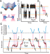
Fig. 1.
Fig. 1.
Measuring pauses within individual transition paths. (A) Transition paths represent the brief portion of a folding trajectory spent crossing the barrier between states (red), in contrast to the majority spent fluctuating within the potential wells (gray). (B) DNA hairpins attached to handles (purple) linked to beads (blue) were held in laser traps (pink) applying tension. (C) End-to-end extension of hairpin 30R50/T4 from ref. fluctuating in equilibrium between folded and unfolded states under conditions of constant trap separation. Locations of folded and unfolded states denoted respectively by xF and xU (orange), boundaries of barrier region by x1 and x2 (cyan). (Inset) Hairpin sequence. (D) Transition paths were identified as those parts of the trajectories (red) crossing between x1 and x2. (E) Transition-path trajectories (Lower, black) showed a wide range of behavior. The velocity profiles of individual trajectories (Upper, blue) were obtained by numerical differentiation of the spline-smoothed trajectories (Lower, gray) of the extension. Pause locations and durations were identified from the portions of the trajectories in which the speed remained under a threshold (Upper, gray) equal to 10% of the average transition speed (magenta bands).
Fig. 2.
Fig. 2.
Pausing within the transition states. (A) Probability density for finding pauses of a given duration at a given location within the barrier region for hairpin 30R50/T4. Extension changes by 0.3 nm per base pair. (B) The distribution of pause locations is the same for folding (black) and unfolding (red). Pauses occurred ubiquitously across the entire transition region but were least likely to occur near the barrier top (). (C) The distribution of pause durations is the same for folding (black) and unfolding (red), dropping subexponentially in each case.
Fig. 3.
Fig. 3.
Pause durations. (A) The distribution of pause durations for hairpin 30R50/T4 (black) was poorly fit by a single-exponential decay reflecting a single rate constant (cyan) but was reasonably well fit by both the double-exponential decay expected if pauses arise from microbarriers associated with A:T and G:C base pair formation (blue) and by the log-normal distribution of rate constants expected from a microscopic theory of folding as a search through nonnative states (red). (Inset) At short durations, the pauses match expectations from the log-normal rate distribution (red) better than double-exponential decay (blue). (B) The pause duration distribution for hairpin 20R0/T4, containing only A:T base pairs, was fit well by a log-normal distribution of rate constants (red) but not by a single-exponential decay (cyan). (Inset) Pauses occurred at the same locations for both folding (black) and unfolding (red). Extension changes by 0.29 nm per base pair. (C) Similar results were found for hairpin 20R100/T4, containing only G:C base pairs. Extension changes by 0.35 nm per base pair.
Fig. 4.
Fig. 4.
Position dependence of D. (A) Calculating D as a function of position along the reaction coordinate from the average pause duration at different positions (red) reveals that D is not constant but rises to a peak near the middle of the barrier region. A similar pattern is seen from Eq. 2 using fits of the position-dependent pause durations (black). (B) D showed a different position dependence for the three hairpins studied: flat for hairpin 20R0/T4 (black), moderately peaked for hairpin 30R50/T4 (red), and strongly peaked for hairpin 20R100/T4 (blue). Error bars represent SEM. Dashed lines indicate boundaries of barrier region at x1 and x2, corresponding respectively to ρ = 1/6 and 5/6.

Similar articles

Cited by

References

    1. Bryngelson J. D., Wolynes P. G., Spin glasses and the statistical mechanics of protein folding. Proc. Natl. Acad. Sci. U.S.A. 84, 7524–7528 (1987). - PMC - PubMed
    1. Bryngelson J. D., Wolynes P. G., Intermediates and barrier crossing in a random energy model (with applications to protein folding). J. Phys. Chem. 93, 6902–6915 (1989).
    1. Buchner J., Kiefhaber T., Protein Folding Handbook (Wiley-VCH, Weinheim, 2005).
    1. Hoffer N. Q., Woodside M. T., Probing microscopic conformational dynamics in folding reactions by measuring transition paths. Curr. Opin. Chem. Biol. 53, 68–74 (2019). - PubMed
    1. Chung H. S., McHale K., Louis J. M., Eaton W. A., Single-molecule fluorescence experiments determine protein folding transition path times. Science 335, 981–984 (2012). - PMC - PubMed

Publication types

LinkOut - more resources