. 2021 Dec 1;12(1):7010.
doi: 10.1038/s41467-021-27131-9.

Counteraction between Astrin-PP1 and Cyclin-B-CDK1 pathways protects chromosome-microtubule attachments independent of biorientation

Affiliations

Counteraction between Astrin-PP1 and Cyclin-B-CDK1 pathways protects chromosome-microtubule attachments independent of biorientation

Xinhong Song et al. Nat Commun. .

Abstract

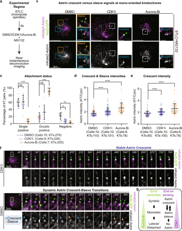
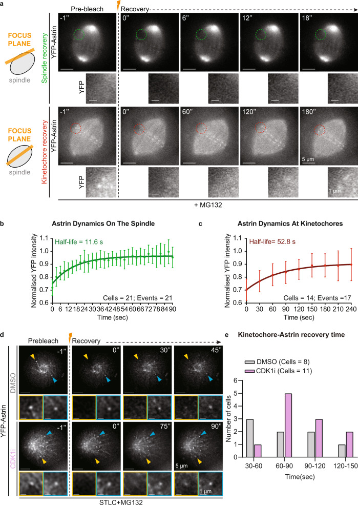
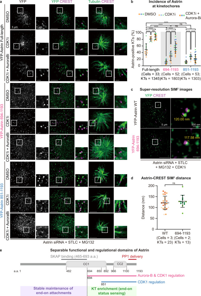
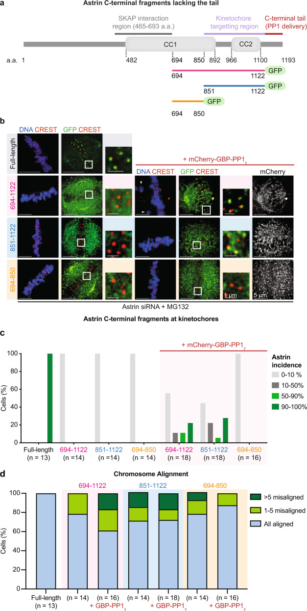
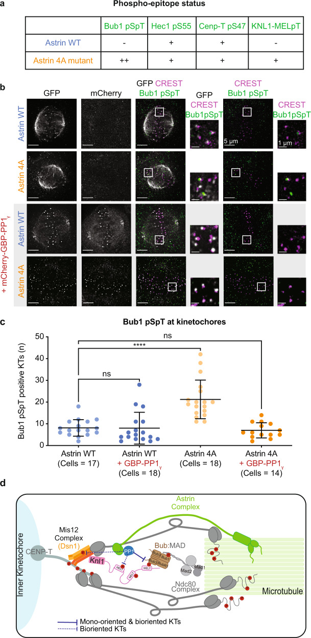
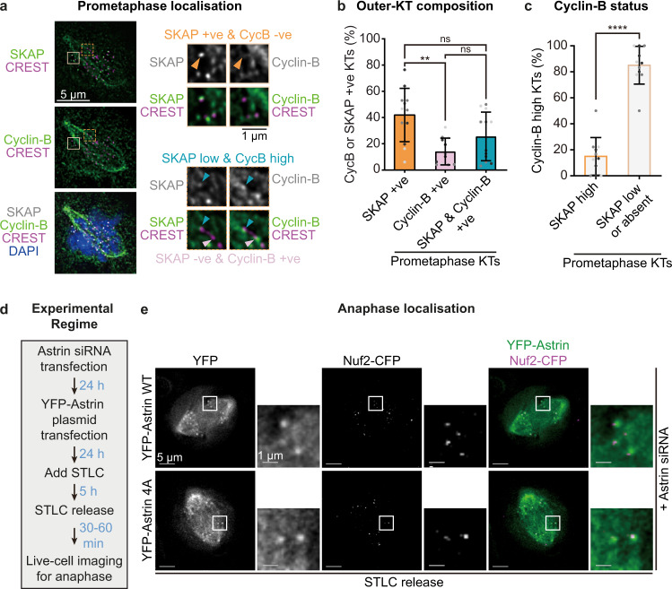
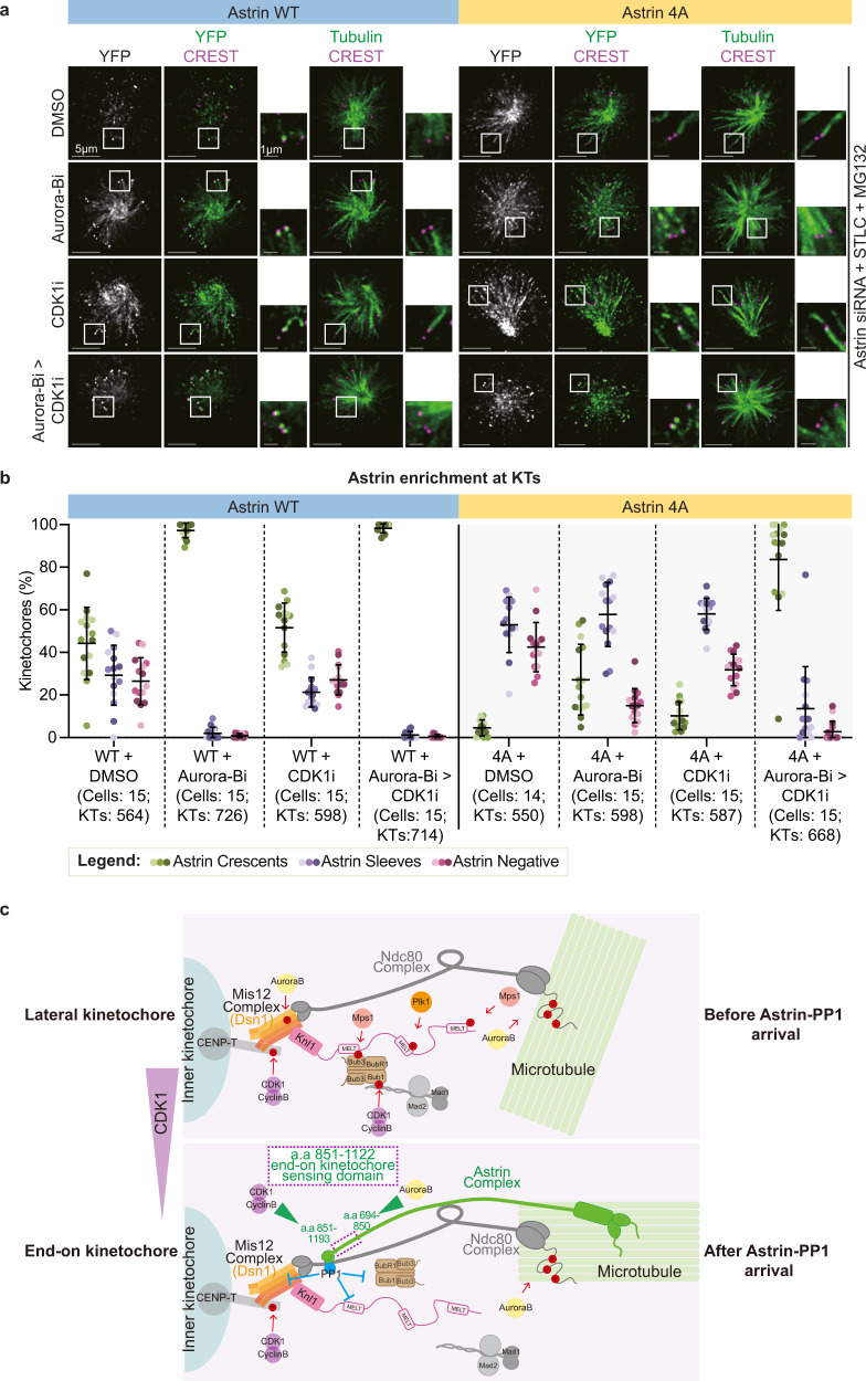
Defects in chromosome-microtubule attachment can cause chromosomal instability (CIN), frequently associated with infertility and aggressive cancers. Chromosome-microtubule attachment is mediated by a large macromolecular structure, the kinetochore. Sister kinetochores of each chromosome are pulled by microtubules from opposing spindle-poles, a state called biorientation which prevents chromosome missegregation. Kinetochore-microtubule attachments that lack the opposing-pull are detached by Aurora-B/Ipl1. It is unclear how mono-oriented attachments that precede biorientation are spared despite the lack of opposing-pull. Using an RNAi-screen, we uncover a unique role for the Astrin-SKAP complex in protecting mono-oriented attachments. We provide evidence of domains in the microtubule-end associated protein that sense changes specific to end-on kinetochore-microtubule attachments and assemble an outer-kinetochore crescent to stabilise attachments. We find that Astrin-PP1 and Cyclin-B-CDK1 pathways counteract each other to preserve mono-oriented attachments. Thus, CIN prevention pathways are not only surveying attachment defects but also actively recognising and stabilising mature attachments independent of biorientation.


Conflict of interest statement

The authors declare no competing interests.

Figures

Fig. 1
Fig. 1. CDK1 activity controls Astrin-mediated sensing of end-on attachment.
a Experimental regime shows near-instantaneous live-imaging of kinetochore fate soon after the addition of kinase inhibitors to understand end-on attachment sensing mechanisms in non-bioriented kinetochores. b Live cell images of monopolar spindles show monotelic kinetochores soon after DMSO or RO3666 treatment and syntelic kinetochores soon after ZM447439 treatment in cells co-expressing Nuf2-GFP and mKate2-Astrin. Cells were exposed to STLC for 5 h before adding CDK1 (RO3306) or Aurora-B (ZM447439) inhibitor or DMSO (control) with the proteasome inhibitor, MG132. Scale bar as indicated. Astrin crescent and sleeves are marked by blue and orange triangles, respectively. c Graph shows the percentage of monotelic (Astrin single-positive) or syntelic (Astrin double-positive) kinetochore pairs following inhibitor treatment as indicated using images as shown in b. d, e, Graph shows intensities of either Astrin crescents and sleeves (d) or Astrin crescents alone (e) in cells treated as in a. Colours in super-plot ce represent independent experimental repeats. Black bars and whiskers in the graph ce mark average value and standard deviation, respectively, across independent experimental repeats (n = 3 CDK1i and 2 Aurora-Bi repeats) independent. ‘*’ and ‘ns’ indicate statistically significant and insignificant differences, respectively, were determined using a Non-parametric two-sided Mann-Whitney test. The exact p-values can be found in Source data. f Time-lapse images of kinetochores show transitions in Astrin crescent versus sleeve at kinetochores of monopolar spindles in cells coexpressing mKate2-Astrin and Nuf2-GFP and treated with DMSO but not CDK1 inhibition. Cells were treated with STLC for 5 h and MG132 was added along with either CDK1 inhibitor or DMSO (control) as indicated and filming started soon after drug addition. The time interval between frames is 45 s. Astrin crescent and sleeves are marked by blue and orange triangles, respectively. N = 8 cells (DMSO) and 7 cells (R03306) from two independent repeats. Scale bar as indicated. g Illustration of separable roles for Aurora-B and CDK1 in regulating Astrin-mediated sensing of end-on attachments. Global reduction in CDK1 increases the amount of Astrin at kinetochores without disrupting error correction that is dependent on Aurora-B.
Fig. 2
Fig. 2. Kinetochore-autonomous regulation of Astrin dynamics at end-on attachments.
a Representative time-lapse images of Fluorescence Recovery After Photobleaching (FRAP) of HeLa cells expressing YFP-Astrin following one hour of MG132 treatment prior to photobleaching. Cropped images show the recovery of YFP intensities after bleaching one spindle area (green circle) or one kinetochore (red circle). Cartoons on the left display a cross-sectional view of the spindle showing the plane of focus (orange bar) for microtubules (top) or kinetochores (middle) of the mitotic spindle (shown as a grey ellipse). Scale bar as indicated. b and c, Graphs show curves of normalised YFP fluorescence intensities on spindle (b) or kinetochores (c), respectively, versus time from FRAP experiments as in a. Lines and whiskers mark the average and standard deviation, respectively, from two independent experiments (kinetochore data) and three independent experiments (microtubules data). The calculated half-life time of recovery is indicated in each graph. d Representative time-lapse images of Fluorescence Recovery After Photobleaching (FRAP) of HeLa cells expressing YFP-Astrin, acquired once every 15 s, following one hour of STLC treatment prior to photobleaching in the presence of CDK1 inhibitor (CDK1i, RO3306) or DMSO (control) as indicated. Cropped images outlined in yellow and blue indicate bleached and unbleached (control) kinetochore, corresponding to yellow and blue arrowheads, respectively. Scale bar as indicated. e Kinetochore-bound Astrin recovery times with or without CDK1 inhibitor obtained using movies as in d. Recovery time was visually scored to indicate the first time point (30 s bin) where Astrin signal at the kinetochore was seen following the bleaching event. The values shown are from two independent experimental repeats.
Fig. 3
Fig. 3. Molecular basis for Astrin’s dual role in maintaining and recognising mono-oriented end-on attachments.
a Representative images of immunostained cells show the localisation of YFP tagged Astrin fragments (694–1193 or 851–1193, as indicated) following 5 h of STLC treatment to increase mono-oriented kinetochores and exposed to CDK1 inhibitor (RO3306) or Aurora-B inhibitor (ZM447439) for 15 min prior to fixation. Cells were immunostained with antibodies against GFP, Tubulin and CREST (as kinetochore marker). Only instances where YFP crescent signals are sandwiched between Tubulin and CREST signals are scored as positive for YFP-Astrin recruitment at the outer-kinetochore. White boxes mark the area of cropped images. Scale bar as indicated. b Graph shows the percentage of YFP-Astrin positive kinetochores in cells expressing YFP-Astrin WT or mutant and treated with CDK1i or Aurora-B inhibitors as indicated in a. c Representative super-resolution microscopy images of cells treated as in a. Measurements in crops indicate intra-kinetochore distances between peak intensities of CREST and YFP signals. d Graph shows the average intra-kinetochore distance between CREST and YFP signals in kinetochores of cells expressing Astrin full-length protein or a 694–1193 a.a fragment. Colours in super-plots b and d represent two (DMSO) or three (CDK1i or CDK1i and Aurora-Bi) independent experimental repeats (b) or cells (d), respectively. Horizontal black bars and whiskers mark average value and standard deviation, respectively, across two (WT) or three (fragment) independent experiments. ‘*’ and ‘ns’ indicate significant and insignificant statistical differences, respectively, as determined by the Non-parametric two-sided Mann-Whitney test. Exact p-values can be found in Source data. e Schematic of Astrin fragments used to dissect Astrin’s dual role in maintaining and sensing end-on attachments. Kinetochore binding C-terminus and microtubule-binding N- terminus of Astrin contribute to the sensing and maintenance of end-on attachments, respectively. The C-terminal region of Astrin that is responsible for sensing end-on kinetochores, bears at least two domains that are differently regulated by Aurora-B and CDK1 pathways.
Fig. 4
Fig. 4. A 273 a.a region of Astrin is sufficient to sense outer-kinetochore changes at end-on attachments.
a Schematic of Astrin fragments was used to ascertain minimal kinetochore targeting domain of Astrin. b Representative images of immunostained cells show the localisation of GFP tagged Astrin fragments (as indicated) in the presence or absence of mCherry-GBP-PP1 following an hour of MG132 treatment prior to fixation. Cells were immunostained with antibodies against GFP, mCherry and CREST (a kinetochore marker) and co-stained with DAPI. White boxes mark the area of cropped images. Scale bar as indicated. White arrowheads mark unaligned chromosomes quantified in d. c, Bar graphs show the percentage of cells displaying GFP-tagged fragments of Astrin, as indicated, in the form of outer-kinetochore crescents at 90–100%, 50–90%, 10–50% or 0–10% of aligned kinetochores in images as in b. Cells coexpressing mCherry-GBP-PP1 are marked separately. d, Bar graph shows the percentage of cells with chromosome alignment defects in cells expressing GFP-C-terminal tagged fragments of Astrin with or without mCherry-GBP-PP1 and treated as in b. To assess the extent of misalignment, cells were segregated into three bins: all chromosomes aligned, 1–5 misaligned chromosomes and >5 misaligned chromosomes. Values in c and d are from three independent experimental repeats.
Fig. 5
Fig. 5. Astrin delivered PP1 lowers Bub1 phosphorylation linked to checkpoint silencing in mono-oriented and bi-oriented kinetochores.
a Table summarises the phosphorylation status of various phospho-epitopes at the outer kinetochore. ‘+’ or ‘−’ refers to phospho-epitope observed in more or less than 20% of kinetochores, respectively, as shown in Supplementary Fig. 5. b Images show the extent of phosphorylated Bub1 (pSpT) at kinetochores of cells depleted of endogenous Astrin and expressing either Astrin WT-GFP or Astrin 4A-GFP mutant alone or along with mCherry-GBP-PP1 as indicated. Cells were treated with MG132 for an hour prior to fixation for immunostaining with antibodies against GFP, Bub1 pSpT phospho-epitope and CREST anti-sera (a centromere marker). Scale bar as indicated. c Graph showing the number of kinetochores positive for phosphorylated Bub1 pSpT, determined using whole spindle Z-stacks of images as shown in b. Each dot represents values from one cell. Bars and whiskers represent mean and standard deviation, respectively, across cells from three independent experimental repeats. “*” and “ns” indicate significant and insignificant statistical differences, respectively, as determined using a Kruskall-Wallis test with Dunn’s multiple comparisons. Mean rank differences are available in the Source data. d Model of dephosphorylation events that depend on the recruitment of Astrin-PP1 at end-on kinetochores. Astrin-PP1 alters the phosphorylation of (i) Bub1 (brown) responsible for positioning Mad1 near KNL1-MELpT, (ii) Dsn1 of Mis12 complex (orange), and (iii) KNL1-MELpT (pink) sites but not those of Ndc80 (S55) or CENP-T (S47) sites (grey), indicating a spatially restricted dephosphorylation of substrates. When PP1 delivery by Astrin is compromised, Bub1 dephosphorylation is impaired on both mono-oriented and bioriented kinetochores, while KNL1 dephosphorylation is only impaired on bioriented kinetochores.
Fig. 6
Fig. 6. CDK1 and Astrin-PP1 counteract each other at the outer-kinetochore.
a Images of RPE1 cells immunostained with antibodies against Cyclin-B and SKAP, and CREST anti-sera, and co-stained with DAPI for DNA. Arrowheads mark kinetochores with low SKAP and high Cyclin-B intensities (blue), SKAP-positive (+ve) and Cyclin-B-negative (−ve) kinetochore (orange), SKAP-negative (-ve) and Cyclin-B positive (+ve) kinetochore (pink). Scale bar as indicated. b Graph of the percentage of kinetochores enriched for either Cyclin-B or SKAP or both, ascertained using images as in a. ‘*’ and ‘ns’ indicates significant and insignificant statistical difference, respectively, determined using Non-parametric Kruskal-Wallis H test combined with Dunn’s multiple comparisons test. c Graph of percentage of Cyclin-B positive kinetochores with SKAP-high or −low intensities as in a. ‘*’ indicates statistical difference tested with two-tailed Mann Whitney test. Height of bars and whiskers (b and c) mark average value and standard deviation, respectively. Dots in the same shade of grey are from the same experimental repeat (n = 723 kinetochores, 12 cells and 3 independent experimental repeats). d Experimental regime to assess anaphase kinetochores: Astrin siRNA transfected cells coexpressing Nuf2-CFP (kinetochore marker) and either YFP-Astrin WT or YFP-Astrin 4A mutant were exposed to STLC for 5 h to synchronise monopolar spindles. After STLC was washed out to release cells into anaphase for live-cell imaging. e Representative anaphase live-cell images of mono-oriented kinetochores displaying YFP-Astrin WT or 4A crescents in cells treated as in d Sample size: WT (4 cells) and 4A (7 cells) from 2 independent experimental repeats. Scale as indicated.
Fig. 7
Fig. 7. Aurora-B and CDK1 pathways separately counteract Astrin at the outer kinetochore.
a Representative images show YFP-Astrin 4A mutant localisation at kinetochores of monopolar spindles sequentially exposed to Aurora-B and CDK1 inhibitors. Cells depleted of endogenous Astrin and expressing YFP-Astrin WT or 4A mutant as indicated were treated with STLC for 45 min followed by MG132 and DMSO or ZM447439 (Aurora-Bi) or Roscovitine (CDK1i) or sequential addition of Roscovitine into ZM447439 (ZM447439 alone for 15 min with a Roscovitine supplementation for an additional 15 min, Aurora-Bi > CDK1i). Cells were immunostained with antibodies against GFP and Tubulin and CREST anti-sera. Scale bar as indicated. b Scatter plot of Astrin recruitment status showing the proportion of Astrin Crescent or Sleeve or no Astrin signal (Astrin Negative) bearing kinetochores in cells expressing YFP-Astrin WT or 4A mutant as indicated and treated with inhibitors as in a. Black bars and whiskers mark average value and standard deviation, respectively, across experimental repeats (n = 4 repeats). Each dot represents a value from one cell. The colours of dots in b represent experimental repeats. c Cartoon of Astrin regulation at end-on kinetochores: Astrin deliver a pool of PP1 that counteracts a subset of spatially-limited phosphorylation events by multiple kinases (CDK1, Aurora-B or PLK1). CDK1 and Aurora-B pathways (green arrowheads) regulate the kinetochore binding of two separable regions at Astrin C-terminus. Inhibiting CDK1 and Aurora-B activities supersedes the need for Astrin-mediated PP1 delivery, revealing a feedback loop that protects non-bioriented end-on attachments from kinase activities.

Similar articles

Cited by

References

    1. Roos U-P. Light and electron microscopy of rat kangaroo cells in mitosis. Chromosoma. 1976;54:363–385. - PubMed
    1. Cassimeris L, Rieder CL, Salmon ED. Microtubule assembly and kinetochore directional instability in vertebrate monopolar spindles: implications for the mechanism of chromosome congression. J. Cell. Sci. 1994;107:285–297. - PubMed
    1. Bajer AS. Functional autonomy of monopolar spindle and evidence for oscillatory movement in mitosis. J. Cell. Biol. 1982;93:33–48. - PMC - PubMed
    1. Hart M, Adams SD, Draviam VM. Multinucleation associated DNA damage blocks proliferation in p53-compromised cells. Commun. Biol. 2021;4:451. - PMC - PubMed
    1. Ben-David U, Amon A. Context is everything: aneuploidy in cancer. Nat. Rev. Genet. 2020;21:44–62. - PubMed

Publication types