. 2021 Dec 1;11(1):23211.
doi: 10.1038/s41598-021-01715-3.

A point-mutation in the C-domain of CMP-sialic acid synthetase leads to lethality of medaka due to protein insolubility

Affiliations

A point-mutation in the C-domain of CMP-sialic acid synthetase leads to lethality of medaka due to protein insolubility

Di Wu et al. Sci Rep. .

Abstract

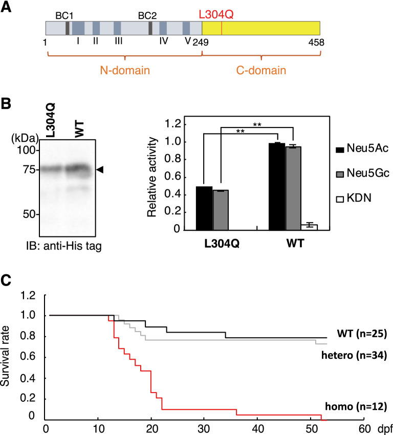
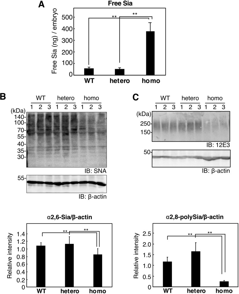
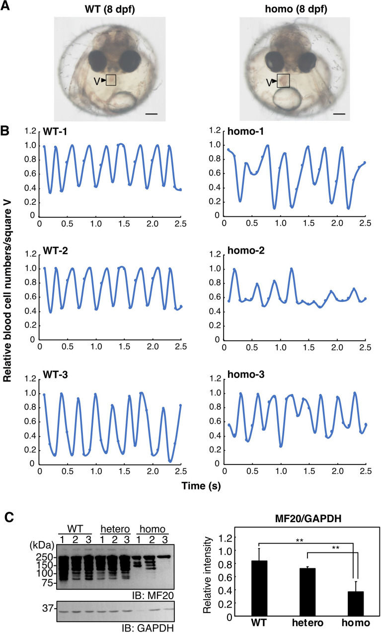
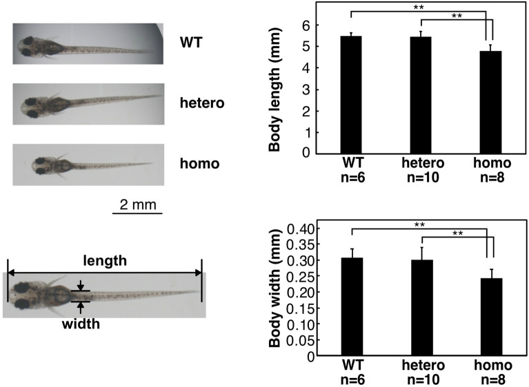
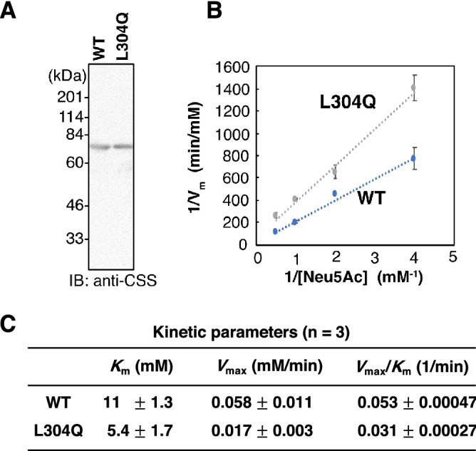
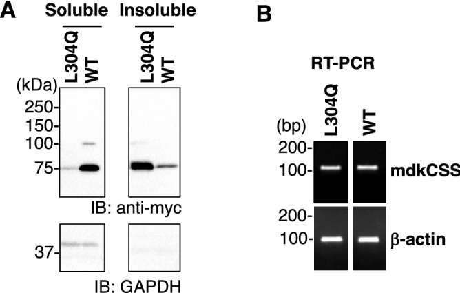
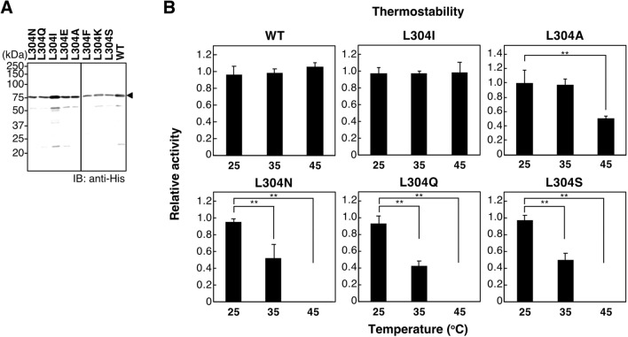
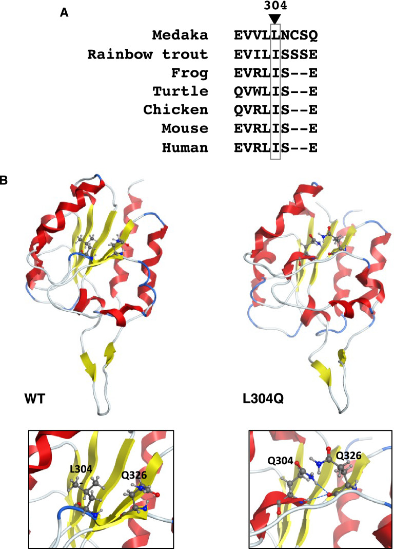
Vertebrate CMP-sialic acid synthetase (CSS), which catalyzes the synthesis of CMP-sialic acid (CMP-Sia), consists of a 28 kDa-N-domain and a 20 kDa-C-domain. The N-domain is known to be a catalytic domain; however, the significance of the C-domain still remains unknown. To elucidate the function of the C-domain at the organism level, we screened the medaka TILLING library and obtained medaka with non-synonymous mutations (t911a), or single amino acid substitutions of CSS, L304Q, in the C-domain. Prominently, most L304Q medaka was lethal within 19 days post-fertilization (dpf). L304Q young fry displayed free Sia accumulation, and impairment of sialylation, up to 8 dpf. At 8 dpf, a marked abnormality in ventricular contraction and skeletal myogenesis was observed. To gain insight into the mechanism of L304Q-induced abnormalities, L304Q was biochemically characterized. Although bacterially expressed soluble L304Q and WT showed the similar Vmax/Km values, very few soluble L304Q was detected when expressed in CHO cells in sharp contrast to the WT. Additionally, the thermostability of various mutations of L304 greatly decreased, except for WT and L304I. These results suggest that L304 is important for the stability of CSS, and that an appropriate level of expression of soluble CSS is significant for animal survival.


Conflict of interest statement

The authors declare no competing interests.

Figures

Figure 1
Figure 1
Properties of L304Q mdkCSS and survival curve of L304Q homo medaka. (A) Schematic drawing of L304Q mdkCSS. The position of L304Q site is shown by red bar. I ~ V, the evolutionarily conserved amino acid sequence motifs; BC1 and BC2 represent basic amino acid clusters that may be functional as nuclear localization signals. (B) Enzyme activity of L304Q mdkCSS. Left, western blotting of the recombinant L304Q and WT using anti-His antibodies was performed for quantifying the enzyme amount. Right, the relative specific activity of L304Q and WT in relation to Neu5Ac, Neu5Gc and KDN. The specific activity of WT in relation to Neu5Ac was set to 1. The bars represent standard deviation from three independent experiments. (C) Survival curve of medaka with the L304Q mutation in the CSS gene. WT, hetero, and L304Q homo medaka were derived from in-crossing of the same L304Q hetero parents. The numbers (n) of fish examined are shown in the parentheses. **indicates p < 0.005. Full-length blots for (B) are presented in Supp_FigS7.
Figure 2
Figure 2
Free and bound Sia in the whole lysate of WT, hetero, and homo medaka of L304Q at 8 dpf. (A) The amount of free Neu5Ac in the lysate was quantified by the fluorometric HPLC analysis, as described in Experimentnal Procedures. The bars represent standard deviations from three independent experiments. (B) The α2,6-Sia epitope evaluated by a lectin blotting with SNA and (C) the α2,8-polySia epitope evaluated by the western blotting with anti-polySia antibody (12E3). The lysate was applied to SDS-PAGE, followed by the SNA lectin-staining (B, upper panel) and 12E3-immuostaining (C, upper panel), respectively. Lanes 1–3 stand for homogenates from three independent fry. Immunoblotting of β-actin was used as a loading control (B,C, middle panels). The relative amounts of the Sia epitope are also shown (lower panels). The bars represent standard deviations from the data obtained from three independent fish. **indicates p < 0.005. Full-length blots for (B,C) are presented in Supp_FigS8 and Supp_FigS9, respectively.
Figure 3
Figure 3
Abnormalities of heart development and function of L304Q homo medaka. (A) Morphological image of WT (left) and L304Q homo medaka (right) at 8 dpf. The scale bars indicate 200 mm. The square marked by V shows the ventricle of heart. (B) Periodic characteristics of ventricle contraction in WT and L304Q homo medaka at 8 dpf. Periodical changes of the blood cell density in the ventricle at the square V (in A) were measured as those of the light and shade intensity using ImageJ. The data from three individual medaka for WT and homo are shown. (C) The expression of myosin heavy chain (MHC) in WT and L304Q homo medaka at 14 dpf. The lysate was analyzed by the SDS-PAGE/western blotting using MF20 antibody, which recognizes the heavy chain of myosin II and have been used to highlight the myocardium of medaka (26)(left upper). GAPDH was also detected by anti-GAPDH antibody as the loading control (left lower). The MHC components were decreased in the homo medaka, which may indicate cardiomyopathy. **indicates p < 0.005. Full-length blots for (C) are presented in Supp_FigS10.
Figure 4
Figure 4
Body length of L304Q medaka at 14 dpf. Images of WT, hetero, and L304Q homo medaka fry at 14 dpf were observed under the microscope (left). The scale bars indicate 2 mm. The body lengths of medaka fry at 14 dpf were measured and the data are summarized in a bar graph (right). n, the number of fry measured. The bars represent standard deviations obtained for the indicated numbers of fry. **indicates p < 0.005.
Figure 5
Figure 5
Kinetic analysis of WT and L304Q mdkCSS. (A) Purified recombinant enzymes used for the kinetic analysis. SDS-PAGE/CBB staining (left). Western blotting using the anti-mdkCSS antibodies (right). Full-length blots for A are presented in Supp_FigS11. (B) Lineweaver–Burk plots for the synthesis of CMP-Neu5Ac by mdkCSSs. (C) Kinetic parameters. Km, Vmax, and Vmax/Km values are expressed as average values ± standard deviations from three independent experiments (n = 3).
Figure 6
Figure 6
Property of WT and L304Q mdkCSS expressed in CHO cells. (A) Distribution of mdkCSS in the soluble and insoluble fractions. The myc-tagged WT and L304Q mdkCSS were transiently expressed in CHO cells, and their homogenates were separated into the soluble and insoluble fractions by the high speed centrifugation, followed by western blotting with the anti-myc antibody. Immunostaining with anti-GAPDH was performed as the loading control for soluble fraction. Note that the GAPDH bands were not detected in the insoluble fraction, which is reasonable because that GAPDH is a soluble protein. The band at 75 kDa stands for mdkCSS. (B) Expression level of the mRNA for mdkCSS. The transfected CHO cells expressing the myc-tagged WT and L304Q mdkCSS were analyzed by RT-PCR using the specific primers. The expression level of β-actin was used as the loading control. Full-length blots/gels for A and B are presented in Supp_FigS12 and Supp_FigS13, respectively.
Figure 7
Figure 7
In vitro properties of the single amino acid-substituted mutants at L304. (A) Western blotting of the recombinant mdkCSS mutants, L304N, L304Q, l304I, L304E, L304A, L304F, L304K, L304S, and WT with the anti-His antibody. The soluble forms were obtained from all the recombinant proteins expressed in E. coli. Full-length blots for A are presented in Supp_FigS14. (B) Thermostability of the L-304 mutants and WT of mdkCSS. Recombinant CSS proteins were pre-heated at 25, 35, or 45 °C for 15 min and measured for in vitro CSS activity. Relative in vitro activity of CSS was obtained as the amount of produced CMP-Neu5Ac divided by the intensity of the recombinant protein, and the value for mdkCSS without pre-heating (i.e., 25 °C) was set to 1.0. The bars represent standard deviations from three independent experiments. **indicates p < 0.005.
Figure 8
Figure 8
Molecular modeling for WT and L304Q mdkCSS. (A) Comparison of the local amino acid sequence around L-304 residue among vertebrates. Medaka, Oryzias latipes; Rainbow trout, Oncorhinchus mykiss; Frog, Xenopus tropicalis; Turtle, Mauremys reevesii; Chicken, Gullus gullus; Mouse, Mus musculus; Human, Homo sapiens. (B) Molecular modeling of the C-domains of WT and L304Q. These models are constructed by the MOE homology modeling based on the crystal structure of the C-domain of mouse CSS (PDB: 3EWI). Magnified views around L-304 residue are shown at lower panels. Two hydrogen bonds newly appear between H of NH group of the Q304 side chain and O of carbonyl group of the Q326 backbone, and between H of NH group of the Q304 backbone and O of carbonyl group of the Q326 backbone. No such interaction is not observed in WT.

Similar articles

Cited by

References

    1. Schauer R. Sialic acids as regulators of molecular and cellular interactions. Curr. Opin. Struct. Biol. 2009;19:507–514. doi: 10.1016/j.sbi.2009.06.003. - DOI - PMC - PubMed
    1. Schauer R, Kamerling JP. Exploration of the sialic acid world. Adv. Carbohydr. Chem. Biochem. 2018;75:1–213. doi: 10.1016/bs.accb.2018.09.001. - DOI - PMC - PubMed
    1. Varki NM, Strobert E, Dick EJ, Benirschke K, Varki A. Biomedical differences between human and nonhuman hominids: Potential roles for uniquely human aspects of sialic acid biology. Annu. Rev. Pathol. 2011;6:365–393. doi: 10.1146/annurev-pathol-011110-130315. - DOI - PubMed
    1. Varki A. Are humans prone to autoimmunity? Implications from evolutionary changes in hominin sialic acid biology. J. Autoimmun. 2017;83:134–142. doi: 10.1016/j.jaut.2017.07.011. - DOI - PubMed
    1. Sato C, Kitajima K. Sialic acids in neurology. Adv. Carbohydr. Chem. Biochem. 2019;76:1–64. doi: 10.1016/bs.accb.2018.09.003. - DOI - PubMed

Publication types

Substances