

Characterization of a G-quadruplex from hepatitis B virus and its stabilization by binding TMPyP4, BRACO19 and PhenDC3
- PMID: 34853392
- PMCID: PMC8636512
- DOI: 10.1038/s41598-021-02689-y
Characterization of a G-quadruplex from hepatitis B virus and its stabilization by binding TMPyP4, BRACO19 and PhenDC3
Abstract
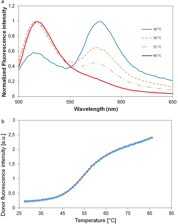
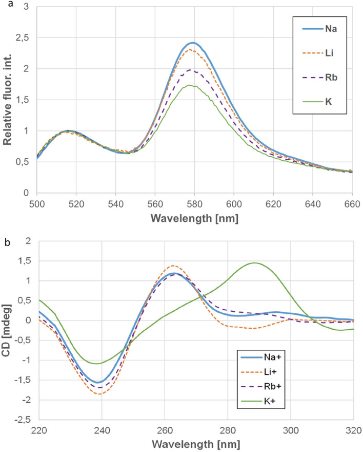
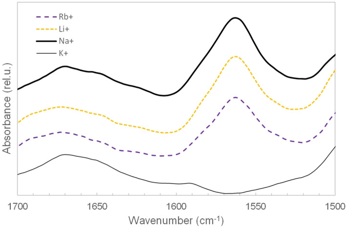
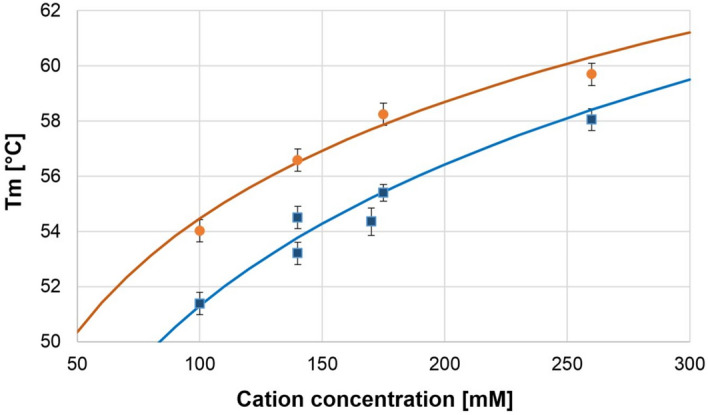
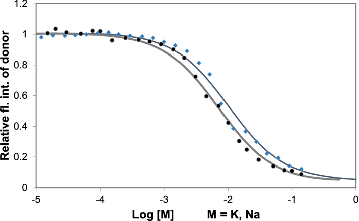
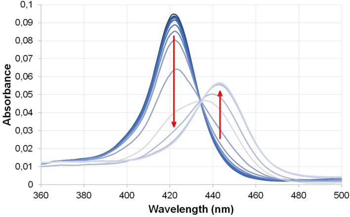
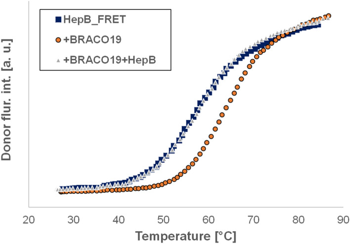
Specific guanine rich nucleic acid sequences can form non-canonical structures, like the four stranded G-quadruplex (GQ). We studied the GQ-forming sequence (named HepB) found in the genome of the hepatitis B virus. Fluorescence-, infrared- and CD-spectroscopy were used. HepB shows a hybrid form in presence of K+, but Na+, Li+, and Rb+ induce parallel structure. Higher concentrations of metal ions increase the unfolding temperature, which was explained by a short thermodynamic calculation. Temperature stability of the GQ structure was determined for all these ions. Na+ has stronger stabilizing effect on HepB than K+, which is highly unusual. The transition temperatures were 56.6, 53.8, 58.5 and 54.4 °C for Na+, K+, Li+, and Rb+ respectively. Binding constants for Na+ and K+ were 10.2 mM and 7.1 mM respectively. Study of three ligands designed in cancer research for GQ targeting (TMPyP4, BRACO19 and PhenDC3) showed unequivocally their binding to HepB. Binding was proven by the increased stability of the bound form. The stabilization was higher than 20 °C for TMPyP4 and PhenDC3, while it was considerably lower for BRACO19. These results might have medical importance in the fight against the hepatitis B virus.
© 2021. The Author(s).
Conflict of interest statement
The authors declare no competing interests.
Figures
References
-
- Fry M. Tetraplex DNA and its interacting proteins. Front. Biosci. 2007;12:4336–4351. - PubMed
-
- Guzman MR, Liquier J, Brahmachari SK, Taillandier E. Characterization of parallel and antiparallel G-tetraplex structures by vibrational spectroscopy. Spectrochim. Acta A Mol. Biomol. Spectrosc. 2006;64:495–503. - PubMed
-
- Takahashi S, Sugimoto N. Stability prediction of canonical and non-canonical structures of nucleic acids in various molecular environments and cells. Chem. Soc. Rev. 2020;49:8439–8468. - PubMed
-
- Matsumoto S, Tateishi-Karimata H, Takahashi S, Ohyama T, Sugimoto N. Effect of molecular crowding on the stability of RNA G-quadruplexes with various numbers of quartets and lengths of loops. Biochemistry. 2020;59:2640–2649. - PubMed
Publication types
MeSH terms
Substances
LinkOut - more resources
Full Text Sources
Other Literature Sources
