

Electroconvulsive therapy, electric field, neuroplasticity, and clinical outcomes
- PMID: 34853404
- PMCID: PMC9095458
- DOI: 10.1038/s41380-021-01380-y
Electroconvulsive therapy, electric field, neuroplasticity, and clinical outcomes
Abstract
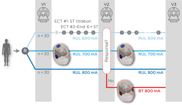
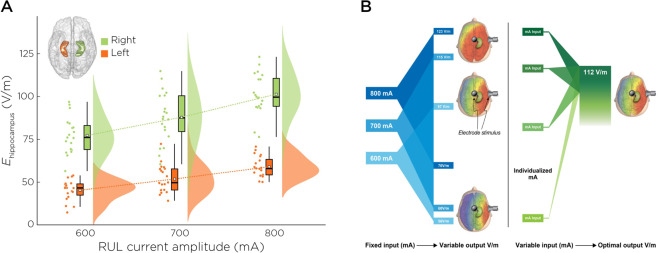
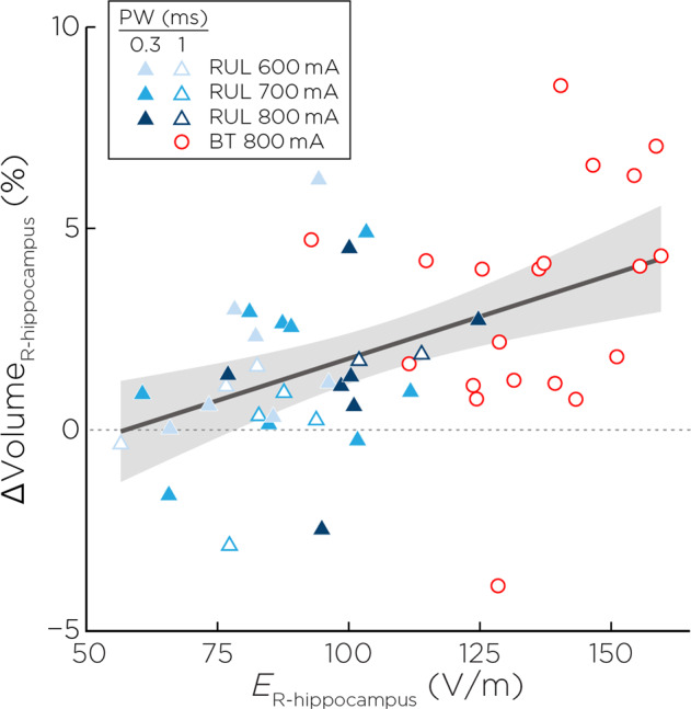
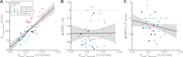
Electroconvulsive therapy (ECT) remains the gold-standard treatment for patients with depressive episodes, but the underlying mechanisms for antidepressant response and procedure-induced cognitive side effects have yet to be elucidated. Such mechanisms may be complex and involve certain ECT parameters and brain regions. Regarding parameters, the electrode placement (right unilateral or bitemporal) determines the geometric shape of the electric field (E-field), and amplitude determines the E-field magnitude in select brain regions (e.g., hippocampus). Here, we aim to determine the relationships between hippocampal E-field strength, hippocampal neuroplasticity, and antidepressant and cognitive outcomes. We used hippocampal E-fields and volumes generated from a randomized clinical trial that compared right unilateral electrode placement with different pulse amplitudes (600, 700, and 800 mA). Hippocampal E-field strength was variable but increased with each amplitude arm. We demonstrated a linear relationship between right hippocampal E-field and right hippocampal neuroplasticity. Right hippocampal neuroplasticity mediated right hippocampal E-field and antidepressant outcomes. In contrast, right hippocampal E-field was directly related to cognitive outcomes as measured by phonemic fluency. We used receiver operating characteristic curves to determine that the maximal right hippocampal E-field associated with cognitive safety was 112.5 V/m. Right hippocampal E-field strength was related to the whole-brain ratio of E-field strength per unit of stimulation current, but this whole-brain ratio was unrelated to antidepressant or cognitive outcomes. We discuss the implications of optimal hippocampal E-field dosing to maximize antidepressant outcomes and cognitive safety with individualized amplitudes.
© 2021. The Author(s).
Conflict of interest statement
The authors declare no competing interests.
Figures

Comment in
-
Electric field distribution models in ECT research.Mol Psychiatry. 2022 Sep;27(9):3571-3572. doi: 10.1038/s41380-022-01516-8. Epub 2022 Mar 18. Mol Psychiatry. 2022. PMID: 35304563 Free PMC article. No abstract available.
References
-
- Gbyl K, Rostrup E, Raghava JM, Andersen C, Rosenberg R, Larsson HBW, et al. Volume of hippocampal subregions and clinical improvement following electroconvulsive therapy in patients with depression. Prog neuro-Psychopharmacol Biol Psychiatry. 2021;104:110048. doi: 10.1016/j.pnpbp.2020.110048. - DOI - PubMed
-
- Gbyl K, Stottrup MM, Mitta Raghava J, Xue Jie S, Videbech P. Hippocampal volume and memory impairment after electroconvulsive therapy in patients with depression. Acta psychiatrica Scandinavica 2021;143:238–52. - PubMed
Publication types
MeSH terms
Substances
Grants and funding
LinkOut - more resources
Full Text Sources
Other Literature Sources
