DNA repair inhibitors sensitize cells differently to high and low LET radiation
- PMID: 34853427
- PMCID: PMC8636489
- DOI: 10.1038/s41598-021-02719-9
DNA repair inhibitors sensitize cells differently to high and low LET radiation
Abstract
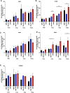
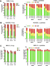
The aim of this study was to investigate effects of high LET α-radiation in combination with inhibitors of DDR (DNA-PK and ATM) and to compare the effect with the radiosensitizing effect of low LET X-ray radiation. The various cell lines were irradiated with α-radiation and with X-ray. Clonogenic survival, the formation of micronuclei and cell cycle distribution were studied after combining of radiation with DDR inhibitors. The inhibitors sensitized different cancer cell lines to radiation. DNA-PKi affected survival rates in combination with α-radiation in selected cell lines. The sensitization enhancement ratios were in the range of 1.6-1.85 in cancer cells. ATMi sensitized H460 cells and significantly increased the micronucleus frequency for both radiation qualities. ATMi in combination with α-radiation reduced survival of HEK293. A significantly elicited cell cycle arrest in G2/M phase after co-treatment of ATMi with α-radiation and X-ray. The most prominent treatment effect was observed in the HEK293 by combining α-radiation and inhibitions. ATMi preferentially sensitized cancer cells and normal HEK293 cells to α-radiation. DNA-PKi and ATMi can sensitize cancer cells to X-ray, but the effectiveness was dependent on cancer cells itself. α-radiation reduced proliferation in primary fibroblast without G2/M arrest.
© 2021. The Author(s).
Conflict of interest statement
The authors have declared conflicts of interest. Kristina Bannik, Sabrina Jarke, Andreas Sutter, Gerhard Siemeister, Christoph Schatz, Dominik Mumberg, Sabine Zitzmann-Kolbe are/were employees of Bayer AG. Balazs Madas is not an employee of Bayer AG.
Figures


Similar articles
-
Nontoxic concentration of DNA-PK inhibitor NU7441 radio-sensitizes lung tumor cells with little effect on double strand break repair.Cancer Sci. 2016 Sep;107(9):1250-5. doi: 10.1111/cas.12998. Epub 2016 Sep 1. Cancer Sci. 2016. PMID: 27341700 Free PMC article.
-
Cisplatin-mediated radiosensitization of non-small cell lung cancer cells is stimulated by ATM inhibition.Radiother Oncol. 2014 May;111(2):228-36. doi: 10.1016/j.radonc.2014.04.001. Epub 2014 May 21. Radiother Oncol. 2014. PMID: 24857596
-
Overcoming hypoxia-induced tumor radioresistance in non-small cell lung cancer by targeting DNA-dependent protein kinase in combination with carbon ion irradiation.Radiat Oncol. 2017 Dec 29;12(1):208. doi: 10.1186/s13014-017-0939-0. Radiat Oncol. 2017. PMID: 29287602 Free PMC article.
-
ATM as a target for novel radiosensitizers.Semin Radiat Oncol. 2001 Oct;11(4):316-27. doi: 10.1053/srao.2001.26030. Semin Radiat Oncol. 2001. PMID: 11677656 Review.
-
Strategizing the clone wars: pharmacological control of cellular sensitivity to radiation.Mol Interv. 2010 Dec;10(6):341-53. doi: 10.1124/mi.10.6.3. Mol Interv. 2010. PMID: 21263160 Free PMC article. Review.
Cited by
-
RYBP Sensitizes Cancer Cells to PARP Inhibitors by Regulating ATM Activity.Int J Mol Sci. 2022 Oct 4;23(19):11764. doi: 10.3390/ijms231911764. Int J Mol Sci. 2022. PMID: 36233063 Free PMC article.
-
DNA damage and repair dependencies of ionising radiation modalities.Biosci Rep. 2023 Oct 31;43(10):BSR20222586. doi: 10.1042/BSR20222586. Biosci Rep. 2023. PMID: 37695845 Free PMC article. Review.
-
Synergistic Activity of DNA Damage Response Inhibitors in Combination with Radium-223 in Prostate Cancer.Cancers (Basel). 2024 Apr 15;16(8):1510. doi: 10.3390/cancers16081510. Cancers (Basel). 2024. PMID: 38672592 Free PMC article.
-
Apatinib enhances the antitumor effects of radiation in HeLa cell line mouse model of invasive cervical cancer.Ann Transl Med. 2022 Apr;10(8):459. doi: 10.21037/atm-22-1442. Ann Transl Med. 2022. PMID: 35571415 Free PMC article.
-
DNA Damage by Radiopharmaceuticals and Mechanisms of Cellular Repair.Pharmaceutics. 2023 Dec 12;15(12):2761. doi: 10.3390/pharmaceutics15122761. Pharmaceutics. 2023. PMID: 38140100 Free PMC article. Review.
References
-
- McGale P, et al. Effect of radiotherapy after mastectomy and axillary surgery on 10-year recurrence and 20-year breast cancer mortality: Meta-analysis of individual patient data for 8135 women in 22 randomised trials. Lancet. 2014;383:2127–2135. doi: 10.1016/S0140-6736(14)60488-8. - DOI - PMC - PubMed
MeSH terms
Substances
LinkOut - more resources
Full Text Sources
Research Materials
Miscellaneous

