. 2021 Dec;600(7889):543-546.
doi: 10.1038/s41586-021-04131-9. Epub 2021 Dec 1.

Accuracy mechanism of eukaryotic ribosome translocation

Affiliations

Accuracy mechanism of eukaryotic ribosome translocation

Muminjon Djumagulov et al. Nature. 2021 Dec.

Abstract

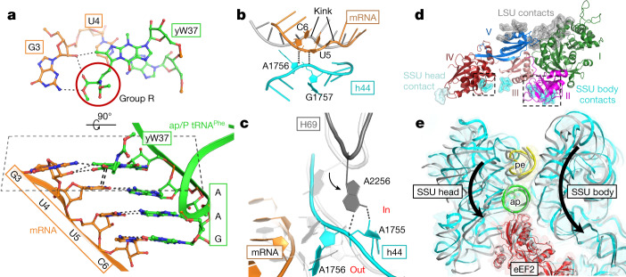
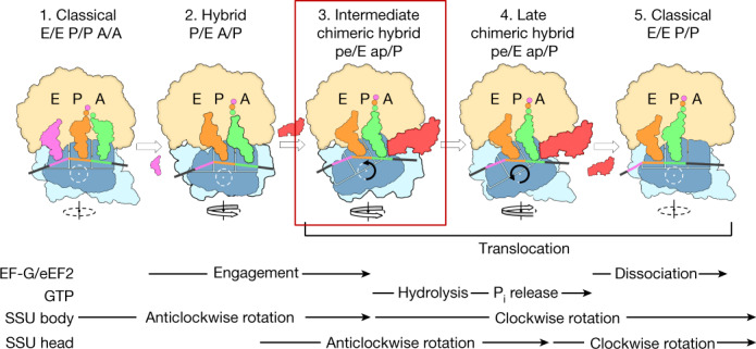
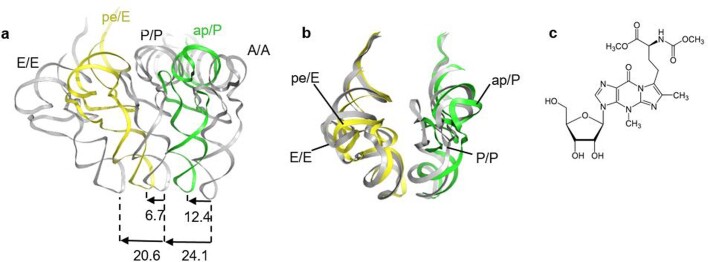
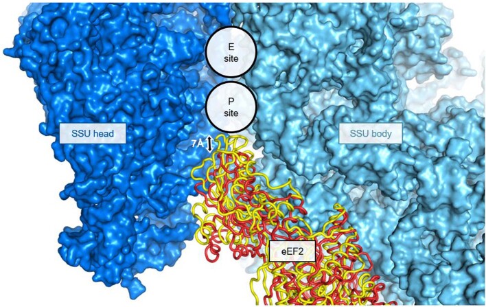
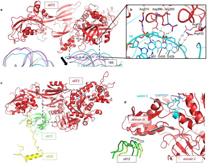
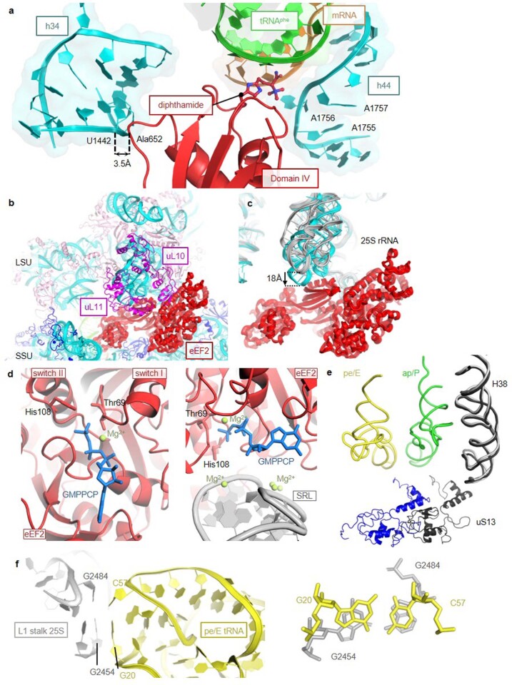
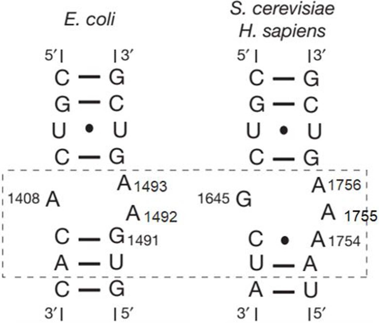
Translation of the genetic code into proteins is realized through repetitions of synchronous translocation of messenger RNA (mRNA) and transfer RNAs (tRNA) through the ribosome. In eukaryotes translocation is ensured by elongation factor 2 (eEF2), which catalyses the process and actively contributes to its accuracy1. Although numerous studies point to critical roles for both the conserved eukaryotic posttranslational modification diphthamide in eEF2 and tRNA modifications in supporting the accuracy of translocation, detailed molecular mechanisms describing their specific functions are poorly understood. Here we report a high-resolution X-ray structure of the eukaryotic 80S ribosome in a translocation-intermediate state containing mRNA, naturally modified eEF2 and tRNAs. The crystal structure reveals a network of stabilization of codon-anticodon interactions involving diphthamide1 and the hypermodified nucleoside wybutosine at position 37 of phenylalanine tRNA, which is also known to enhance translation accuracy2. The model demonstrates how the decoding centre releases a codon-anticodon duplex, allowing its movement on the ribosome, and emphasizes the function of eEF2 as a 'pawl' defining the directionality of translocation3. This model suggests how eukaryote-specific elements of the 80S ribosome, eEF2 and tRNAs undergo large-scale molecular reorganizations to ensure maintenance of the mRNA reading frame during the complex process of translocation.


Conflict of interest statement

The authors declare no competing interests

Figures

Fig. 1
Fig. 1. The translocation-intermediate state of the eukaryotic 80S ribosome with eEF2–GMPPCP, mRNA and tRNAs,, showing diphthamide of eEF2 is involved in stabilizing codon–anticodon interactions early in translocation.
a, Overview of the translocation-intermediate complex with two tRNAs trapped in chimeric hybrid ap/P and pe/E states. b, Close-up view of ap/P and pe/E tRNA anticodon stem loops in the context of elements of the SSU body and head and domain IV of eEF2. Position of wybutosine (yW) of ap/P tRNAPhe is indicated by the asterisk. c, Position of diphthamide (Diph699) at the conjunction of the AAG anticodon of ap/P tRNAPhe, mRNA codon UUC and decoding adenosines 1755–1756 of helix 44 (h44). d, Stabilization networks around codon–anticodon interactions in the translocation-intermediate complex (left) and the decoding centre of the bacterial ribosome in the cognate classical state, (right). Top, middle and bottom panels depict stabilization around the first, second and third base pairs (BP1–3) of a codon–anticodon duplex, respectively. Conserved adenosines 1755 and 1756 in yeast 18S rRNA correspond to adenosines 1492 and 1493 in 16S rRNA of the bacterial decoding centre. eEF2 is shown in red, mRNA is in orange, chimeric hybrid ap/P tRNA is in green and chimeric hybrid pe/E is in yellow. LSU rRNA and proteins are shown in grey and purple; SSU rRNA and proteins are in cyan and in deep blue. Degrees of SSU body and head rotations are indicated and were obtained by superimposing with the non-rotated 80S ribosome (PDB ID 3J78).
Fig. 2
Fig. 2. Stabilization of mRNA–tRNA by wybutosine modification of tRNAPhe, rearrangements of the decoding centre and depiction of a pawl function for eEF2 during translocation.
a, Wybutosine cross-strand stacks on the first codon–anticodon base pair formed by mRNA codon UUC and tRNAPhe at the ap/P state and stabilizes the last position of the adjacent upstream AUG codon (G3). An alternative view (bottom) of the same pattern clearly demonstrates how wybutosine enhances stabilization of mRNA to prevent a frame shift during translocation. b, Interactions between the backbones of translocating mRNA and decoding A1756 and A1757 of h44; mRNA in classical state is in grey. c, A shift of helix 69 (H69) of 25S rRNA is coupled to rearrangements of the decoding centre as compared to the bacterial classical state in white (PDB ID 4V6F). d, Contacts of eEF2 (coloured by domain) with LSU (grey), body (cyan) and head (pale cyan) of SSU; dashed squares indicate contacts that are disrupted at the late stage of translocation, allowing eEF2 to uncouple from the SSU body. e, Superposition of intermediate- and late (grey) (TI-POST-1, PDB ID 6GZ3)-translocation structures relative to LSU reveals eEF2 acting as a pawl anchoring and decoupling ap/P tRNA from the SSU head and body. The reverse direction of the SSU head and body rotation to a classical post-translocation state is indicated with arrows.
Fig. 3
Fig. 3. Integrating kinetic and structural studies of translocation.
Top, translocation scheme based on the crystal structure of the intermediate translocation complex reported here (in frame) and on cryo-EM structures of late translocation (PDB ID: 6GZ3 and 6GZ5), as well as hybrid and classical post-translocation states (PDB ID: 3J77 and 3J78). A proposed sequence of events based on kinetic studies is shown at the bottom. Steps 1 and 2, thermally driven intersubunit rotations lead to tRNAs adopting hybrid A/P and P/E states and eEF2–GTP binding to the 80S ribosome. Steps 2 and 3, concomitant changes of LSU H69 composing intersubunit bridge B2a and the decoding centre, and insertion of the eEF2 diphthamide to the SSU A-site induce unlocking of the decoding centre. The released codon–anticodon duplex becomes stabilized by direct interactions with diphthamide. Detachment of tRNA ASLs from the SSU body and further insertion of the eEF2 domain IV into the A-site cause initial anticlockwise rotation of the head and movement of the second tRNA from the SSU P-site towards the E-site where it binds to L1 stalk. Steps 3 and 4, eEF2 remains anchored to LSU via domains I and V but is released from SSU where domain IV uncouples tRNA–mRNA from rearrangements of the SSU body and head. What is perceived as an additional large swivelling of the head is actually a result of the body back-rotation while the head remains fixed to tRNAs. Step 4 and 5, this body rotation increases the strain in the SSU neck and leads to uncoupling of the head from tRNAs. Formation of contacts between rRNA of the head and domain IV of eEF2 restrain the head position. The last steps of translocation are achieved when the head, owing to the increasing strain on the neck, snaps back to a non-rotated state and tRNA–mRNA binds to the SSU P-site environment.
Extended Data Fig. 1
Extended Data Fig. 1. Examples of the electron density maps of the 80S ribosome translocation intermediate complex.
FEM map for a representative part of 25S rRNA (a) and for eEF2 (b) contoured at 2 sigma. 2Fo-Fc electron density map contoured at 1.5 sigma for 25S rRNA (c) and at 1 sigma for the diphthamide loop of eEF2 (d).
Extended Data Fig. 2
Extended Data Fig. 2. Comparison of various states of tRNAs on the ribosome.
a, Positions of tRNAs of the intermediate translocation complex relative to tRNAs in the classical A, P and E states (grey, PDB ID 4V6F) aligned on the SSU body; indicated distances are in Å. b, Positions of tRNAs relative to tRNAs in the classical nonrotated state (PDB ID 3J78) aligned on LSU. c, Chemical structure of the wybutosine tRNAPhe modification at position 37.
Extended Data Fig. 3
Extended Data Fig. 3. Insertion of eEF2 into the SSU A site in the late translocation complex TI-POST1 (yellow, PDB ID 6GZ3) in comparison to the position of eEF2 in the early translocation intermediate state that we determined in this study (red).
View from the LSU side. Alignment is performed using the SSU body.
Extended Data Fig. 4
Extended Data Fig. 4. Interactions of eEF2 with the SSU body.
a, Comparison of the current X-ray structure of an early translocation intermediate (18S rRNA in cyan) with cryo-EM models of late translocation steps TI-POST1 (magenta, PDB ID 6GZ3) and TI-POST3 (grey, PDB ID 6GZ3). Superimposition of eEF2 in the two structures shows that there is a noticeable shift of 18S rRNA of the SSU body away from eEF2 at later stages of translocation. b, Contacts between eEF2 and h5 of SSU of the rotated ribosome in the reported intermediate translocation complex. c, Interactions between eEF2 and the SSU shoulder proteins eS30 and uS12. Stabilization of the eEF2 domain IV by the N-terminus of eukaryote-specific protein eS30 that itself interacts with conserved decoding protein uS12. Presumably, eS30 co-evolved with eEF2, whose domain IV has 65 additional amino acids compared to its bacterial counterpart EF-G, to provide supplementary stabilization as well as to enhance propagation of conformational changes at the decoding site,. d, A close up view of the dashed region from (c). Movement of uS12 (arrow 1) induced by the SSU body back-rotation can propagate to switch II (in cyan) of eEF2 through domain III (arrow 2) and can trigger GTP hydrolysis or Pi release. Regions of uS12 adjacent to eEF2 are shown and coloured dark- to light-green based on the rotational state where dark-green is the most rotated state (intermediate translocation complex) and medium (TI-POST1) and light (TI-POST3) greens represent the least rotated states. Alignment was done using eEF2.
Extended Data Fig. 5
Extended Data Fig. 5. Rearrangement of h34 and h31 of SSU with ASL movement of tRNAs in the translocation intermediate complex.
a, A new position of conserved C1274 in h34 of 18S rRNA (C1054 of 16S rRNA in bacteria, Fig. 1d) relative to ASL of the A site tRNA and mRNA. By establishing a contact with mRNA nucleotide in position (+7) C1274 can contribute to maintaining mRNA reading frame during translocation. b, In the early intermediate state of translocation h31 of 18S rRNA moves together with pe/E tRNA. In this state, the U1191-C1637 interaction is broken, however, U1191 remains in contact with C35 of pe/E tRNA. A similar situation was described for the bacterial ribosome with the pe/E tRNA in a structure modelling spontaneous translocation without EF-G. Compared to the intermediate eukaryotic state reported here, the bacterial ap/P tRNA coupled to mRNA is shifted towards the P-site indicating translational reading frame slippage in the factor-free system. This comparison shows the importance of role that translocase eEF2 (or EF-G) fulfils in the coordinated movement of tRNA-mRNA during translocation.
Extended Data Fig. 6
Extended Data Fig. 6. Contacts between eEF2, tRNAs and the 80S ribosome in the crystal structure of the translocation intermediate complex.
a, Position of diphthamide at the conjunction of ap/P tRNAPhe, mRNA and decoding adenines of h44 and interactions of eEF2 (Ala652) with rRNA of the SSU head (U1442 of 18S). b, A solvent view on the P stalk region. c, Movement of the P stalk upon eEF2 binding seen when comparing our structure to that of the vacant 80S ribosome structure (PDB ID 4V88). Alignment was performed using 25S rRNA as a reference. d, The G domain of eEF2 with ordered switch I and II regions indicating that this is a pre-hydrolysis state (left panel). Close-up view of the GTP pocket and sarcin-ricin loop (SRL) of 25S rRNA (right panel). e, Disruption of the B1a bridge consisting of helix 38 of 25S rRNA (A-site finger) and protein uS13 induced by rotation of the SSU head and body. Non-rotated 80S (PDB ID 3J78) is coloured in black. f, Stacking interactions of rRNA elements (magnified in the right panel) of the L1 stalk with the elbow region of pe/E tRNA.
Extended Data Fig. 7
Extended Data Fig. 7. Comparison of secondary structures of the decoding loop in bacteria (left) and eukaryotes (right).
In bacteria the internal loop of h44 of 16S rRNA consists of two nucleotides (A1492-A1493) on the 3’-side and has one nucleotide on the 5’-side (A1408). In contrast, eukaryotic 18S rRNA contains at least one additional nucleotide on each side is included (dashed box). Secondary structure diagrams of helix 44 from bacteria (16S rRNA, left) and from yeast and human (18S rRNA, right).

Similar articles

Cited by

References

    1. Ortiz PA, Ulloque R, Kihara GK, Zheng H, Kinzy TG. Translation elongation factor 2 anticodon mimicry domain mutants affect fidelity and diphtheria toxin resistance. J. Biol. Chem. 2006;281:32639–32648. doi: 10.1074/jbc.M607076200. - DOI - PubMed
    1. Waas WF, Druzina Z, Hanan M, Schimmel P. Role of a tRNA base modification and its precursors in frameshifting in eukaryotes. J. Biol. Chem. 2007;282:26026–26034. doi: 10.1074/jbc.M703391200. - DOI - PubMed
    1. Noller HF, Lancaster L, Mohan S, Zhou J. Ribosome structural dynamics in translocation: yet another functional role for ribosomal RNA. Q. Rev. Biophys. 2017;50:e12. doi: 10.1017/S0033583517000117. - DOI - PubMed
    1. Murray, J. et al. Structural characterization of ribosome recruitment and translocation by type IV IRES. Elife5, e13567 (2016). - PMC - PubMed
    1. Jørgensen R, Merrill AR, Andersen GR. The life and death of translation elongation factor 2. Biochem. Soc. Trans. 2006;34:1–6. doi: 10.1042/BST0340001. - DOI - PubMed

Publication types

MeSH terms