. 2021 Nov 22:2021:6338722.
doi: 10.1155/2021/6338722. eCollection 2021.

Human Cerebral Organoid Implantation Alleviated the Neurological Deficits of Traumatic Brain Injury in Mice

Affiliations

Human Cerebral Organoid Implantation Alleviated the Neurological Deficits of Traumatic Brain Injury in Mice

Zhongyuan Bao et al. Oxid Med Cell Longev. .

Abstract

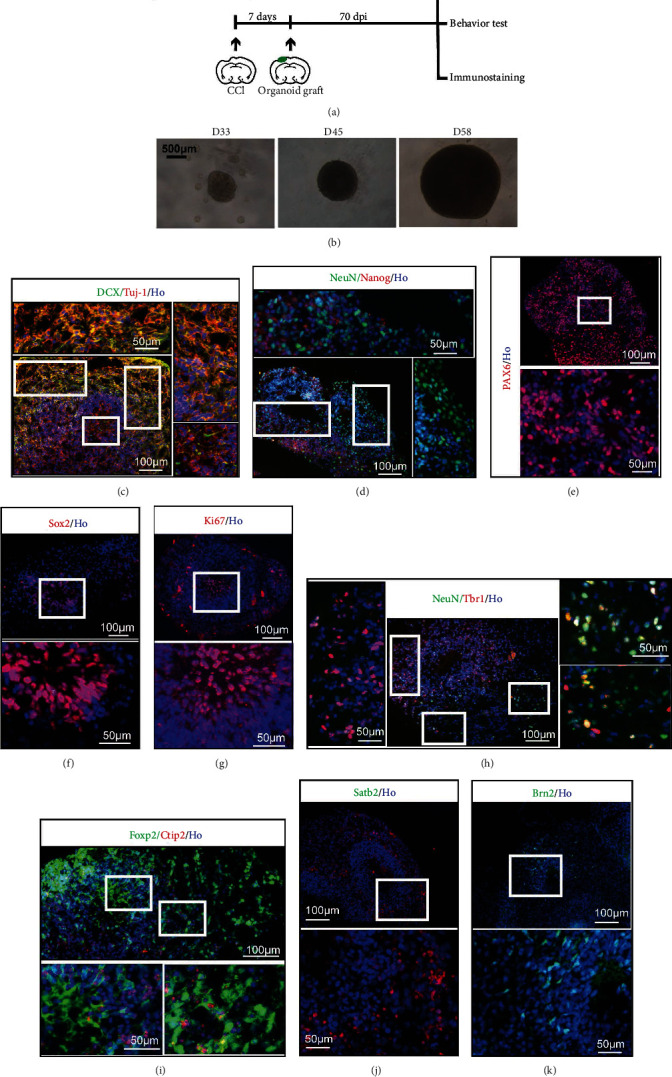
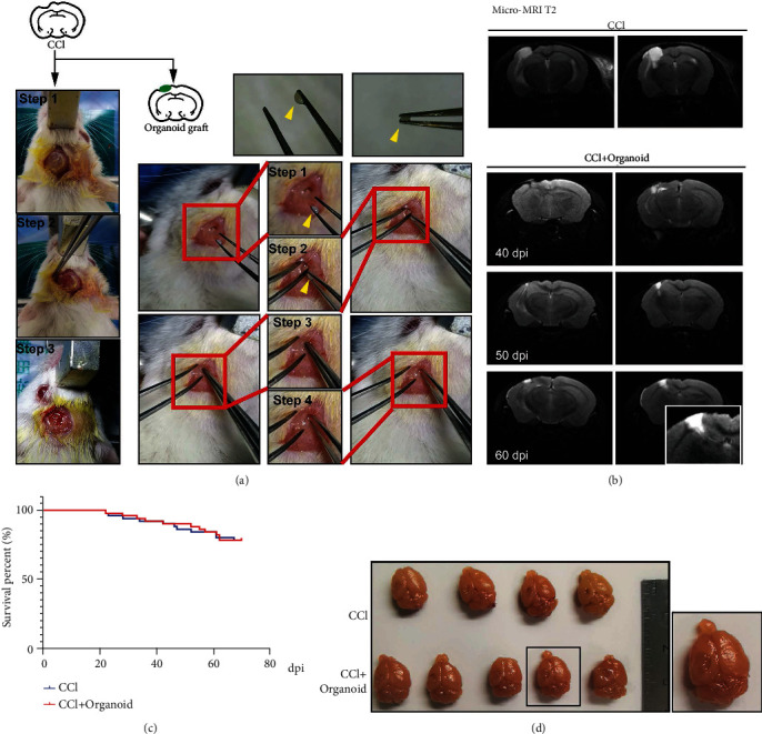
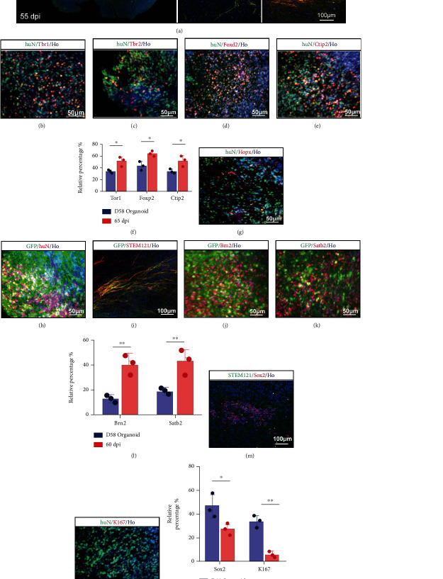
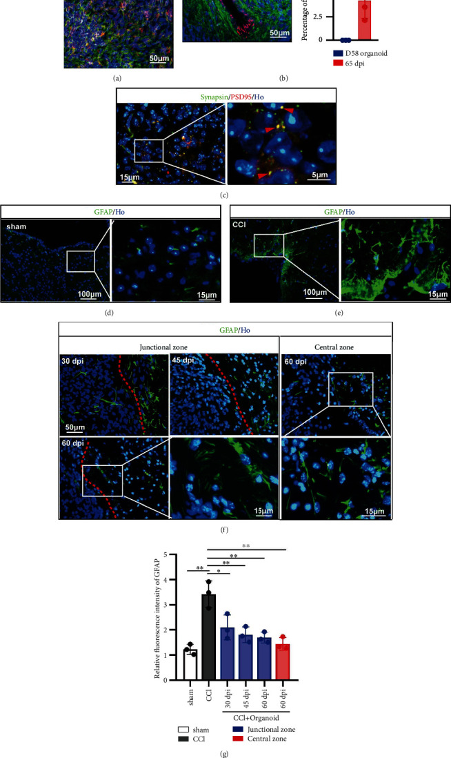
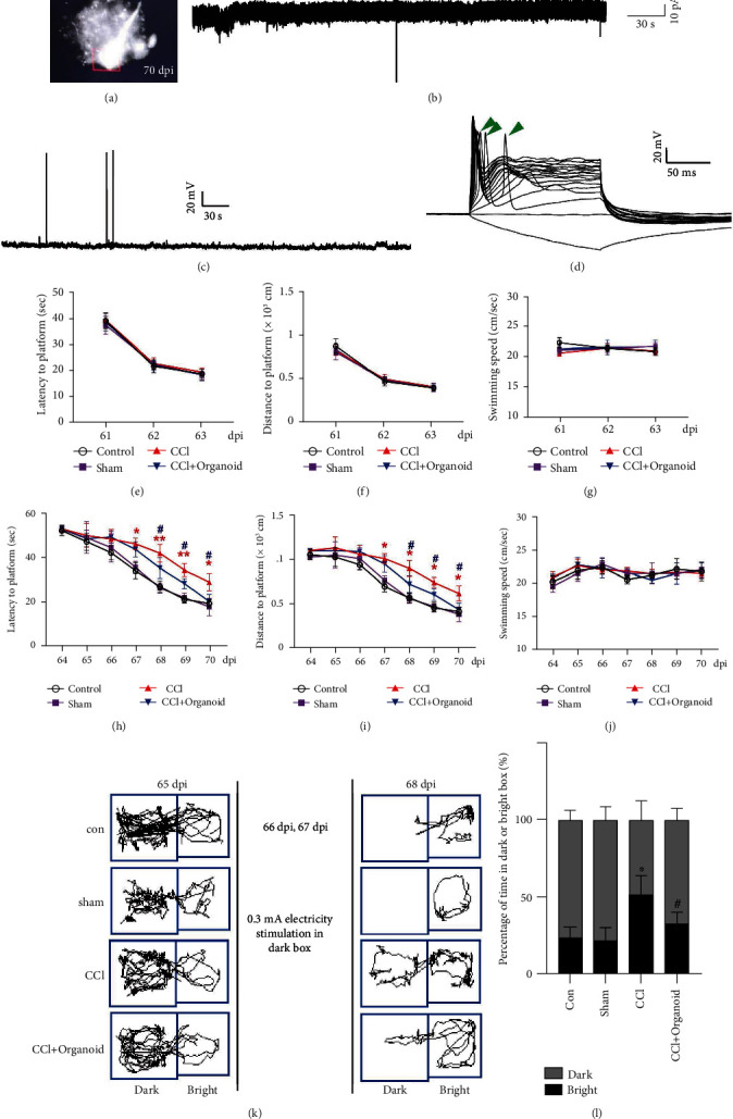
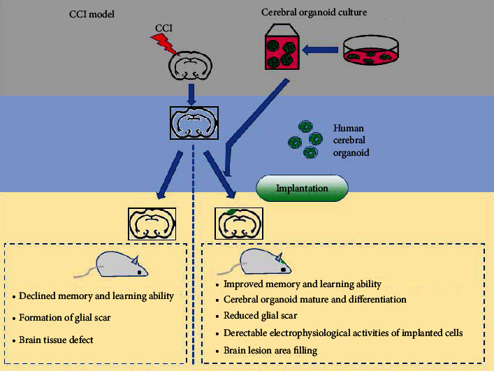
Traumatic brain injury (TBI) causes a high rate of mortality and disability, and its treatment is still limited. Loss of neurons in damaged area is hardly rescued by relative molecular therapies. Based on its disease characteristics, we transplanted human embryonic stem cell- (hESC-) derived cerebral organoids in the brain lesions of controlled cortical impact- (CCI-) modeled severe combined immunodeficient (SCID) mice. Grafted organoids survived and differentiated in CCI-induced lesion pools in mouse cortical tissue. Implanted cerebral organoids differentiated into various types of neuronal cells, extended long projections, and showed spontaneous action, as indicated by electromyographic activity in the grafts. Induced vascularization and reduced glial scar were also found after organoid implantation, suggesting grafting could improve local situation and promote neural repair. More importantly, the CCI mice's spatial learning and memory improved after organoid grafting. These findings suggest that cerebral organoid implanted in lesion sites differentiates into cortical neurons, forms long projections, and reverses deficits in spatial learning and memory, a potential therapeutic avenue for TBI.


Conflict of interest statement

The authors declare that there is no conflict of interest regarding the publication of this paper.

Figures

Figure 1
Figure 1
Human cerebral organoids cultured in vitro. (a) Experimental procedure of this study including cerebral organoid implantation and schedule. (b) Generation of organoids from human ESCs. The images were taken at 33 days, 45 days, and 58 days. (c) Immunohistochemistry for neurons (Tuj-1, red) and the new development of the cerebral cortex (DCX, green). Scale bars are 100 μm and 50 μm. (d) Staining for mature neurons (NeuN, green) and embryonic stem cell (Nanog, red). Scale bars are 100 μm and 50 μm. (e) Immunohistochemistry in the section for the forebrain marker Pax6. Scale bars are 100 μm and 50 μm. (f) Staining for embryonic stem cell pluripotency (Sox2, red). Scale bars are 100 μm and 50 μm. (g) Staining for the cell proliferation marker Ki67. Scale bars are 100 μm and 50 μm. (h) Staining for the preplate marker Tbr1 (red) and neuronal marker NeuN (green) revealing organoid maturity. Scale bars are 100 μm and 50 μm. (i) Staining for deep-layer subcortical neuron marker Ctip2 (red) and developing (Foxp2, green) neurons. Scale bars are 100 μm and 50 μm. (j) Staining for Satb2 (red). Scale bars are 100 μm and 50 μm. (k) Staining for Brn2 (green). Scale bars are 100 μm and 50 μm. All cell nuclei were stained by Hoechst (Ho).
Figure 2
Figure 2
Organoids grafted into SCID CCI-model mice exhibited appropriated growth and survival. (a) Schematic image of CCI (3 steps) and organoid graft (4 steps). Organoids could be picked up by tweezers (yellow arrow). (b) Micro-MRI T2 assay was used to display tissue lesions in CCI mice and the growth situation of grafted organoids at 40 dpi, 50 dpi, and 60 dpi. (c) Survival percentage of SCID mice in CCI group and CCI+organoid group. (d) Image of mouse brain tissues in the CCI and CCI plus organoid groups.
Figure 3
Figure 3
Degree of differentiation and maturity of grafted human cerebral organoids. (a) Immunohistochemistry for human-derived cells (STEM121, green) and the new development of neuronal cells (DCX, red). The grafted cells could migrate to the contralateral brain region via the callosum. Scale bar is 200 μm. (b, c) Staining for the preplate marker Tbr1 (red) and intermediate progenitor marker Tbr2 (red). Grafted human-derived cells were both stained for huN (green). Scale bars are 50 μm. (d, e) Staining for developing neuronal cells (Foxp2, red) and early-born neurons (Ctip2, red). Grafted human-derived cells were both stained for huN (green). Scale bars are 50 μm. (f) Compared to organoids in vitro, the percentage of grafted neurons expressing Tbr1, Foxp2, and Ctip2 increased. Error bars represent S.D. (n = 3 experiments). P < 0.05 versus relative organoid group in vitro. (g) Staining for neural progenitor (Hopx, red) involved in differentiation of neurons in OSVZ. Grafted human-derived cells were stained for huN (green). Scale bar is 50 μm. (h, i) Development of organoid conjunct with GFP. (h) Staining for GFP (green) and huN (red). Scale bar is 50 μm. (i) Staining for GFP (green) and STEM121 (red). Scale bar is 100 μm. (j) Staining for neuronal subtype progression in cortex (Brn2, red). GFP was used to label grafted cells. Scale bar is 50 μm. (k) Staining for late-born neurons (Satb2, red). GFP was used to label grafted cells. Scale bar is 50 μm. (l) Compared to organoids in vitro, the percentage of grafted neurons expressing Brn2 and Stab2 increased. Error bars represent S.D. (n = 3 experiments). ∗∗P < 0.01 versus relative organoid group in vitro. (m) Staining for embryonic stem cell pluripotency (Sox2, red). Scale bar is 100 μm. STEM121 was used to display human-derived cells. (n) Staining for Ki67 to reveal cell proliferation level. huN was used to display human-derived cells. Scale bar is 50 μm. (o) The percentage of grafted neurons expressing Sox2 or Ki67 decreased in organoids 65 dpi. Error bars represent S.D. (n = 3 experiments). P < 0.05 or ∗∗P < 0.01 versus relative organoid group in vitro. All cell nuclei were stained by Hoechst.
Figure 4
Figure 4
Grafted organoid improved local situation contributing to neural repair. (a) Staining for glutamate (red). Grafted human-derived cells were stained for STEM121 (green). Scale bar is 50 μm. (b) Vascular endothelial cell was labeled by CD31 (red). Grafted human-derived cells were stained for STEM121 (green). Scale bar is 50 μm. All cell nuclei were stained by Hoechst (Ho). The percentage of CD31 was displayed in the bar graph (right part). (c) Double immunofluorescence staining for presynaptic marker Synapsin and the postsynaptic marker PSD95 at 60 dpi, showing a coassociation between pre- and postsynaptic compartments and the formation of synaptic connections in the graft. Scale bar is 15 μm and 5 μm. (d, e) The formations of glial scar in sham and CCI groups were detected by staining for GFAP. Scale bar is 100 μm and 15 μm. (f) In grafted model, GFAP was detected in junctional (left) and central zone (right). GFAP in junctional zone was recorded in 30 dpi, 45 dpi, and 60 dpi. Scale bar is 50 μm and 15 μm. GFAP in central zone was displayed in 60 dpi. Scale bar is 50 μm and 15 μm. (g) Compared to sham group, GFAP was significantly increased around lesion area. Grafted organoid alleviated the GFAP expression in junctional or central area, suggesting a reduced formation of glial scar. Error bars represent S.D. (n = 3 experiments). P < 0.05 versus CCI group; P < 0.05 versus sham or CCI group in vivo.
Figure 5
Figure 5
Neuroprotective effects of organoid implantation. (a) Cells carrying GFP were eligible for electrophysiological examination. (b) Spontaneous potential and (c, d) action potential were recorded. Green arrows indicate stimulated action. Training for mice to locate visible (e–g) or hidden (h–j) platforms 61–70 dpi is shown in Morris water maze. n = 8–10. Error bars represent s.d. (n = 8–10 mice per group). One-way ANOVA followed by Tukey's post hoc test was used to analyze the difference between groups at each time point ( or # labels the relative time points). P < 0.05 and ∗∗P < 0.01 versus sham group; #P < 0.05 versus CCI group. A two-way ANOVA with repeated measures followed by Tukey's post hoc test was used for the whole groups, revealing the group-by-day interaction effect in latency to platform (F18,217 = 6.405, P < 0.0001), swimming distance (F18,21 = 5.265, P = 0.0145), and swimming speed (F18,217 = 2.200, P = 0.0043) during the hidden test. (k, l) Passive avoidance assay was performed. Course and time in the dark and bright boxes were recorded and analyzed, respectively. Error bars represent s.d. (n = 8 mice per group). One-way ANOVA plus Tukey's test was used to measure the time in the bright box after electrical stimulation. P < 0.05 versus sham group; #P < 0.05 versus CCI group.
Figure 6
Figure 6
A schematic diagram showing the neuroprotective effects of human cerebral organoid implantation.

Similar articles

Cited by

References

    1. Pearn M. L., Niesman I. R., Egawa J., et al. Pathophysiology associated with traumatic brain injury: current treatments and potential novel therapeutics. Cellular and Molecular Neurobiology . 2017;37(4):571–585. doi: 10.1007/s10571-016-0400-1. - DOI - PMC - PubMed
    1. Shiho F. The burden of traumatic brain injury in children. Lancet . 2018;391(10123):p. 813. doi: 10.1016/S0140-6736(18)30547-6. - DOI - PubMed
    1. Chao H., Anthonymuthu T. S., Kenny E. M., et al. Disentangling oxidation/hydrolysis reactions of brain mitochondrial cardiolipins in pathogenesis of traumatic injury. JCI insight . 2018;3(21) doi: 10.1172/jci.insight.97677. - DOI - PMC - PubMed
    1. Stocchetti N., Carbonara M., Citerio G., et al. Severe traumatic brain injury: targeted management in the intensive care unit. The Lancet Neurology . 2017;16(6):452–464. doi: 10.1016/S1474-4422(17)30118-7. - DOI - PubMed
    1. Rosenfeld J. V., Maas A. I., Bragge P., Morganti-Kossmann M. C., Manley G. T., Gruen R. L. Early management of severe traumatic brain injury. Lancet . 2012;380(9847):1088–1098. doi: 10.1016/S0140-6736(12)60864-2. - DOI - PubMed

LinkOut - more resources