. 2021 Oct 21:26:1159-1172.
doi: 10.1016/j.omtn.2021.10.020. eCollection 2021 Dec 3.

11C-radiolabeled aptamer for imaging of tumors and metastases using positron emission tomography- computed tomography

Affiliations

11C-radiolabeled aptamer for imaging of tumors and metastases using positron emission tomography- computed tomography

Anastasia V Ozerskaya et al. Mol Ther Nucleic Acids. .

Abstract

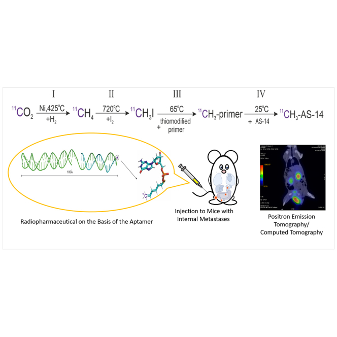
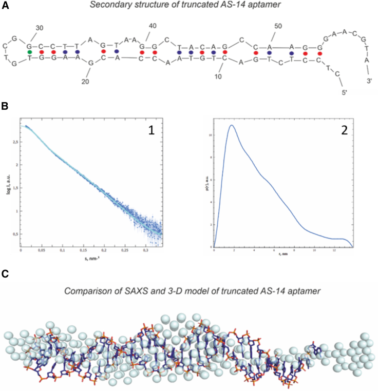
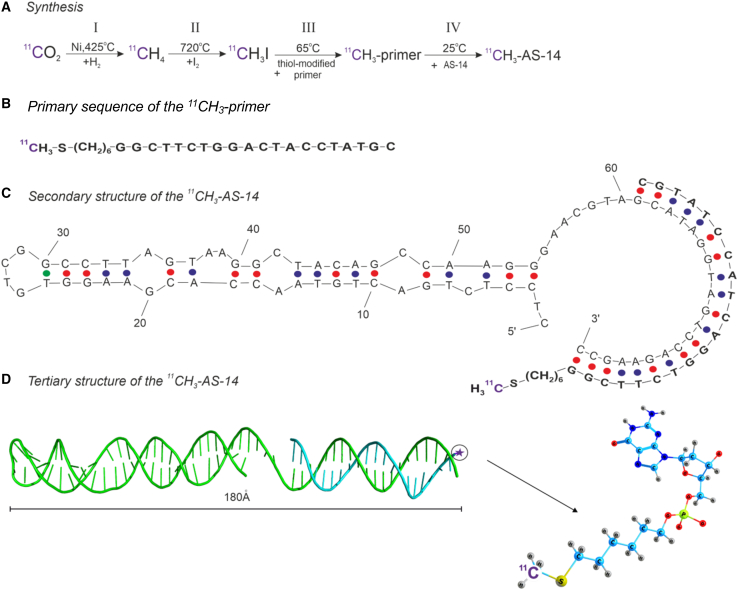
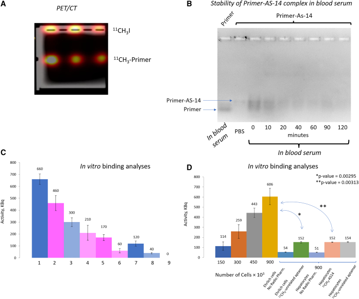
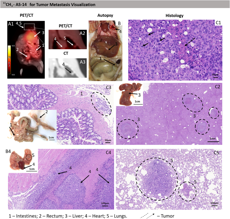
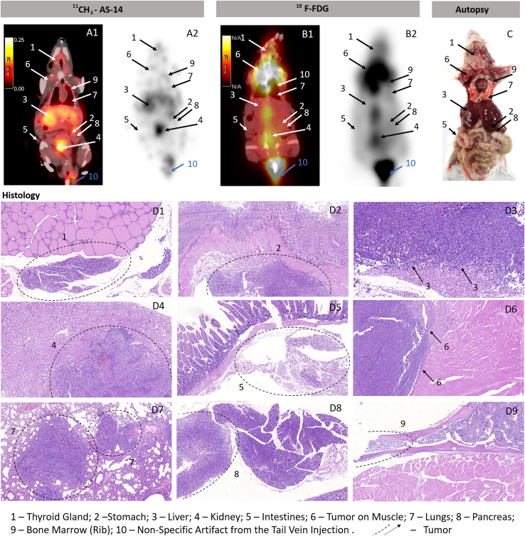
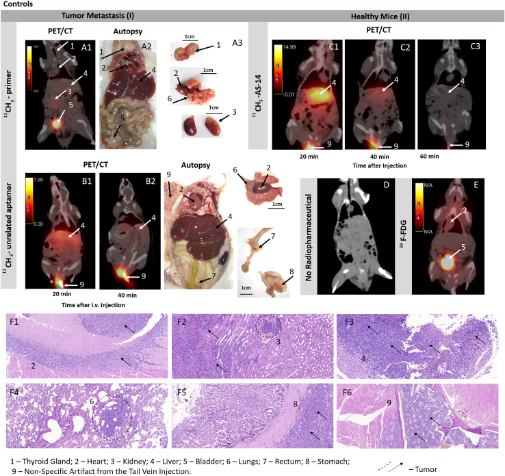
Identification of primary tumors and metastasis sites is an essential step in cancer diagnostics and the following treatment. Positron emission tomography-computed tomography (PET/CT) is one of the most reliable methods for scanning the whole organism for malignancies. In this work, we synthesized an 11C-labeled oligonucleotide primer and hybridized it to an anti-cancer DNA aptamer. The 11C-aptamer was applied for in vivo imaging of Ehrlich ascites carcinoma and its metastases in mice using PET/CT. The imaging experiments with the 11C-aptamer determined very small primary and secondary tumors of 3 mm2 and less. We also compared 11C imaging with the standard radiotracer, 2-deoxy-2-[fluorine-18]fluoro-D-glucose (18F-FDG), and found better selectivity of the 11C-aptamer to metastatic lesions in the metabolically active organs than 18F-FDG. 11C radionuclide with an ultra-short (20.38 min) half-life is considered safest for PET/CT imaging and does not cause false-positive results in heart imaging. Its combination with aptamers gives us high-specificity and high-contrast imaging of cancer cells and can be applied for PET/CT-guided drug delivery in cancer therapies.

Keywords: 11C radiolabeling; DNA aptamers; Ehrlich ascites carcinoma; PET/CT; in vivo imaging; metastasis; radiopharmaceuticals.


Conflict of interest statement

Authors declare no competing interests.

Figures

None
Graphical abstract
Figure 1
Figure 1
Finding the 3D structure of the truncated AS-14 aptamer (A) The secondary structure of AS-14t. (B) (1) Experimental SAXS data (dark blue dots) fitted by the theoretical SAXS curve (a light blue line). (2) Pair distance distribution function p (r) of AS-14t. The maximal value of r is the Dmax (the size of the molecule). (C) Comparing the modeled 3D structures with the experimental SAXS model. Dark blue sticks are from molecular modeling; blue color spheres are from SAXS data.
Figure 2
Figure 2
Synthesis of the 11C-labeled aptamer (A) Synthesis of the 11CH3-labeled primer. (B) The primary sequence of the 11CH3 primer. (C) Secondary structure of 11CH3-AS-14. (D) The tertiary structure of 11СH3- AS-14 (green ribbon, AS-14 aptamer; cyan ribbon, 11CH3 primer).
Figure 3
Figure 3
Degradation and binding analyses of 11C-labeled oligonucleotides (A) Agarose gel electrophoresis of the 11C-labeled primer after synthesis. (B) Agarose gel electrophoresis of the FAM-labeled primer AS-14 in mouse serum at different times. (C) Dependence of the sample's radioactivity on the content of 11CH3 primer or 11CH3-AS-14, where sample (1) corresponds to 0.6 nmol of 11CH3 primer; (2) 0.6 nmol of 11CH3 primer hybridized with 0.6 nmol of AS-14; (3) 0.3 nmol of 11CH3 primer; (4) 0.3 nmol of 11CH3 primer hybridized with 0.3 nmol of AS-14 (11CH3-AS-14); (5) 0.6 nmol of 11CH3 primer hybridized with 0.6 nmol of AS-14 (11CH3-AS-14) incubated with Ehrlich cells; (6) 0.3 nmol of 11CH3 primer hybridized with 0.3 nmol of AS-14 (11CH3-AS-14) incubated with Ehrlich cells; (7) 0.6 nmol of 11CH3 primer incubated with Ehrlich cells; (8) 0.3 nmol of 11CH3 primer incubated with Ehrlich cells; (9) Ehrlich cells only. (D) Correlation of the radioactivity of the samples versus the number of ascites cells or hepatocytes bound with 11CH3-AS-14 or the 11CH3-unrelated aptamer. Errors bars are one SD of three measurements.
Figure 4
Figure 4
Tumor and metastases localization analysis using the 11CH3-AS-14 probe and PET/CT (A–C) The representative PET/CT images of mice with metastases (A) were confirmed by autopsy (B) and histological analyses of metastatic tumor lesions (C) in different organs: intestines (2); liver (3); heart (4); and lung spleen (5). Cytological characteristics of the tumor tissue (C). Hematoxylin-eosin staining. Magnification С1, ×400; С2, ×200; C3, ×30; С4 and 5, ×100. Arrows and dashed circles indicate the tumor sites in different organs at PET/CT, autopsy, and corresponding tissue sections.
Figure 5
Figure 5
Comparative PET/CT imaging using 11CH3-AS-14 with 18F-FDG (A–D) Images with 11CH3-AS-14 (A1 and 2) and 18F-FDG (B1 and 2) on the same mice scanned on the next day. Accuracy of PET/CT results was confirmed by autopsy (C) and histological analyses of metastatic tumor lesions (D) in different organs: thyroid gland (1); stomach (2); liver (3); kidney (4); intestines (5); muscle (6); lung (7); pancreas (8); rib bone marrow (9). Arrows and dashed circles indicate the tumor sites in different organs at PET/CT, autopsy, and corresponding tissue sections.
Figure 6
Figure 6
Specific recognition of the tumor sites by 11CH3-AS-14 proved by control experiments (A–F) The representative PET/CT analysis of mice with tumor metastasis (I) using the 11CH3 primer (A) and 11CH3-unrelated aptamer (B) as probes. Metastasis formation was confirmed by autopsy (A2, B3). PET/CT images of physiological distribution and excretion of 11CH3-AS-14 (C) and 18F-FDG (E) in healthy mice (II). PET/CT image of a healthy mouse without the injected radiopharmaceutical (D). Histological examination with the staining of tissue sections with hematoxylin-eosin (F). Arrows indicate the tumor sites in different organs at PET/CT and autopsy.

References

    1. Seyfried T.N., Huysentruyt L.C. On the origin of cancer metastasis. Crit. Rev. Oncog. 2013;18:43–73. - PMC - PubMed
    1. Wafaie A.M., Moussa K.M., Ebeid E.M. Cancer of unknown primary origin: can FDG PET/CT have a role in detecting the site of primary? Egypt. J. Radiol. Nucl. Med. 2018;49:190–195.
    1. Peck M., Pollack H.A., Friesen A., Muzi M., Shoner S.C., Shankland E.G., Fink J.R., Armstrong J.O., Link J.M., Krohn K.A. Applications of PET imaging with the proliferation marker [18F]-FLT. Q. J. Nucl. Med. Mol. Imaging. 2015;59:95–104. - PMC - PubMed
    1. Bollineni V.R., Kramer G.M., Jansma E.P., Liu Y., Oyen W.J.G. A systematic review on [18F]FLT-PET uptake as a measure of treatment response in cancer patients. Eur. J. Cancer. 2016;55:81–97. - PubMed
    1. Positron Emission Tomography: A Guide for Clinicians (2014). (Springer Berlin Heidelberg)

LinkOut - more resources