

Emergent Management of Traumatic Posterior Sternoclavicular Joint Dislocation: A Case Report and Literature Review
- PMID: 34853739
- PMCID: PMC8608401
- DOI: 10.7759/cureus.18996
Emergent Management of Traumatic Posterior Sternoclavicular Joint Dislocation: A Case Report and Literature Review
Abstract
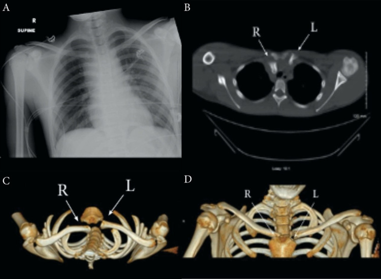
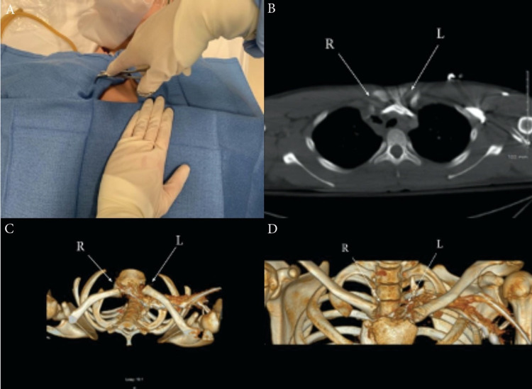
Sternoclavicular joint (SCJ) dislocation is a rare but serious orthopedic injury. Posterior dislocations are more concerning due to the SCJ's proximity to visceral structures such as the trachea, esophagus, subclavian vessels, and brachial plexus. Due to the potential long-term sequelae of missed diagnosis, clinical suspicion should be high when a patient presents with a compression-type injury to the shoulder girdle and pain or deformity to the SCJ. Here we present a case of a 15-year-old soccer player who presented to the emergency department (ED) after a fall onto his right shoulder with additional compound injuries. A posterior SCJ dislocation diagnosis was suspected and confirmed after a computed tomography scan. A successful closed reduction was done in the ED after consultation with cardiothoracic and orthopedic surgery. This case adds to the body of literature describing diagnosis and management of posterior SCJ dislocations.
Keywords: case report; closed reduction; computed tomography; sternoclavicular joint; traumatic sternoclavicular joint dislocation.
Copyright © 2021, Chen et al.
Conflict of interest statement
The authors have declared that no competing interests exist.
Figures
References
-
- Pediatric posterior sternoclavicular joint injuries. Chaudhry S. J Am Acad Orthop Surg. 2015;23:468–475. - PubMed
-
- Instability of the sternoclavicular joint: current concepts in classification, treatment and outcomes. Sewell MD, Al-Hadithy N, Le Leu A, Lambert SM. Bone Joint J. 2013;95-B:721–731. - PubMed
-
- Sternoclavicular joint dislocation: a systematic review and meta-analysis. Sernandez H, Riehl J. J Orthop Trauma. 2019;33:0–5. - PubMed
-
- Posterior dislocation of the sternoclavicular joint and epiphyseal disruption of the medial clavicle with posterior displacement in sports participants. Laffosse JM, Espié A, Bonnevialle N, et al. J Bone Joint Surg Br. 2010;92:103–109. - PubMed
Publication types
LinkOut - more resources
Full Text Sources