. 2020 Dec 21;8(2):ofaa631.
doi: 10.1093/ofid/ofaa631. eCollection 2021 Feb.

Clinical Assessment and Validation of a Rapid and Sensitive SARS-CoV-2 Test Using Reverse Transcription Loop-Mediated Isothermal Amplification Without the Need for RNA Extraction

Affiliations

Clinical Assessment and Validation of a Rapid and Sensitive SARS-CoV-2 Test Using Reverse Transcription Loop-Mediated Isothermal Amplification Without the Need for RNA Extraction

Melis N Anahtar et al. Open Forum Infect Dis. .

Abstract

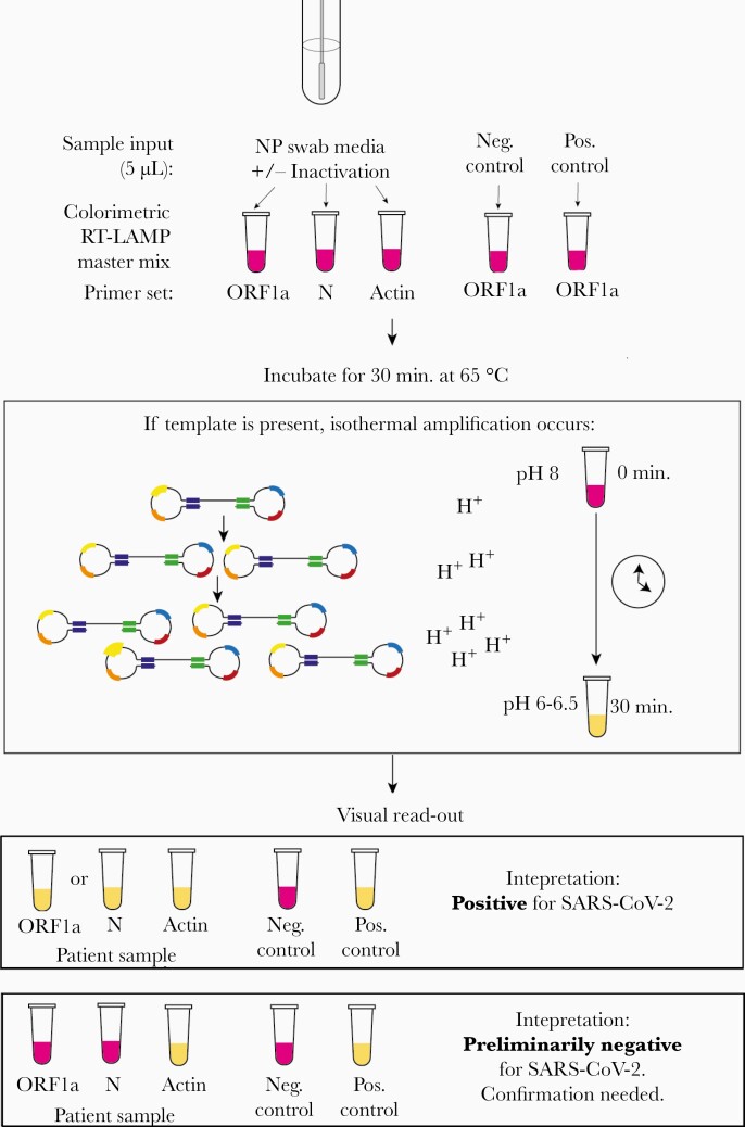
Background: Amid the enduring pandemic, there is an urgent need for expanded access to rapid, sensitive, and inexpensive coronavirus disease 2019 (COVID-19) testing worldwide without specialized equipment. We developed a simple test that uses colorimetric reverse transcription loop-mediated isothermal amplification (RT-LAMP) to detect severe acute resrpiratory syndrome coronavirus 2 (SARS-CoV-2) in 40 minutes from sample collection to result.

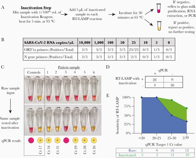
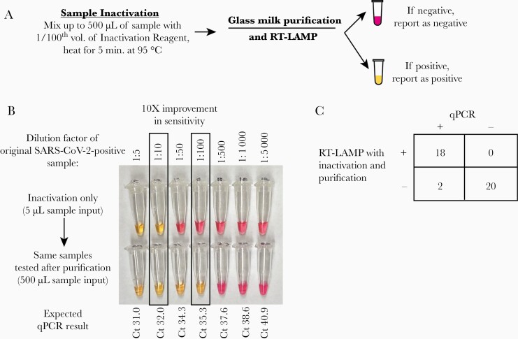
Methods: We tested 135 nasopharyngeal specimens from patients evaluated for COVID-19 infection at Massachusetts General Hospital. Specimens were either added directly to RT-LAMP reactions, inactivated by a combined chemical and heat treatment step, or inactivated then purified with a silica particle-based concentration method. Amplification was performed with 2 SARS-CoV-2-specific primer sets and an internal specimen control; the resulting color change was visually interpreted.

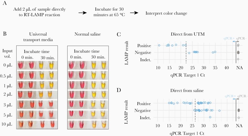
Results: Direct RT-LAMP testing of unprocessed specimens could only reliably detect samples with abundant SARS-CoV-2 (>3 000 000 copies/mL), with sensitivities of 50% (95% CI, 28%-72%) and 59% (95% CI, 43%-73%) in samples collected in universal transport medium and saline, respectively, compared with quantitative polymerase chain reaction (qPCR). Adding an upfront RNase inactivation step markedly improved the limit of detection to at least 25 000 copies/mL, with 87.5% (95% CI, 72%-95%) sensitivity and 100% specificity (95% CI, 87%-100%). Using both inactivation and purification increased the assay sensitivity by 10-fold, achieving a limit of detection comparable to commercial real-time PCR-based diagnostics.

Conclusions: By incorporating a fast and inexpensive sample preparation step, RT-LAMP accurately detects SARS-CoV-2 with limited equipment for about US$6 per sample, making this a potentially ideal assay to increase testing capacity, especially in resource-limited settings.

Keywords: COVID-19; LAMP; SARS-CoV-2; diagnostics; isothermal amplification; nucleic acid technology; rapid tests.


Figures

Figure 1.
Figure 1.
Schematic for the use of RT-LAMP directly from NP specimen using 2 SARS-CoV-2 specific primers, which target the ORF1a and N genes, and 1 internal specimen control targeting the human actin gene. Before sample addition to the RT-LAMP reaction, the NP specimen can undergo a 5-minute heat and chemical inactivation step to destroy endogenous RNases and lyse viral particles and human cells. The RT-LAMP reaction occurs at 65°C for 30 minutes, during which the amplification of SARS-CoV-2 RNA generates protons that decrease the pH of the reaction mix and result in a color change due to the media’s colorimetric pH indicator. Samples are removed from the heat block, immersed in ice to enhance the color brightness, and color change is visually determined. If the controls are valid, a yellow color change with the ORF1a and/or N gene primers indicates the presence of SARS-CoV-2 RNA in the sample. Abbreviations: NP, nasopharyngeal; RT-LAMP, reverse transcription loop-mediated isothermal amplification; SARS-CoV-2, severe acute respiratory syndrome coronavirus 2.
Figure 2.
Figure 2.
Detection of SARS-CoV-2 directly from nasopharyngeal samples collected in VTM or 0.9% normal saline. A, Schematic for the 35-minute protocol of direct-from-sample testing. B, Determination of the optimal sample input volume for VTM and saline using a standardized 1000-copy/µL synthetic SARS-CoV-2 input. Samples are pictured before and after the 30-minute amplification step. C, Comparison of the sensitivity of qPCR with RT-LAMP with direct addition of 1 µL of clinical NP specimen collected in universal transport media (16 qPCR-positive samples, 15 qPCR-negative samples) and testing using the N gene primers alone. D, Comparison of the sensitivity of qPCR to RT-LAMP with direct addition of 5 µL of clinical NP samples collected in saline (40 qPCR-positive samples, 45 qPCR-negative samples), using both the N gene and ORF1a primer sets. One invalid result occurred from a sample that had a negative human actin control and was noted to be bloody. The approximate clinical limit of detection is shown with a dotted line. Abbreviations: NA, no amplification; NP, nasopharyngeal; qPCR, quantitative polymerase chain reaction; RT-LAMP, reverse transcription loop-mediated isothermal amplification; SARS-CoV-2, severe acute respiratory syndrome coronavirus 2; VTM, viral transport media.
Figure 3.
Figure 3.
Detection of SARS-CoV-2 after inactivation of RNases and viral lysis from clinical nasopharyngeal swabs collected in saline. A, Schematic for a 40-minute rule-in protocol where samples are first treated with an inactivation reagent (TCEP/EDTA), and heated to 95°C for 5 minutes before sample addition to the RT-LAMP reaction. B, Determination of the analytic sensitivity of the RT-LAMP assay with each primer set, as determined by synthetic SARS-CoV-2 RNA spiked into inactivated SARS-CoV-2-negative nasopharyngeal samples collected in saline. C, Representative clinical samples illustrating the improvement of RT-LAMP sensitivity after inactivation, with corresponding qPCR results and Ct values. Amplification reactions using the ORF1a primer set are shown. D, Overall performance of RT-LAMP in 62 inactivated clinical samples. RT-LAMP results were categorized as positive or negative using the criteria outlined in Table 1. E, Sensitivity of RT-LAMP with or without inactivation as a function of the input SARS-CoV-2 RNA concentration, as determined by qPCR. The total number of samples tested within each Ct range is shown in the table. Abbreviations: Ct, cycle threshold; qPCR, quantitative polymerase chain reaction; RT-LAMP, reverse transcription loop-mediated isothermal amplification; SARS-CoV-2, severe acute respiratory syndrome coronavirus 2; TCEP, tris(2-carboxyethyl)phosphine.
Figure 4.
Figure 4.
Further improvement in sensitivity by reflexing RT-LAMP-negative inactivated samples to a glass milk purification procedure. A, Schematic for combining inactivation with glass milk purification. B, Demonstration that purification improves the assay detection limit by ~10-fold using a serially diluted SARS-CoV-2-positive patient’s nasopharyngeal specimen. Using the ORF1a primer set, the lowest dilution detected from the inactivated sample was 1:10. After purifying 500 µL of each dilution, the 1:100 dilution could be detected by the same primer set. C, Overall performance of RT-LAMP in 40 clinical samples that were inactivated and then purified, including 4 specimens that were falsely negative with inactivation alone. Abbreviations: RT-LAMP, reverse transcription loop-mediated isothermal amplification.

Similar articles

Cited by

References

    1. Zhen W, Smith E, Manji R, et al. Clinical evaluation of three sample-to-answer platforms for the detection of SARS-CoV-2. J Clin Microbiol 2020; 58:e00783–20. - PMC - PubMed
    1. Zhen W, Manji R, Smith E, Berry GJ. Comparison of four molecular in vitro diagnostic assays for the detection of SARS-CoV-2 in nasopharyngeal specimens. J Clin Microbiol 2020; 58:e00743–20. - PMC - PubMed
    1. Bruning AHL, Leeflang MMG, Vos JMBW, et al. Rapid tests for influenza, respiratory syncytial virus, and other respiratory viruses: a systematic review and meta-analysis. Clin Infect Dis 2017; 65:1026–32. - PMC - PubMed
    1. Lambert-Niclot S, Cuffel A, Le Pape S, et al. Evaluation of a rapid diagnostic assay for detection of SARS CoV-2 antigen in nasopharyngeal swab. J Clin Microbiol 20202; 58:e00977–20. - PMC - PubMed
    1. Nagamine K, Hase T, Notomi T. Accelerated reaction by loop-mediated isothermal amplification using loop primers. Mol Cell Probes 2002; 16:223–9. - PubMed