Review
. 2022 Feb;143(2):179-224.
doi: 10.1007/s00401-021-02384-2. Epub 2021 Dec 1.

Microglia and monocytes in inflammatory CNS disease: integrating phenotype and function

Affiliations
Review

Microglia and monocytes in inflammatory CNS disease: integrating phenotype and function

Alanna G Spiteri et al. Acta Neuropathol. 2022 Feb.

Abstract

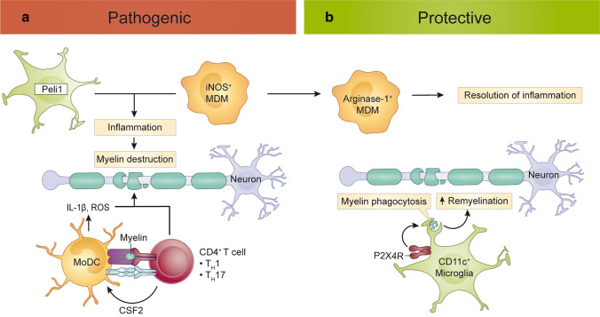
In neurological diseases, the actions of microglia, the resident myeloid cells of the CNS parenchyma, may diverge from, or intersect with, those of recruited monocytes to drive immune-mediated pathology. However, defining the precise roles of each cell type has historically been impeded by the lack of discriminating markers and experimental systems capable of accurately identifying them. Our ability to distinguish microglia from monocytes in neuroinflammation has advanced with single-cell technologies, new markers and drugs that identify and deplete them, respectively. Nevertheless, the focus of individual studies on particular cell types, diseases or experimental approaches has limited our ability to connect phenotype and function more widely and across diverse CNS pathologies. Here, we critically review, tabulate and integrate the disease-specific functions and immune profiles of microglia and monocytes to provide a comprehensive atlas of myeloid responses in viral encephalitis, demyelination, neurodegeneration and ischemic injury. In emphasizing the differential roles of microglia and monocytes in the severe neuroinflammatory disease of viral encephalitis, we connect inflammatory pathways common to equally incapacitating diseases with less severe inflammation. We examine these findings in the context of human studies and highlight the benefits and inherent limitations of animal models that may impede or facilitate clinical translation. This enables us to highlight common and contrasting, non-redundant and often opposing roles of microglia and monocytes in disease that could be targeted therapeutically.

Keywords: Encephalitis; Immune-mediated pathology; Microglia; Monocyte-derived cells; Neurodegeneration; Neuroinflammation.


Conflict of interest statement

The authors have no conflicts of interest to declare that are relevant to the content of this article.

Figures

Fig. 1
Fig. 1
Macrophage/microglial CD68 tissue staining in human CNS pathologies. Ameboid (solid arrows, likely macrophages and/or reactive microglia) and ramified (open arrows, likely microglia) CD68+ myeloid cells are shown in various neuropathologies: a West Nile virus (WNV) [247]. Macrophage/microglia engulfing a degenerating neuron (arrowheads) in the substantia nigra in a patient with fulminant WNV encephalitis (400X magnification). b Cortical stroke [94]. Foamy macrophages/microglia are present in a cerebral infarct (several weeks old) (scale bar represents 50 μm). c Parkinson disease (PD) [65]. Ramified microglia and macrophages with enlarged cytoplasm and short stout processes are present in the substantia nigra (400X magnification). d–f Amyotrophic lateral sclerosis (ALS) [38]. The three images show the variable extent of microglia activation in the corticospinal tract in patients with ALS assessed as either mild (d), moderate (e) or severe (f) (scale bar represents 250 μm). g–i Alzheimer disease (AD) [148]. The three images show the rounded ameboid microglia (g), ramified microglia (h) or foamy macrophages (i) that can be seen in AD brains (scale bar represents 10 μm). j–l Multiple sclerosis (MS) [193]. Three images show variable inflammatory activity in MS, with either numerous foamy macrophages within a demyelinating plaque (j), macrophages at the rim of a plaque (arrowheads) (k), and an inactive plaque with only a few ramified microglia (l). All images reproduced with permission
Fig. 2
Fig. 2
Protective and pathogenic roles of monocytes and microglia in viral encephalitis. a Protective functions. In viral encephalitis, microglia enhance viral clearance by phagocytosing virus-infected cells. Both microglia and MDMs stimulate anti-viral T cell responses, which is optimized by microglia-mediated regulation of Treg infiltration. b Pathogenic functions. NO- and IL-6-producing MDMs exacerbate neuronal damage and contribute to immunopathology. MDM, monocyte-derived macrophage; MHC, major histocompatibility complex; NO nitric oxide; IL interleukin; Treg regulatory T cell
Fig. 3
Fig. 3
Roles of monocytes and microglia in ischemic stroke. In stroke, microglia migrate towards neurons with high intracellular calcium levels to reduce excitotoxicity and neuronal damage. Microglia also prevent bystander tissue damage by inhibiting reactive astrocytes and phagocytosing infiltrating neutrophils. CXCR4+ MDM are recruited to the site of injury, where they produce microglia-activating mediators, IL-1β and ROS, and stimulate microglia proliferation and the glial scar formation. These inflammatory mediators may also injure neurons and contribute to secondary damage. CNS-infiltrating Ly6Chi monocytes may also help repair the damaged BBB via the production of collagen-4 and TGF-β1. BBB, blood–brain barrier; CXCR4, CXC chemokine receptor type 4; CXCL12, CXC Motif Chemokine Ligand 12; IL, interleukin; MDM, monocyte-derived macrophage; ROS, reactive oxygen species; TGF-β1, transforming growth factor beta 1
Fig. 4
Fig. 4
Protective and pathogenic roles of monocytes and microglia in neurodegeneration. a Protective functions. In AD, microglia and Ly6Clo monocytes reduce parenchymal and vascular Aβ spread, respectively. b Pathogenic functions. Microglia phagocytose C3-tagged synaptic termini via C3R, leading to neurodegeneration and cognitive dysfunction in post-infectious encephalitis, AD, EAE and MS. amyloid beta; AD Alzheimer’s disease; C3 complement component 3; C3R complement component 3 receptor; EAE experimental autoimmune encephalomyelitis; MS multiple sclerosis; ZIKV Zika virus
Fig. 5
Fig. 5
Protective and pathogenic roles of monocytes and microglia in autoimmune neuroinflammation. a Pathogenic functions. Peli-mediated microglial expression of pro-inflammatory cytokine and chemokines can exacerbate inflammatory damage to myelin. GM-CSF-stimulated moDC activate myelin-reactive T cells and exacerbate myelin damage via the production of ROS and IL-1β. iNOS+ MDM producing ROS and NO contribute to myelin destruction but may also transition to arginase-1+ MDM and facilitate the resolution of inflammation (b). b Protective functions. Microglia may stimulate re-myelination via P2X4R-mediated phagocytosis of myelin debris. GM-CSF granulocyte–macrophage stimulating factor; IL interleukin; iNOS inducible nitric oxide synthase; MDM monocyte-derived macrophage; MHC major histocompatibility complex; moDC monocyte-derived dendritic cell; NO nitric oxide; P2X4R P2X purinoceptor 4; Peli Pellino E3 Ubiquitin Protein Ligase; ROS reactive oxygen species; TH T helper cell

Similar articles

Cited by

References

    1. Afagh A, Cummings BJ, Cribbs DH, Cotman CW, Tenner AJ. Localization and cell association of C1q in Alzheimer's disease brain. Exp Neurol. 1996;138:22–32. doi: 10.1006/exnr.1996.0043. - DOI - PubMed
    1. Ajami B, Bennett JL, Krieger C, McNagny KM, Rossi FM. Infiltrating monocytes trigger EAE progression, but do not contribute to the resident microglia pool. Nat Neurosci. 2011;14:1142–1149. doi: 10.1038/nn.2887. - DOI - PubMed
    1. Ajami B, Bennett JL, Krieger C, Tetzlaff W, Rossi FM. Local self-renewal can sustain CNS microglia maintenance and function throughout adult life. Nat Neurosci. 2007;10:1538–1543. - PubMed
    1. Ajami B, Samusik N, Wieghofer P, Ho PP, Crotti A, Bjornson Z, et al. Single-cell mass cytometry reveals distinct populations of brain myeloid cells in mouse neuroinflammation and neurodegeneration models. Nat Neurosci. 2018;21:541–551. doi: 10.1038/s41593-018-0100-x. - DOI - PMC - PubMed
    1. Akiyama H, McGeer PL. Brain microglia constitutively express beta-2 integrins. J Neuroimmunol. 1990;30:81–93. doi: 10.1016/0165-5728(90)90055-r. - DOI - PubMed

Publication types