FAM3A Ameliorates Brain Impairment Induced by Hypoxia-Ischemia in Neonatal Rat
- PMID: 34853925
- PMCID: PMC9813043
- DOI: 10.1007/s10571-021-01172-6
FAM3A Ameliorates Brain Impairment Induced by Hypoxia-Ischemia in Neonatal Rat
Abstract
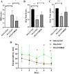
Hypoxia-ischemia (HI) during crucial periods of brain formation can lead to changes in brain morphology, propagation of neuronal stimuli, and permanent neurodevelopmental impairment, which can have profound effects on cognitive function later in life. FAM3A, a subgroup of family with sequence similarity 3 (FAM3) gene family, is ubiquitously expressed in almost all cells. Overexpression of FAM3A has been evidenced to reduce hyperglycemia via the PI3K/Akt signaling pathway and protect mitochondrial function in neuronal HT22 cells. This study aims to evaluate the protective role of FAM3A in HI-induced brain impairment. Experimentally, maternal rats underwent uterine artery bilateral ligation to induce neonatal HI on day 14 of gestation. At 6 weeks of age, cognitive development assessments including NSS, wire grip, and water maze were carried out. The animals were then sacrificed to assess cerebral mitochondrial function as well as levels of FAM3A, TNF-α and IFN-γ. Results suggest that HI significantly reduced FAM3A expression in rat brain tissues, and that overexpression of FAM3A through lentiviral transduction effectively improved cognitive and motor functions in HI rats as reflected by improved NSS evaluation, cerebral water content, limb strength, as well as spatial learning and memory. At the molecular level, overexpression of FAM3A was able to promote ATP production, balance mitochondrial membrane potential, and reduce levels of pro-inflammatory cytokines TNF-α and IFN-γ. We conclude that FAM3A overexpression may have a protective effect on neuron morphology, cerebral mitochondrial as well as cognitive function. Created with Biorender.com.
Keywords: Brain; FAM3A; Hypoxia–ischemia; IFN-γ; Mitochondria; TNF-α.
© 2021. The Author(s).
Conflict of interest statement
The authors declare no competing interests.
Figures


Similar articles
-
FAM3A Protects HT22 Cells Against Hydrogen Peroxide-Induced Oxidative Stress Through Activation of PI3K/Akt but not MEK/ERK Pathway.Cell Physiol Biochem. 2015;37(4):1431-41. doi: 10.1159/000438512. Epub 2015 Oct 23. Cell Physiol Biochem. 2015. PMID: 26492522
-
Sevoflurane postconditioning improves long-term learning and memory of neonatal hypoxia-ischemia brain damage rats via the PI3K/Akt-mPTP pathway.Brain Res. 2016 Jan 1;1630:25-37. doi: 10.1016/j.brainres.2015.10.050. Epub 2015 Nov 2. Brain Res. 2016. PMID: 26541582
-
FAM3A promotes vascular smooth muscle cell proliferation and migration and exacerbates neointima formation in rat artery after balloon injury.J Mol Cell Cardiol. 2014 Sep;74:173-82. doi: 10.1016/j.yjmcc.2014.05.011. Epub 2014 May 23. J Mol Cell Cardiol. 2014. PMID: 24857820
-
FGF21 promotes functional recovery after hypoxic-ischemic brain injury in neonatal rats by activating the PI3K/Akt signaling pathway via FGFR1/β-klotho.Exp Neurol. 2019 Jul;317:34-50. doi: 10.1016/j.expneurol.2019.02.013. Epub 2019 Feb 23. Exp Neurol. 2019. PMID: 30802446
-
FAM3A mediates PPARγ's protection in liver ischemia-reperfusion injury by activating Akt survival pathway and repressing inflammation and oxidative stress.Oncotarget. 2017 Jul 25;8(30):49882-49896. doi: 10.18632/oncotarget.17805. Oncotarget. 2017. PMID: 28562339 Free PMC article.
Cited by
-
The circular RNA Rap1b promotes Hoxa5 transcription by recruiting Kat7 and leading to increased Fam3a expression, which inhibits neuronal apoptosis in acute ischemic stroke.Neural Regen Res. 2023 Oct;18(10):2237-2245. doi: 10.4103/1673-5374.369115. Neural Regen Res. 2023. PMID: 37056143 Free PMC article.
-
FAM3A reshapes VSMC fate specification in abdominal aortic aneurysm by regulating KLF4 ubiquitination.Nat Commun. 2023 Sep 2;14(1):5360. doi: 10.1038/s41467-023-41177-x. Nat Commun. 2023. PMID: 37660071 Free PMC article.
-
FAM3A mediates the phenotypic switch of human aortic smooth muscle cells stimulated with oxidised low-density lipoprotein by influencing the PI3K-AKT pathway.In Vitro Cell Dev Biol Anim. 2023 Jun;59(6):431-442. doi: 10.1007/s11626-023-00775-1. Epub 2023 Jul 20. In Vitro Cell Dev Biol Anim. 2023. PMID: 37474885
-
FAM3 family genes are associated with prognostic value of human cancer: a pan-cancer analysis.Sci Rep. 2023 Sep 13;13(1):15144. doi: 10.1038/s41598-023-42060-x. Sci Rep. 2023. PMID: 37704682 Free PMC article.
-
FAM3A plays a key role in protecting against tubular cell pyroptosis and acute kidney injury.Redox Biol. 2024 Aug;74:103225. doi: 10.1016/j.redox.2024.103225. Epub 2024 Jun 8. Redox Biol. 2024. PMID: 38875957 Free PMC article.
References
-
- Ames A III (2000) CNS energy metabolism as related to function. Brain Res Rev 34(1–2):42–68 - PubMed
-
- Andreollo NA, Santos EF, Araújo MR, Lopes LR (2012) Rat’s age versus human’s age: what is the relationship? ABCD Arquivos Brasileiros De Cirurgia Digestiva 25:49–51 - PubMed
-
- Babenko O, Kovalchuk I, Metz GA (2015) Stress-induced perinatal and transgenerational epigenetic programming of brain development and mental health. Neuro Biobehav Rev 48:70–91 - PubMed
-
- Charil A, Laplante DP, Vaillancourt C, King S (2010) Prenatal stress and brain development. Brain Res Rev 65(1):56–79 - PubMed
MeSH terms
Substances
Grants and funding
LinkOut - more resources
Full Text Sources

