. 2022 Jan;26(1):151-162.
doi: 10.1111/jcmm.17065. Epub 2021 Dec 2.

Exosomal hsa_circ_0125310 promotes cell proliferation and fibrosis in diabetic nephropathy via sponging miR-422a and targeting the IGF1R/p38 axis

Affiliations

Exosomal hsa_circ_0125310 promotes cell proliferation and fibrosis in diabetic nephropathy via sponging miR-422a and targeting the IGF1R/p38 axis

Yingchun Zhu et al. J Cell Mol Med. 2022 Jan.

Abstract

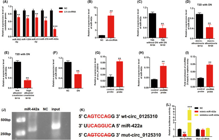
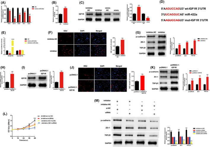
Diabetic nephropathy (DN) is still on the rise worldwide, and millions of patients have to be treated through dialysis or transplant because of kidney failure caused by DN. Recent reports have highlighted circRNAs in the treatment of DN. Herein, we aimed to investigate the mechanism by which high glucose-induced exo-circ_0125310 promotes diabetic nephropathy progression. circ_0125310 is highly expressed in diabetic nephropathy and exosomes isolated from high glucose-induced mesangial cells (MCs). High glucose-induced exosomes promote the proliferation and fibrosis of MCs. However, results showed that the effects of exosomes on MCs can be reversed by the knockdown of circ_0125310. miR-422a, which targets IGF1R, was the direct target of circ_0125310. circ_0125310 regulated IGF1R/p38 axis by sponging miR-422a. Exo-circ_0125310 increased the luciferase activity of the WT-IGF1R reporter in the dual-luciferase reporter gene assays and upregulated the expression level of IGF1R and p38. Finally, in vivo research indicated that the overexpression of circ_0125310 promoted the diabetic nephropathy progression. Above results demonstrated that the high glucose-induced exo-circ_0125310 promoted cell proliferation and fibrosis in diabetic nephropathy via sponging miR-422a and targeting the IGF1R/p38 axis.

Keywords: MCs; diabetic nephropathy; fibrosis; hsa_circ_0125310; proliferation.


Conflict of interest statement

The authors declare that there are no conflicts of interest.

Figures

FIGURE 1
FIGURE 1
Expression patterns of exo‐circ_0125310 in DN. A, TEM image of exosomes isolated from culture media (scale bar, 100 nm). B, Exosomes were isolated from MCs with or without high glucose. Western blotting of three representative exosome specific markers: CD63, Alix and Tsg101. C, The expression level of circ_0125310 was detected in exosomes from MCs with or without high glucose. D, The expression level of circ_0125310 was detected in MCs treated by exosomes, which were secreted from normal or high glucose. E, The expression level of circ_0125310 was detected in kidney tissues with T2D or without DN (T2D without DN = 24, T2D with DN = 32). F, Expression levels of circ_0125310 was detected in kidney tissues with microalbuminuria or with macroalbuminuria (microalbuminuria group = 12, macroalbuminuria group = 20). G, Based on the average value of eGFR, expression levels of circ_0125310 were detected in kidney tissues with low or high eGFR (low eGFR group = 14, high eGFR group = 18). H, H&E staining results of the DN rat model and negative control. Results indicated that the epithelium cells of tubule were edematous. Glomerular mesangial proliferation is shown in DN (n = 6). I, Blood glucose was detected in the DN rat model and negative control (n = 6). J, Western blotting of two representative specific markers of renal fibrosis: p‐cadherin and ZO‐1. K, The expression level of circ_0125310 was detected in kidney tissues of NC and DN rat model. L, Validation of circ_0125310 by RNase R treatment and reverse transcription PCR (RT‐PCR) analysis
FIGURE 2
FIGURE 2
Exo‐circ_0125310 promotes growth and fibrosis of MCs cells. A, Proliferating mesangial cells were labelled with EdU. MCs were treated with NC, exosomes and exosomes‐circRNA del. B, Flow cytometric assay showed that G2/M phase of MCs was increased in MCs treated with exosomes, but this promotion was eliminated when circ_0125310 was knocked down in exosomes. C, Western blotting results showed that the expression level of p‐cadherin and ZO‐1 was upregulated in MCs treated with exosomes, but this promotion was eliminated when circ_0125310 was knocked down in exosomes. D and E, Efficiency of LV‐circ_0125310 and sh‐circ_0125310 were detected by qPCR. F and G, Proliferating mesangial cells were labelled with EdU. MCs were transfected with LV‐circ_0125310 and sh‐circ_0125310. H and I, Flow cytometric assay showed that G2/M phase of MCs was increased in MCs treated with LV‐circ_0125310, but G2/M phase was decreased when circ_0125310 was knocked down in MCs. J and K, LV‐circ_0125310 upregulated the expression level of p‐cadherin and ZO‐1 and sh‐circ_0125310 downregulated the expression level of p‐cadherin and ZO‐1
FIGURE 3
FIGURE 3
miR‐422a was the directly target of circ_0125310. A, Five miRNAs were screened in MCs transfected with LV‐circ_0125310. B, The expression level of miR‐422a was upregulated in MCs transfected with sh‐circ_0125310. C, The expression level of miR‐422a was decreased in kidney tissues with DN (T2D without DN=24, T2D with DN = 32). D, Expression levels of miR‐422a was downregulated in kidney tissues with macroalbuminuria group (microalbuminuria group = 12, macroalbuminuria group = 20). E, Expression levels of miR‐422a was downregulated in kidney tissues with high eGFR group (low eGFR group = 14, high eGFR group = 18). F, The expression level of miR‐422a was decreased in kidney tissues of the DN rat model. G, circ_0125310 in cell lysis was pulled down and enriched with circ_0125310 specific probe in MCs. H, miR‐422a was pulled down and enriched with circ_0125310 specific probe in MCs. I and J, Biotin‐coupled miR‐422a captured a fold change of circ_0125310 in the complex as compared with biotin‐coupled NC in biotin‐coupled miRNA capture. Then products were detected by agarose gel electrophoresis. K, The direct binding sites between circ_0125310 and miR‐422a were presented. L, Luciferase reporter assay was performed to confirm the direct binding relationship between circ_0125310 and miR‐422a
FIGURE 4
FIGURE 4
miR‐422a suppressed proliferation and fibrosis of MCs by targeting IGF1R. A, Five genes were screened in MCs transfected with mimic‐miR‐422a. B, The expression level of miR‐422a was upregulated in MCs transfected with inhibitor‐miR‐422a. C, The protein expression level of IGF1R was increased by inhibitor‐miR‐422a and decreased by mimic‐miR‐422a. D, The direct binding sites between miR‐422a and IGF1R were presented. E, Luciferase reporter assay was performed to confirm the direct binding relationship between miR‐422a and IGF1R. F, Proliferating mesangial cells were labelled with EdU. Inhibitor‐miR‐422a enhanced the growth of MCs. G, Inhibitor‐miR‐422a enhanced the expression level of markers of fibrosis: p‐cadherin, ZO‐1 and TGF‐β1. H, and I, Efficiency of pcDNA3.1‐IGF1R were detected by qPCR and western blotting. J, and K, Overexpression of IGF1R enhanced the growth and fibrosis of MCs. L, The CCK8 assays indicated that the growth of MCs was promoted by inhibitor‐miR‐422a, but this promotion was eliminated when IGF1R was knocked down in MCs. M, Western blotting results showed that the expression level of p‐cadherin, ZO‐1 and TGF‐β1 was upregulated in MCs treated with inhibitor‐miR‐422a, but this promotion was restored when IGF1R was downregulated in MCs
FIGURE 5
FIGURE 5
circ_0125310 regulates IGF1R/p38 expression and promoted proliferation and fibrosis of MCs cells by acting as miR‐422a sponge. A, B, and C, The expression level of IGF1R was upregulated by LV‐circ_0125310, but the expression level of IGF1R was downregulated by sh‐circ_0125310. D, The mutant‐type reporter gene (Mut‐IGF1R reporter) was established, which binds sites of miR‐422a and IGF1R was knockout. Luciferase reporter assay was performed to confirm the relationship between circ_0125310 and IGF1R. E, F and G, The expression level of p38 was upregulated by LV‐circ_0125310, but the expression level of p38 was downregulated by sh‐circ_0125310. H and I, Western blotting results showed that the expression level of p38 was upregulated in MCs treated with LV‐circ_0125310, but this promotion was restored when IGF1R was downregulated in MCs. J, The CCK8 assays indicated that the proliferation of MCs was promoted by LV‐circ_0125310, but this promotion was restored when MCs was transfected by mimic‐miR‐422a. K, The protein expression level of p‐cadherin and ZO‐1 was upregulated in MCs treated with LV‐circ_0125310, but this upregulation was restored when MCs were transfected by mimic‐miR‐422a
FIGURE 6
FIGURE 6
Effects of exo‐circ_0125310 on miR‐422a and IGF1R/p38 axis in MCs cells. A, The expression level of miR‐422a was detected in MCs treated with NC or exosomes. B, C and D, The expression level of IGF1R and p38 was detected in MCs treated with NC or exosomes. E, The Mut‐IGF1R reporter was established, which binds sites of circ_0125310 and IGF1R was knockout. Luciferase reporter assay was performed to confirm the relationship between exosomes and IGF1R. F, The expression level of miR‐422a was detected in MCs treated with exosomes or exo‐circRNA del. G, H, and I, The expression level of IGF1R and p38 was detected in MCs treated with exosomes or exo‐circRNA del. J Luciferase reporter assay was performed to confirm the relationship between exo‐circRNA del and IGF1R
FIGURE 7
FIGURE 7
Overexpression of circ_0125310 promoted DN progression in vivo. A, Graphical representation of DN model treated with PBS, LV‐NC or LV‐circ_0125310. B, Blood glucose was detected in the DN+PBS group, DN+LV‐NC group and DN+LV‐circ_0125310 group. C, H&E staining results of the DN+PBS group, DN+LV‐NC group and DN+LV‐circ_0125310 group. Results indicated that the epithelium cells of tubule were more edematous in DN+LV‐circ_0125310 group. Then, glomerular mesangial proliferation was enhanced in DN+LV‐circ_0125310 group than DN+PBS group or DN+LV‐NC group. D, Urinary albumin excretion rate was measured in the DN+PBS group, DN+LV‐NC group and DN+LV‐circ_0125310 group. E, The expression level of p‐cadherin and ZO‐1 was measured in DN+PBS group, DN+LV‐NC group and DN+LV‐NC group. F, G, H, and I, The expression level of p‐cadherin and ZO‐1 was measured in DN+PBS group, DN+LV‐NC group and DN+LV‐circ_0125310 group tissues by IHC or IF assays. J, The expression level of miR‐422a was measured in DN+PBS group, DN+LV‐NC group and DN+LV‐NC group. K, L, and M, The expression levels of IGF1R/p38 axis were detected in DN+PBS group, DN+LV‐NC group and DN+LV‐NC group. N, Schematic diagram of mechanism on this research was showed

Similar articles

Cited by

References

    1. Dewanjee S, Bhattacharjee N. MicroRNA: A new generation therapeutic target in diabetic nephropathy. Biochem Pharmacol. 2018;155:32‐47. - PubMed
    1. Bjornstad P, Lanaspa MA, Ishimoto T, et al. Fructose and uric acid in diabetic nephropathy. Diabetologia. 2015;58:1993‐2002. - PMC - PubMed
    1. Lv M, Chen Z, Hu G, Li Q. Therapeutic strategies of diabetic nephropathy: recent progress and future perspectives. Drug Discovery Today. 2015;20:332‐346. - PubMed
    1. Zha D, Wu X, Gao P. Adiponectin and Its Receptors in Diabetic Kidney Disease: Molecular Mechanisms and Clinical Potential. Endocrinology. 2017;158:2022‐2034. - PubMed
    1. Reidy K, Kang HM, Hostetter T, Susztak K. Molecular mechanisms of diabetic kidney disease. J Clin Investig. 2014;124:2333‐2340. - PMC - PubMed

Publication types