Proportionality of Clinical Outcome and Placental Changes to the Increasing Severity of Maternal Hypertension- An Observational Study
- PMID: 34854472
- PMCID: PMC10518212
- DOI: 10.5146/tjpath.2021.01563
Proportionality of Clinical Outcome and Placental Changes to the Increasing Severity of Maternal Hypertension- An Observational Study
Abstract
Objective: Preeclampsia and eclampsia remain the major causes of maternal and perinatal mortality and morbidity worldwide, causing 12-15% of direct maternal deaths. Although preeclampsia and related hypertensive disorders of pregnancy continue to affect 8% of all pregnancies, the incidence of preeclampsia has increased 40% in recent years. This study was carried out to analyse the different placental lesions and fetal outcome in different grades of maternal hypertension and to see if there is a linear relationship of the same.
Material and method: A total of 539 placenta specimens received at the department of Pathology from October 2017 to March 2019 were collected after obtaining informed consent. Of the 539 placentas, 87 hypertensive cases were graded and grouped according to the severity as gestational hypertension, mild preeclampsia, severe preeclampsia, eclampsia, and chronic hypertension and compared with 88 normotensive cases. The gross and microscopic findings were tabulated and analysed using the Statistical Package for the Social Sciences (SPSS) software.
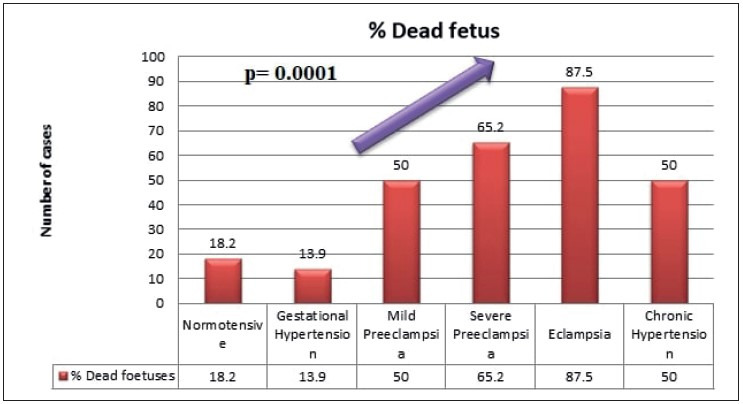
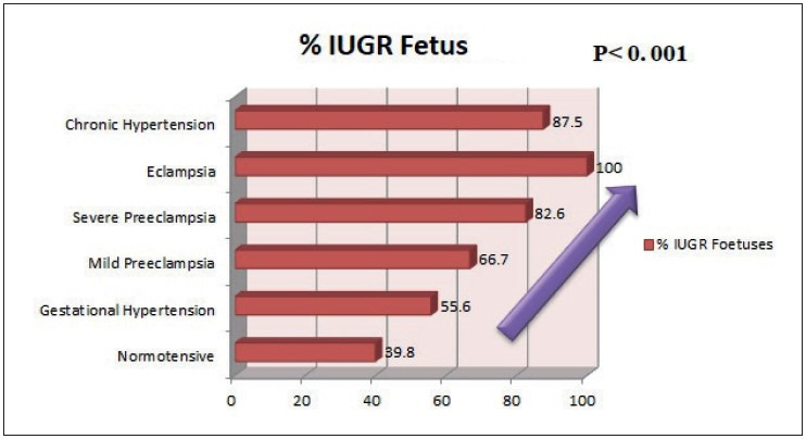
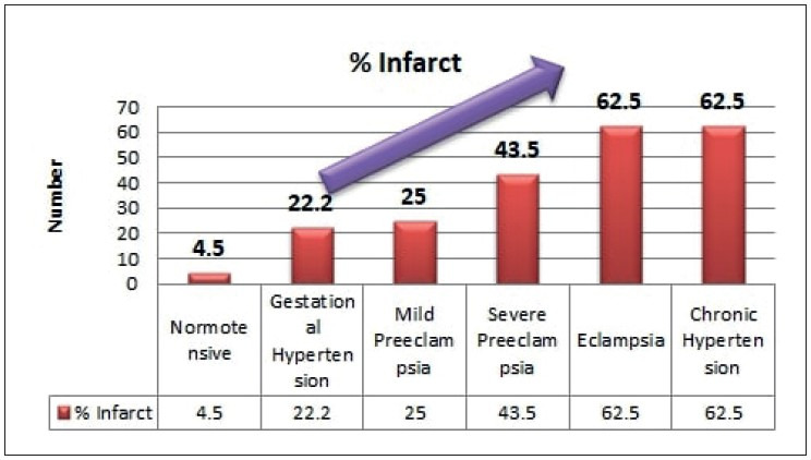
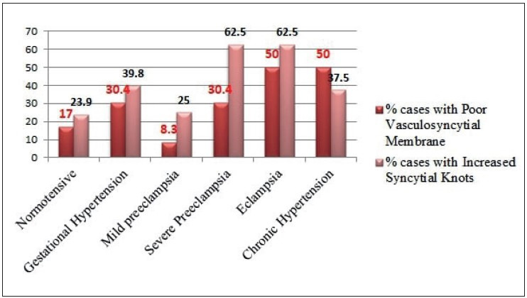
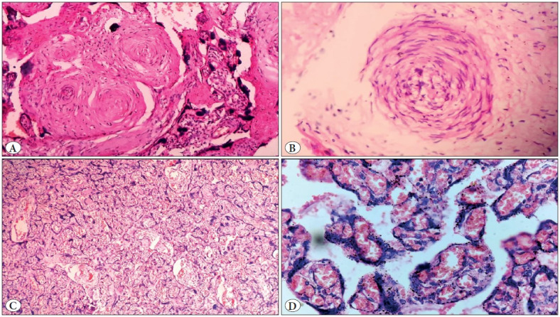
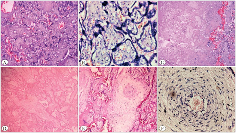
Results: Incidence of fetal death and growth restriction increased with increasing grade of maternal hypertension (p= 0.001). Abnormal shape of placenta (p= 0.034) and abnormal umbilical cord insertion (p= 0.028) were seen significantly more in the hypertensive group than in the normotensive group. Infarct and abnormal vasculo-syncytial membrane (p < 0.05) and abnormal villous maturation (p= 0. 039) were significantly increased in the hypertensive group than the normotensive group.
Conclusion: The incidence of adverse fetal outcome and placental changes suggestive of feto-maternal malperfusions shows a proportional trend with the increasing grade of maternal hypertension.
Conflict of interest statement
All authors declare that they have no conflict of interest.
Figures
Similar articles
-
Maternal and fetal vascular lesions of malperfusion in the placentas associated with fetal and neonatal death: results of a prospective observational study.Am J Obstet Gynecol. 2021 Dec;225(6):660.e1-660.e12. doi: 10.1016/j.ajog.2021.06.001. Epub 2021 Jun 8. Am J Obstet Gynecol. 2021. PMID: 34111407
-
Placental transcriptional and histologic subtypes of normotensive fetal growth restriction are comparable to preeclampsia.Am J Obstet Gynecol. 2019 Jan;220(1):110.e1-110.e21. doi: 10.1016/j.ajog.2018.10.003. Epub 2018 Oct 9. Am J Obstet Gynecol. 2019. PMID: 30312585
-
Placental pathology in early intrauterine growth restriction associated with maternal hypertension.Placenta. 2014 Sep;35(9):696-701. doi: 10.1016/j.placenta.2014.06.375. Epub 2014 Jul 9. Placenta. 2014. PMID: 25052232
-
Early Fetal Growth Restriction with or Without Hypertensive Disorders: a Clinical Overview.Reprod Sci. 2024 Mar;31(3):591-602. doi: 10.1007/s43032-023-01330-9. Epub 2023 Sep 8. Reprod Sci. 2024. PMID: 37684516 Review.
-
Placental vascular pathology as a mechanism of disease in pregnancy complications.Thromb Res. 2013 Jan;131 Suppl 1:S18-21. doi: 10.1016/S0049-3848(13)70013-6. Thromb Res. 2013. PMID: 23452733 Review.
References
-
- Lambert G., Brichant J. F., Hartstein G., Bonhomme V., Dewandre P. Y. Preeclampsia: an update. 2014Acta Anaesthesiol Belg. 65:137–149. - PubMed
-
- Salgado Sujatha S., M K R Salgado null. Structural changes in pre-eclamptic and eclamptic placentas--an ultrastructural study. Aug;2011 J Coll Physicians Surg Pak. 21:482–486. - PubMed
-
- Roberts James M., Pearson Gail, Cutler Jeff, Lindheimer Marshall, NHLBI Working Group on Research on Hypertension During Pregnancy Summary of the NHLBI Working Group on Research on Hypertension During Pregnancy. Mar;2003 Hypertension. 41:437–445. doi: 10.1161/01.HYP.0000054981.03589.E9. - DOI - PubMed
-
- Weiner Eran, Mizrachi Yossi, Grinstein Ehud, Feldstein Ohad, Rymer-Haskel Noa, Juravel Elchanan, Schreiber Letizia, Bar Jacob, Kovo Michal. The role of placental histopathological lesions in predicting recurrence of preeclampsia. Oct;2016 Prenat Diagn. 36:953–960. doi: 10.1002/pd.4918. - DOI - PubMed
-
- Khong T. Yee, Mooney Eoghan E., Ariel Ilana, Balmus Nathalie C. M., Boyd Theonia K., Brundler Marie-Anne, Derricott Hayley, Evans Margaret J., Faye-Petersen Ona M., Gillan John E., Heazell Alex E. P., Heller Debra S., Jacques Suzanne M., Keating Sarah, Kelehan Peter, Maes Ann, McKay Eileen M., Morgan Terry K., Nikkels Peter G. J., Parks W. Tony, Redline Raymond W., Scheimberg Irene, Schoots Mirthe H., Sebire Neil J., Timmer Albert, Turowski Gitta, Voorn J. Patrick, Lijnschoten Ineke, Gordijn Sanne J. Sampling and Definitions of Placental Lesions: Amsterdam Placental Workshop Group Consensus Statement. Jul;2016 Arch Pathol Lab Med. 140:698–713. doi: 10.5858/arpa.2015-0225-CC. - DOI - PubMed
Publication types
MeSH terms
LinkOut - more resources
Full Text Sources
Medical