. 2021 Dec 1;62(15):3.
doi: 10.1167/iovs.62.15.3.

EphA2 and Ephrin-A5 Guide Eye Lens Suture Alignment and Influence Whole Lens Resilience

Affiliations

EphA2 and Ephrin-A5 Guide Eye Lens Suture Alignment and Influence Whole Lens Resilience

Catherine Cheng. Invest Ophthalmol Vis Sci. .

Abstract

Purpose: Fine focusing of light by the eye lens onto the retina relies on the ability of the lens to change shape during the process of accommodation. Little is known about the cellular structures that regulate elasticity and resilience. We tested whether Eph-ephrin signaling is involved in lens biomechanical properties.

Methods: We used confocal microscopy and tissue mechanical testing to examine mouse lenses with genetic disruption of EphA2 or ephrin-A5.

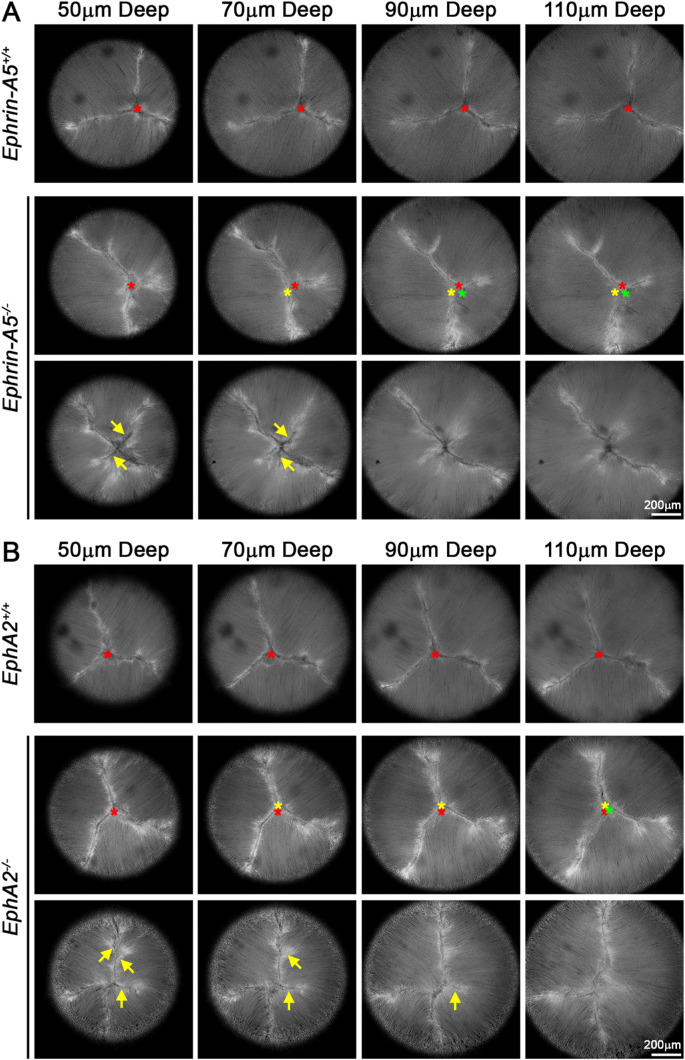
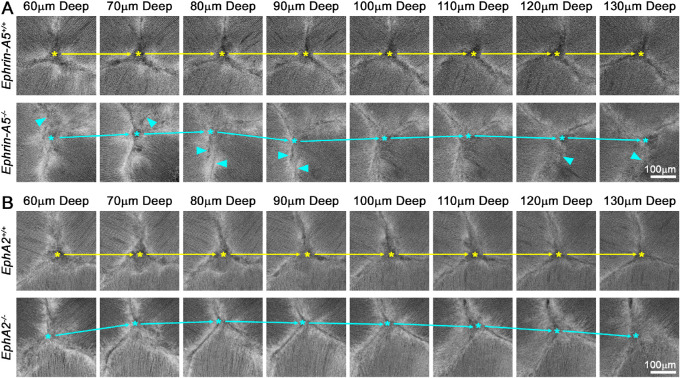
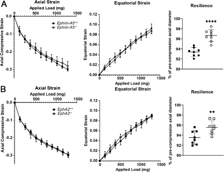
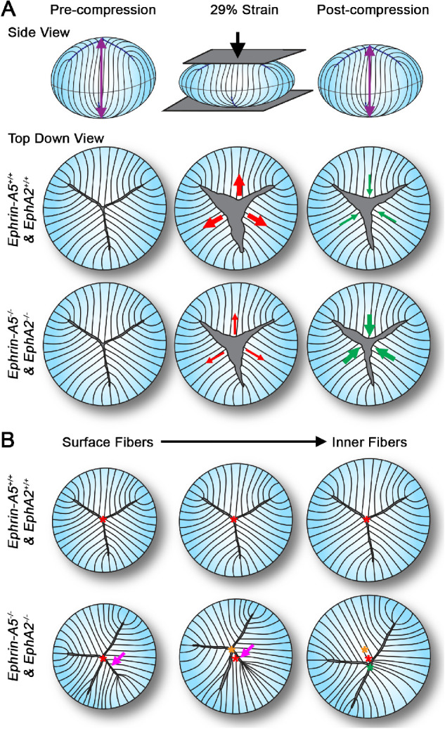
Results: Confocal imaging revealed misalignment of the suture between each shell of newly added fiber cells in knockout lenses. Despite having disordered sutures, loss of EphA2 or ephrin-A5 did not affect lens stiffness. Surprisingly, knockout lenses were more resilient and recovered almost completely after load removal. Confocal microscopy and quantitative image analysis from live lenses before, during, and after compression revealed that knockout lenses had misaligned Y-sutures, leading to a change in force distribution during compression. Knockout lenses displayed decreased separation of fiber cell tips at the anterior suture at high loads and had more complete recovery after load removal, which leads to improved whole-lens resiliency.

Conclusions: EphA2 and ephrin-A5 are needed for normal patterning of fiber cell tips and the formation of a well-aligned Y-suture with fiber tips stacked on top of previous generations of fiber cells. The misalignment of lens sutures leads to increased resilience after compression. The data suggest that alignment of the Y-suture may constrain the overall elasticity and resilience of the lens.


Conflict of interest statement

Disclosure: C. Cheng, None

Figures

Figure 1.
Figure 1.
Single optical images through the anterior of the lens revealed abnormal suture apex localization and increased suture branching in ephrin-A5/ and EphA2/ lenses. (A) Images of the lens suture at various depths (50–110 µm) from the anterior pole from representative lenses showed that the control lens had a normal Y-shaped suture and that the suture apex (marked with a red asterisk) remained in the same location between shells of fiber cells. In contrast, in the ephrin-A5/ lens (middle row), the suture apex changed location between shells of lens fibers (red, yellow, and green asterisks). The KO lens also showed extra branches in the Y-suture in the outer layers of the lens (arrows) that disappeared in deeper fiber layers. (B) Similar to ephrin-A5/ lenses, EphA2/ lenses also displayed wandering suture apex locations between shells of fiber cells (red, yellow, and green asterisks) and often displayed extra branches in newly differentiating layers of lens fibers (arrows). Scale bars: 200 µm.
Figure 2.
Figure 2.
Magnified single optical images of the anterior suture region showed the misalignment of the suture apex in ephrin-A5/ and EphA2/ lenses. (A) Images of the center of the lens suture at various depths (60–130 µm) from the anterior pole. Asterisks mark the apex of the lens suture, and arrows highlight the movement of the apex in the ephrin-A5/ lens compared with the control lens. The suture branches of the ephrin-A5/ lens were often indistinct (arrowheads), indicating that fiber cells tips were also misaligned. (B) In the EphA2/ lens, the suture apex was misaligned and moved significantly between layers. The suture branches in the EphA2/ lens were distinct, like the control lens. Scale bars: 100 µm.
Figure 3.
Figure 3.
EphA2/ and ephrin-A5/ lenses displayed increased resilience after compression. (A) Compression testing of 8-week-old ephrin-A5+/+ and ephrin-A5/ lenses revealed no differences in lens stiffness (axial compression and equatorial expansion strains). The ephrin-A5/ lenses had increased resilience, calculated as the ratio of the post-compression over pre-compression axial diameter. KO lenses recovered to 96.64% ± 1.18% of the pre-compression axial diameter, whereas control lenses recovered to 93.39% ± 0.87% of the pre-loading axial diameter. (B) Similarly, although there was no change in lens stiffness between EphA2+/+ and EphA2/ lenses, the KO lenses recovered to 95.55% ± 1.30% of the pre-compression axial diameter compared with 93.53% ± 1.35% of the pre-loading axial diameter in littermate controls (n = 8 lenses for each genotype). **P < 0.01; ****P < 0.0001.
Figure 4.
Figure 4.
Under maximum compressive load and post-compression, the suture gap area was smaller in ephrin-A5/ and EphA2/ lenses. (A) Maximum-intensity projections of the z-stack through the anterior 130 µm of representative lenses before, during, and after compression. Yellow overlays outline the suture gap area. In ephrin-A5/ lenses, the suture gap appeared smaller at 29% strain (maximum compression) and post-compression after load removal. (B) Quantification of the suture gap area revealed a statistically significant decrease in suture gap area of the ephrin-A5/ lenses under 29% strain and after compression. (C, D) Similar decreases in suture gap area of EphA2/ lenses can be observed in representative images and through area quantification (n = 8 or 9 lenses for each genotype). *P < 0.05; **P < 0.01; ****P < 0.0001. Scale bars: 200 µm.
Figure 5.
Figure 5.
(A) Our data show that, under compressive load, ephrin-A5/ and EphA2/ lenses had smaller suture gap areas, and after loading these KO lenses had better recovery and closure of the lens suture gap, leading to increased lens resilience. (B) Compared with control lenses, the ephrin-A5/ and EphA2/ lenses had misaligned suture apex locations between fiber shells, and ephrin-A5/ and EphA2/ lenses often had extra branches of the lens suture present in the surface (outer) layers of lens fibers. These extra branches often disappeared in deeper (inner) fiber layers. The branching defect was also present in some control lenses, but the control lenses did not display the suture apex misalignment defect. Illustration not drawn to scale.

Similar articles

Cited by

References

    1. Heys KR, Cram SL, Truscott RJ.. Massive increase in the stiffness of the human lens nucleus with age: the basis for presbyopia? Mol Vis. 2004; 10: 956–963. - PubMed
    1. Glasser A, Campbell MC.. Biometric, optical and physical changes in the isolated human crystalline lens with age in relation to presbyopia. Vision Res. 1999; 39: 1991–2015. - PubMed
    1. Weeber HA, Eckert G, Soergel F, Meyer CH, Pechhold W, van der Heijde RG.. Dynamic mechanical properties of human lenses. Exp Eye Res. 2005; 80: 425–434. - PubMed
    1. Heys KR, Friedrich MG, Truscott RJ.. Presbyopia and heat: changes associated with aging of the human lens suggest a functional role for the small heat shock protein, alpha-crystallin, in maintaining lens flexibility. Aging Cell. 2007; 6: 807–815. - PubMed
    1. Pierscionek BK. Age-related response of human lenses to stretching forces. Exp Eye Res. 1995; 60: 325–332. - PubMed

Publication types