. 2022 Jan 1;135(1):jcs258694.
doi: 10.1242/jcs.258694. Epub 2022 Jan 12.

Downregulation of collagen XI during late postnatal corneal development is followed by upregulation after injury

Affiliations

Downregulation of collagen XI during late postnatal corneal development is followed by upregulation after injury

Mei Sun et al. J Cell Sci. .

Abstract

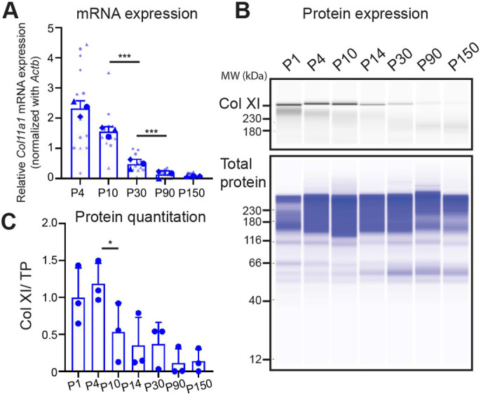
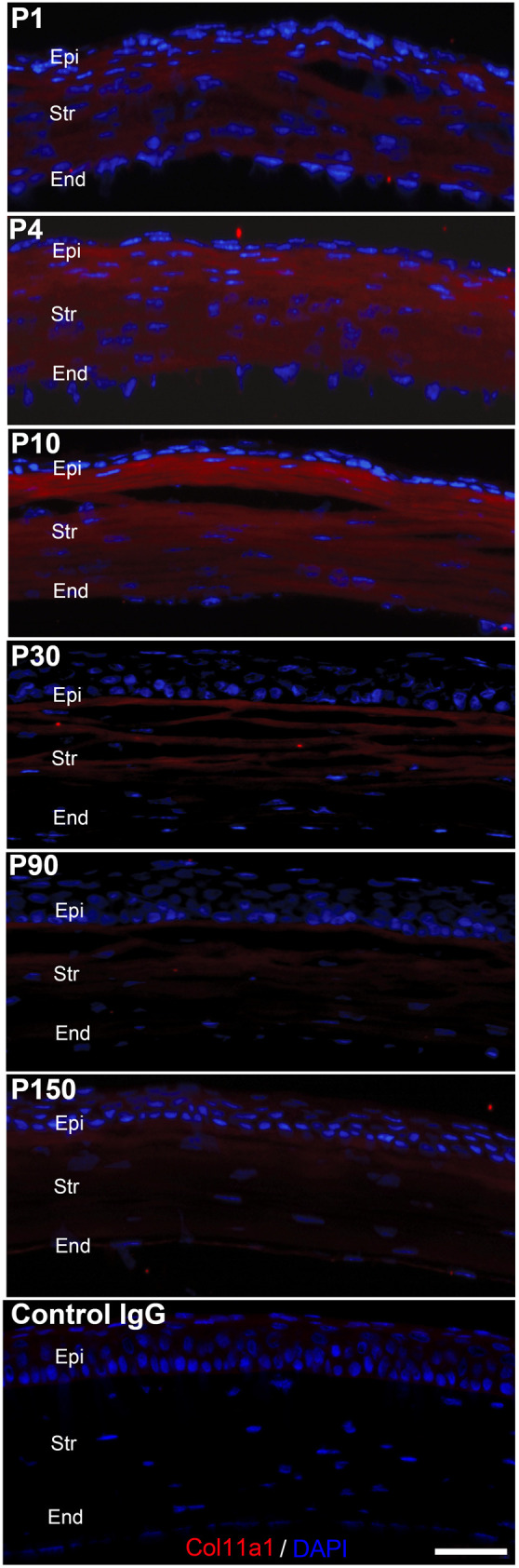
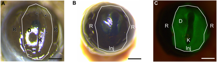
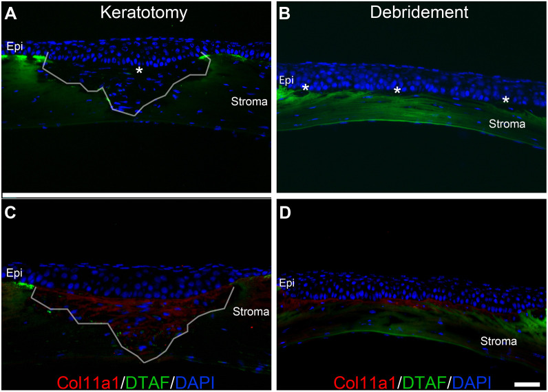
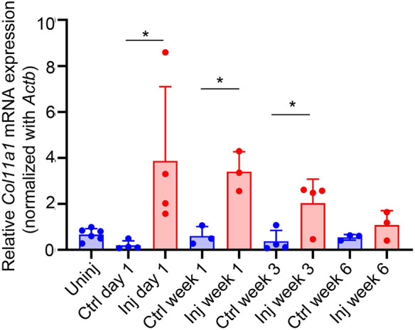
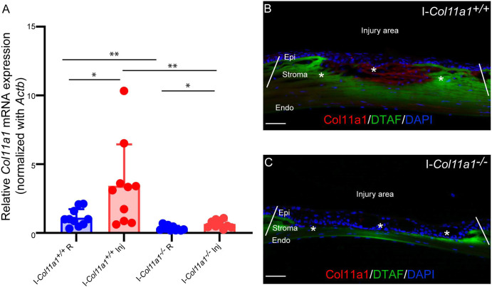
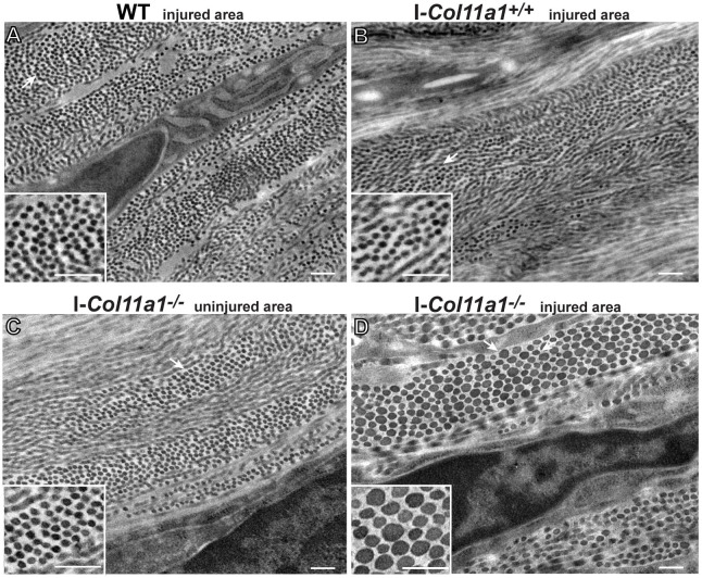
Collagen XI plays a role in nucleating collagen fibrils and in controlling fibril diameter. The aim of this research was to elucidate the role that collagen XI plays in corneal fibrillogenesis during development and following injury. The temporal and spatial expression of collagen XI was evaluated in C57BL/6 wild-type mice. For wound-healing studies in adult mice, stromal injuries were created using techniques that avoid caustic chemicals. The temporal expression and spatial localization of collagen XI was studied following injury in a Col11a1 inducible knockout mouse model. We found that collagen XI expression occurs during early maturation and is upregulated after stromal injury in areas of regeneration and remodeling. Abnormal fibrillogenesis with new fibrils of heterogeneous size and shape occurs after injury in a decreased collagen XI matrix. In conclusion, collagen XI is expressed in the stroma during development and following injury in adults, and is a regulator of collagen fibrillogenesis in regenerating corneal tissue.

Keywords: Collagen XI; Fibroblasts; Regeneration; Stroma; Wound.


Conflict of interest statement

Competing interests The authors declare no competing or financial interests.

Figures

Fig. 1.
Fig. 1.
Temporal expression of collagen XI in wild-type (WT) mice. (A) Col11a1 mRNA expression was higher during stromal development and decreased sharply after postnatal day (P)10. Circles, triangles and diamonds represent sets of replicates. Each replicate consists of three to six individual corneas from different animals. Relative Col11a1 levels normalized to β-actin (Actb). (B) Collagen XI protein was present in the maturing stroma. Representative image shows that collagen XI was present early in the maturing stroma but declined after P10. Total protein was used as a protein loading control. (C) Relative collagen XI protein quantitation normalized to total protein (TP). The standard deviation was obtained by Wes analysis of three sets of independent protein samples. Each set was obtained from pooling tissue from three to 12 corneas from different animals. *P<0.05; ***P<0.005 (Cuzick's test).
Fig. 2.
Fig. 2.
Spatial expression of collagen XI in WT mice. Immunofluorescence localization images showed expression of collagen XI restricted to stromal maturation only (red) in the WT cornea. No corneal epithelial or endothelial staining was noted. Negative controls showed no reactivity. Nuclei were stained with DAPI (blue). Scale bar: 50 µm. Corneal tissue was obtained from three to four animals per age. Epi, epithelium; Str, stroma; End, endothelium.
Fig. 3.
Fig. 3.
Injury model and scar formation as well as DTAF labeling seen 3 weeks post-injury. (A) Surgical microscope image obtained after creating an injury. Injury area delimitated by the white line is divided into the debridement area of the wound (D) and the keratotomy (K), delineated by asterisks. (B) Photographs obtained 3 weeks after injury show the area of injury (Inj) and the remote area (R) that was not injured. (C) Corneal stroma DTAF labeling in the injured area is demonstrated by fluorescence microscopy. Scale bars: 500 µm.
Fig. 4.
Fig. 4.
Collagen XI re-expression following injury as shown in histology slides 3 weeks post-injury. (A) DTAF-labeled stroma and area of newly formed matrix after keratotomy injury not labeled by DTAF, demarcated by the white line and marked with an asterisk. (B) DTAF-labeled stroma and area of newly formed matrix after debridement injury not labeled by DTAF and marked with asterisks. (C,D) There is re-expression of collagen XI in both types of injuries (C, keratotomy; D, debridement) shown in red. Scale bar: 50 µm. Re-expression was noted in seven different eyes in which injury was created and tested. Epi, epithelium.
Fig. 5.
Fig. 5.
Collagen XI mRNA expression is upregulated following injury. Uninj, normal adult corneal stroma; Ctrl, contralateral uninjured stroma; Inj, injured stroma. A significant trend for decreasing Col11a1 mRNA expression over time was observed in the injured group (P=0.0264). Relative Col11a1 levels normalized to Actb. n=36. *P<0.05 (Cuzick's test).
Fig. 6.
Fig. 6.
An inducible mouse model with decreased collagen XI expression following injury. (A) Col11a1 mRNA expression was decreased significantly after Tamoxifen (Tm) injection compared to a control group injected with intraperitoneal vehicle (corn oil). R, remote area; Inj, injured area. (B) Immunofluorescence microscopy shows collagen XI (red) expression in the stroma at 3 weeks in control cornea. (C) Decreased to absent collagen XI expression is observed in the injured model after induction with Tm. Tissue was procured 3 weeks after injury. Injury area demarcated by white lines and asterisks. Relative Col11a1 levels normalized to Actb. n=10 per condition, experiments were performed three times. *P<0.05; **P<0.01 (one-tailed unpaired Student's t-test). Scale bars: 50 µm.
Fig. 7.
Fig. 7.
Collagen XI is a regulator of corneal fibrillogenesis 6 weeks after injury. (A) Microphotographs from control WT mice injected with intraperitoneal Tm show typical small corneal fibrils of homogenous size in the injured area. (B) A second control group, RosaCre-ERT2/Cre-ERT2/Col11a1flox/flox mice, injected with intraperitoneal oil show typical small corneal fibrils of homogenous size in the injured area. (C) Similarly, I-Col11a1−/− mice injected with intraperitoneal Tm show typical small corneal fibrils of homogenous size in the uninjured area. (D) I-Col11a1−/− mice injected with intraperitoneal Tm show an abnormal group of regenerated fibrils of different size and shape in the injured area. Insets show fibrils at higher magnification. Three to five corneas were studied per condition. Scale bars: 200 nm.

Similar articles

Cited by

References

    1. Birk, D. E. (2001). Type V collagen: heterotypic type I/V collagen interactions in the regulation of fibril assembly. Micron 32, 223-237. 10.1016/S0968-4328(00)00043-3 - DOI - PubMed
    1. Blaschke, U. K., Eikenberry, E. F., Hulmes, D. J. S., Galla, H.-J. and Bruckner, P. (2000). Collagen XI nucleates self-assembly and limits lateral growth of cartilage fibrils. J. Biol. Chem. 275, 10370-10378. 10.1074/jbc.275.14.10370 - DOI - PubMed
    1. Cintron, C. and Kublin, C. L. (1977). Regeneration of corneal tissue. Dev. Biol. 61, 346-357. 10.1016/0012-1606(77)90304-9 - DOI - PubMed
    1. Cogswell, D., Sun, M., Greenberg, E., Margo, C. E. and Espana, E. M. (2021). Creation and grading of experimental corneal scars in mice models. Ocul. Surf. 19, 53-62. 10.1016/j.jtos.2020.11.008 - DOI - PMC - PubMed
    1. Donovan, C., Koudouna, E., Margo, C. E., Avila, M. Y. and Espana, E. M. (2021). Genipin delays corneal stromal enzymatic digestion. Transl. Vis. Sci. Technol. 10, 25. 10.1167/tvst.10.9.25 - DOI - PMC - PubMed

Publication types