. 2022 Apr;227(3):925-941.
doi: 10.1007/s00429-021-02432-3. Epub 2021 Dec 2.

Subcellular localization of D2 receptors in the murine substantia nigra

Affiliations

Subcellular localization of D2 receptors in the murine substantia nigra

Joseph J Lebowitz et al. Brain Struct Funct. 2022 Apr.

Abstract

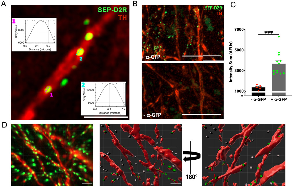
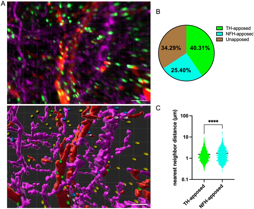
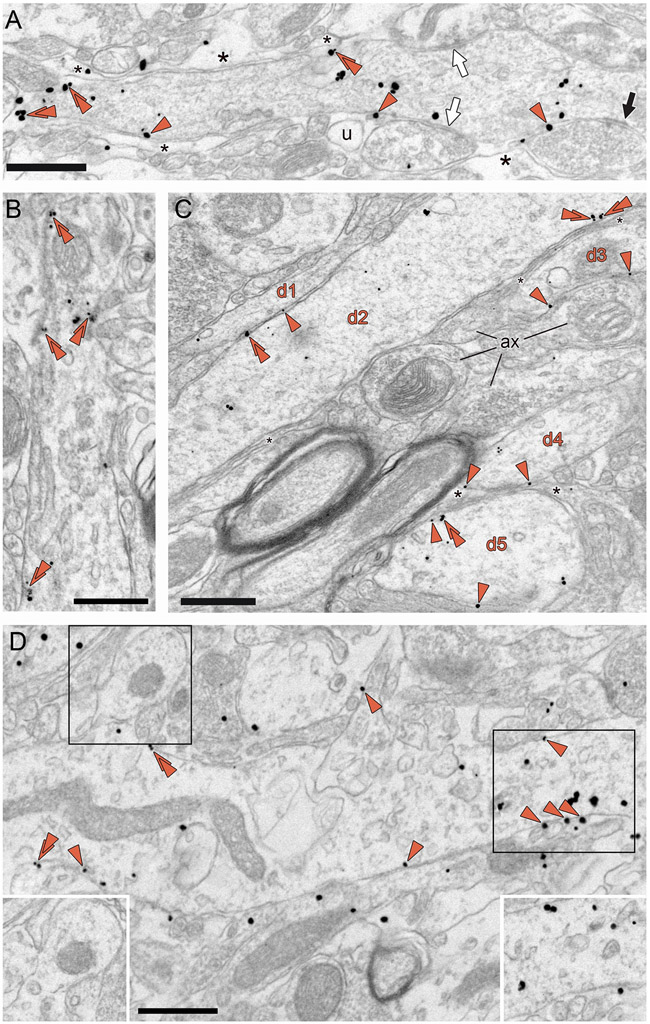
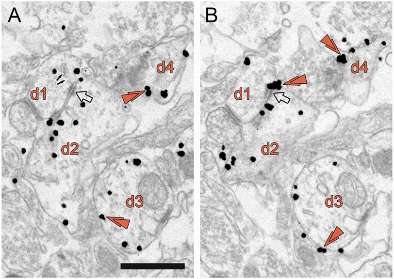
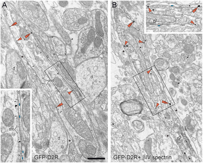
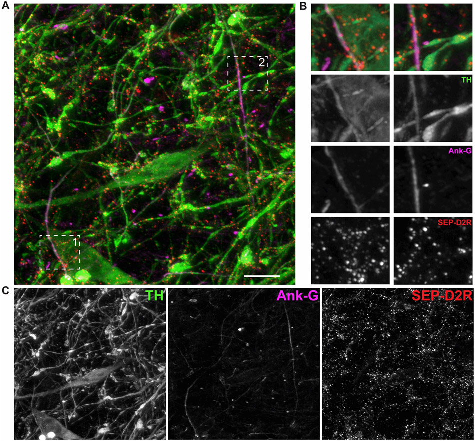
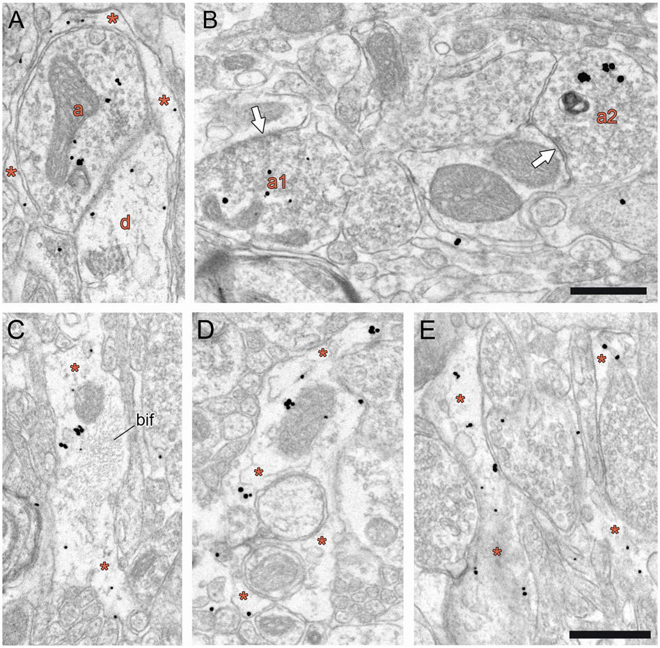
G-protein-coupled D2 autoreceptors expressed on dopamine neurons (D2Rs) inhibit transmitter release and cell firing at axonal endings and somatodendritic compartments. Mechanistic details of somatodendritic dopamine release remain unresolved, partly due to insufficient information on the subcellular distribution of D2Rs. Previous studies localizing D2Rs have been hindered by a dearth of antibodies validated for specificity in D2R knockout animals and have been limited by the small sampling areas imaged by electron microscopy. This study utilized sub-diffraction fluorescence microscopy and electron microscopy to examine D2 receptors in a superecliptic pHlourin GFP (SEP) epitope-tagged D2 receptor knockin mouse. Incubating live slices with an anti-SEP antibody achieved the selective labeling of plasma membrane-associated receptors for immunofluorescent imaging over a large area of the substantia nigra pars compacta (SNc). SEP-D2Rs appeared as puncta-like structures along the surface of dendrites and soma of dopamine neurons visualized by antibodies to tyrosine hydroxylase (TH). TH-associated SEP-D2Rs displayed a cell surface density of 0.66 puncta/µm2, which corresponds to an average frequency of 1 punctum every 1.50 µm. Separate ultrastructural experiments using silver-enhanced immunogold revealed that membrane-bound particles represented 28% of total D2Rs in putative dopamine cells within the SNc. Structures immediately adjacent to dendritic membrane gold particles were unmyelinated axons or axon varicosities (40%), astrocytes (19%), other dendrites (7%), or profiles unidentified (34%) in single sections. Some apposed profiles also expressed D2Rs. Fluorescent and ultrastructural analyses also provided the first visualization of membrane D2Rs at the axon initial segment, a compartment critical for action potential generation. The punctate appearance of anti-SEP staining indicates there is a population of D2Rs organized in discrete signaling sites along the plasma membrane, and for the first time, a quantitative estimate of spatial frequency is provided.

Keywords: Autoreceptor; D2 receptor; Dopamine; Substantia nigra; Ultrastructure.


Conflict of interest statement

Conflicts of interest/Competing interests

None of the authors report a conflict of interest. One author (SRS) acknowledges being a Co-Editor-In-Chief of this journal.

Figures

Figure 1.
Figure 1.. D2 autoreceptors localize to discrete sites in SNc DA neurons.
A. Single optical plane of an Airyscan image showing labeled SEP-D2Rs (green) on the soma and dendritic processes of SNc dopamine neurons labeled for TH (red). Inset graphs are the the spatial distribution of intensity (gray values, AFUs) along the central axis of annotated puncta in the X/Y plane. Scale: 2.5μM B. Representative image of labeled dendrites in SEP-D2R animals with (top) or without (bottom) antibody amplification. Scale: 10 μm C. Average intensity of IMARIS-detected puncta in images with or without antibody amplification (p < 0.001, unpaired t-test). D. Maximum intensity projection (left) and IMARIS rendering (middle, right) of SEP-D2R puncta along the surface of SNc dendrites. Scale: 2 μm
Figure 2.
Figure 2.. Clustered organization of D2Rs in the SNc is cell-autonomous.
A. Maximum intensity projection (top) and IMARIS 3D-render of SEP-D2R puncta on SNc dendrites (TH, red) and axonal profiles (NFH, magenta). In the 3D render, green spots are apposed to dendrites, blue apposed to axons, yellow opposed to neither, and white apposed to both a TH(+) and a NFH(+) surface; scale: 5 μm B. Relative distribution of SEP-D2R puncta within the midbrain. Spots apposed to both TH(+) and NFH(+) surfaces were excluded from this analysis. C. Nearest neighbor distribution of SEP-D2R puncta associated with SNc dendrites (TH-apposed) or afferent terminals (NFH-apposed). A slight but significant increase in average distance between nearest neighboring puncta is seen on NFH(+) surfaces compared to that measure on TH(+) surfaces. (p<0.0001, unpaired t-test).
Figure 3.
Figure 3.. Subcellular distribution of SEP-D2RS in SNc dendrites.
A,B. SEP-D2R immunogold is commonly associated with the cell membrane (orange arrowheads; double arrowheads show doublet or triplet particles), where it is randomly dispersed and typically adjacent to unlabeled axons or axon varicosities, astrocytes (asterisks), or unidentified profiles (u). In panel A, the dense accumulation of gold particles at the left of the dendrite did not recur when examined in serial sections. This dendrite receives symmetric synapses (white arrows) and asymmetric synapses (black arrow) from unlabeled axon varicosities. C. Electron micrograph of SEP-D2R immunogold-labeled dendrites (d1-d5) in apposition or separated by axons and astrocytes in the SNc. The d1 and d2 dendrites share a direct apposition of their membranes, while the remaining dendrites are separated from each other by astrocytic leaflets (asterisks between d2 and d3, d4 and d5) or axon varicosities (ax) in the adjacent neuropil. D. A dendrite gives rise to a spine (upper left black boxed area) that contains an immunogold particle within the neck. Analysis of serial sections indicates this spine is otherwise unlabeled for SEP-D2R (lower left inset). In addition to individual gold particles associated with the plasma membrane, a grouping of membrane gold is found in one specific location (right black boxed area). Serial sections through this region indicate the membrane cluster does not continue far into the z-plane, even though immunoreactivity for SEP-D2R is continuous (lower right inset). All scale bars, 0.6 μm.
Figure 4.
Figure 4.. D2Rs are present at putative dendro-dendritic synapses in the SNc.
Serial electron micrographs of SEP-D2R immunogold-labeled intermediate dendrites (d1-d4) in the SNc. Membrane-associated doublet or triplet gold particles are shown by double arrowheads. A putative dendrodendritic synapse (transparent arrow) is visible between dendrites d1 and d2. The small arrows in panel A indicate a few vesicles in close proximity to this site. Membrane-associated SEP-D2R is found adjacent to this synapse, especially as depicted in panel B. Dendrites d3 and d4 show preferential membrane-associated SEP-D2R. Scale bar, 0.6 μm
Figure 5.
Figure 5.. D2R expression at the axon initial segment of SNc cells.
A,B. Electron micrographic images showing immunogold-silver labeling for SEP-D2R in structures with morphological features indicative of the AIS: dense undercoating of the plasma membrane (small black arrows in panel A), apparent microtubules (mt in panel A) near the membrane, and enlargements of the extracellular space (small white arrows in panels A,B) sometimes containing granules (small blue arrows in panels A,B). Unlabeled astrocytes (*) appear in proximity. In panel B, the immunoreactive structure is verified as an AIS by immunoperoxidase labeling for beta IV-spectrin; transparent arrowheads indicate accumulations of peroxidase product near the plasma membrane that obscure the dense undercoating and microtubules. Orange arrowheads in panels A and B indicate gold particles for SEP-D2R that are close to the plasma membrane; double arrowheads show doublet or triplet particles. The boxed region in panel A, shown at higher magnification and contrast in the inset, demonstrates that some gold particles are displaced from the plasma membrane by roughly 40 nm and may be associated with microtubules. The boxed region in panel B, shown at higher magnification and contrast in the inset, shows that some gold particles are more immediately adjacent to the membrane and even just outside the plasmalemma. Scale bar 0.6 μm for panels A and B, 0.38 μm for panel A inset, 0.47 μm for panel B inset.
Figure 6.
Figure 6.. D2 autoreceptor expression at the axon initial segment of SNc dopamine cells.
Immunofluorescent labeling of SEP-D2R in the AIS of SNc neurons labeled for tyrosine hydroxylase (TH). A. Sub-diffraction limited (Airyscan) image displaying the axon initial segments of TH (+) dopamine processes (green) as marked by Ank-G localization (magenta). SEP-D2Rs are present at the membrane of both processes as red puncta. B. Specified insets from panel A displayed at 2.2x zoom. Both inset panels correspond to regions of AIS profiles where SEP-D2Rs appear enriched. C. Individual channels of the merged image shown in panel A. Scale bar, 10 μm
Figure 7.
Figure 7.. Subcellular distribution of D2Rs in midbrain axon varicosities and astrocytes.
A. Electron micrograph of the SNc showing immunogold-silver labeling for SEP-D2R in a representative axon varicosity (a). As was typical, gold particles are contained within the intracellular environment more consistently than being found on the plasmalemma. Often immunoreactive axons do not form synaptic contacts in single sections, although this labeled axon varicosity closely apposes a labeled dendrite (d) and is surrounded by an immunoreactive astrocyte (orange asterisks). B. When synapses are formed by immunolabeled axon varicosities (a1 and a2), they are most commonly symmetric (white arrows) and onto immunonegative dendrites. C-E. Electron micrographs of the SNc from SEP-D2R transgenic mice showing immunogold-silver labeling in glial profiles (orange asterisks). These are likely astrocytes as evidenced by their electron lucent cytoplasm, convoluted shape, and presence of bundles of intermediate filaments (bif). Scale bars, 0.6 μm

Similar articles

Cited by

References

    1. Bal A, Bachelot T, Savasta M, Manier M, Verna JM, Benabid AL, Feuerstein C, 1994. Evidence for dopamine D2 receptor mRNA expression by striatal astrocytes in culture: in situ hybridization and polymerase chain reaction studies. Mol. Brain Res 23, 204–212. 10.1016/0169-328X(94)90227-5 - DOI - PubMed
    1. Bayer VE, Pickel VM, 1990. Ultrastructural localization of tyrosine hydroxylase in the rat ventral tegmental area: relationship between immunolabeling density and neuronal associations. J. Neurosci 10, 2996–3013. 10.1523/JNEUROSCI.10-09-02996.1990 - DOI - PMC - PubMed
    1. Beaulieu J-M, Gainetdinov RR, 2011. The Physiology, Signaling, and Pharmacology of Dopamine Receptors. Pharmacol. Rev 63, 182–217. 10.1124/pr.110.002642 - DOI - PubMed
    1. Beckstead MJ, Grandy DK, Wickman K, Williams JT, 2004. Vesicular Dopamine Release Elicits an Inhibitory Postsynaptic Current in Midbrain Dopamine Neurons. Neuron 42, 939–946. 10.1016/j.neuron.2004.05.019 - DOI - PubMed
    1. Beeler JA, Dreyer JK, 2019. Synchronicity: The Role of Midbrain Dopamine in Whole-Brain Coordination. eNeuro 6. 10.1523/ENEURO.0345-18.2019 - DOI - PMC - PubMed

LinkOut - more resources