. 2022 Jun;35(3):421-440.
doi: 10.1007/s10334-021-00976-3. Epub 2021 Dec 2.

Combined acquisition of diffusion and T2*-weighted measurements using simultaneous multi-contrast magnetic resonance imaging

Affiliations

Combined acquisition of diffusion and T2*-weighted measurements using simultaneous multi-contrast magnetic resonance imaging

Nora-Josefin Breutigam et al. MAGMA. 2022 Jun.

Abstract

Object: In this work, we present a technique called simultaneous multi-contrast imaging (SMC) to acquire multiple contrasts within a single measurement. Simultaneous multi-slice imaging (SMS) shortens scan time by allowing the repetition time (TR) to be reduced for a given number of slices. SMC imaging preserves TR, while combining different scan types into a single acquisition. This technique offers new opportunities in clinical protocols where examination time is a critical factor and multiple image contrasts must be acquired.

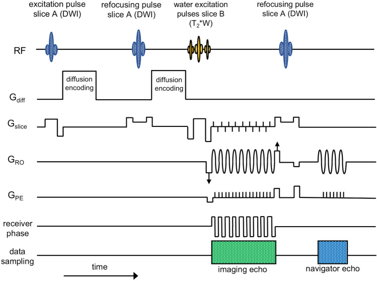
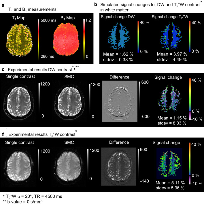
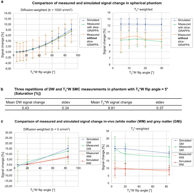
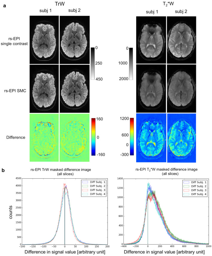
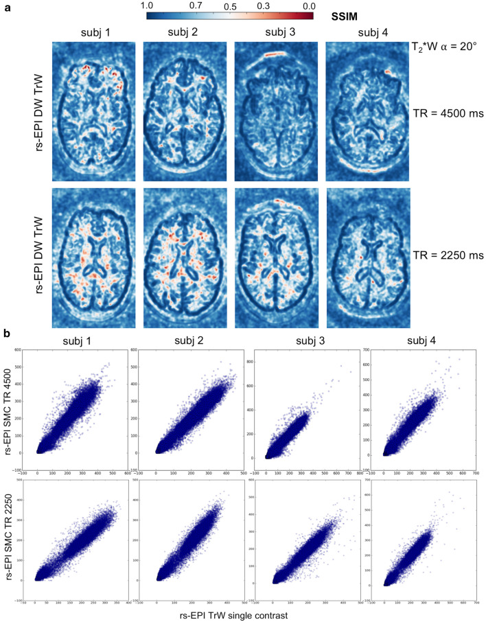
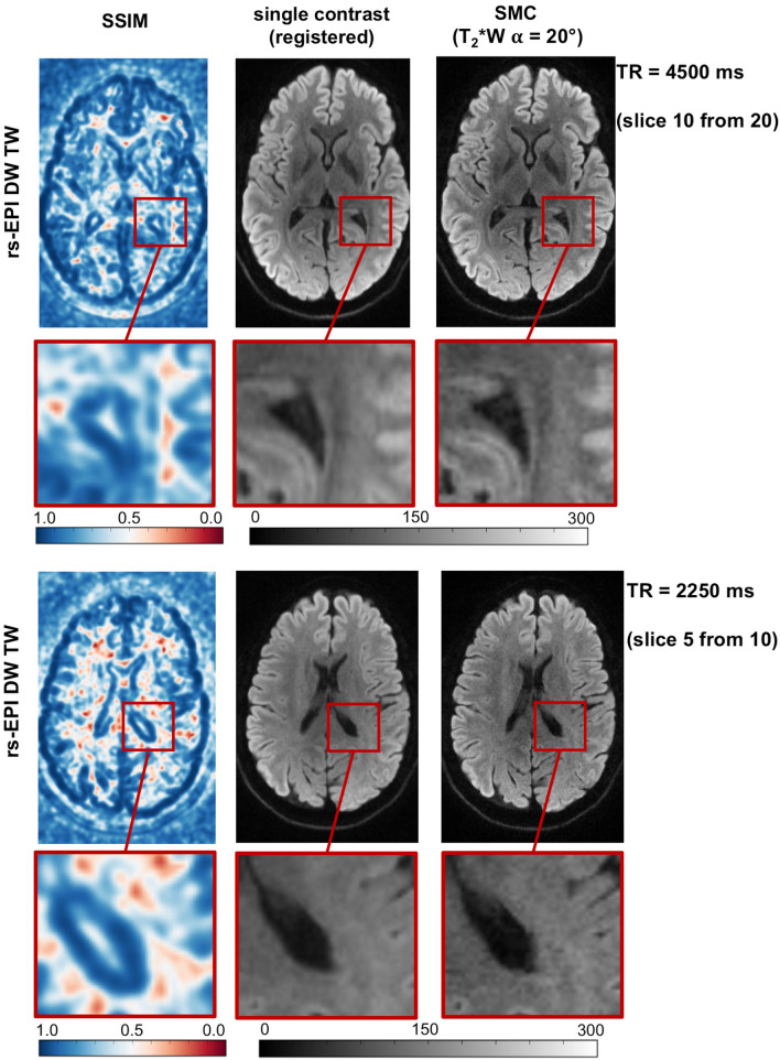
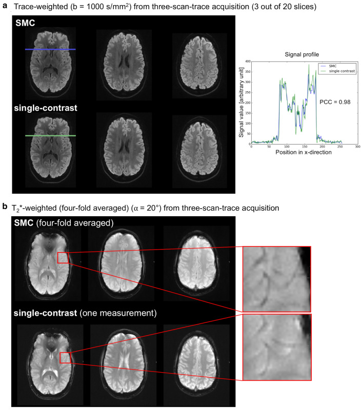
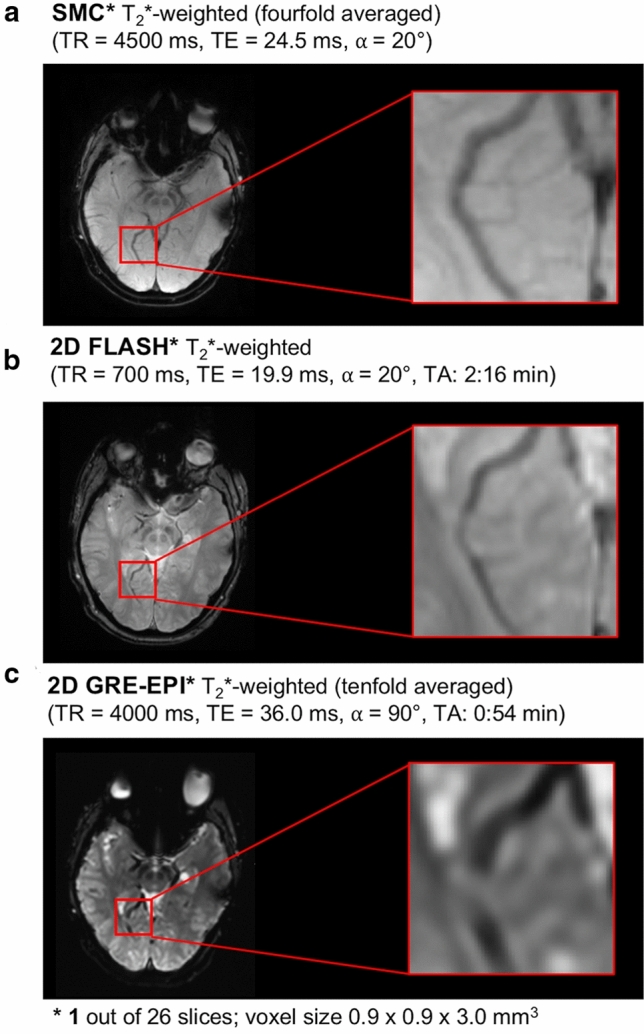
Materials and methods: High-resolution, navigator-corrected, diffusion-weighted imaging was performed simultaneously with T2*-weighted acquisition at 3 T in a phantom and in five healthy subjects using an adapted readout-segmented EPI sequence (rs-EPI).

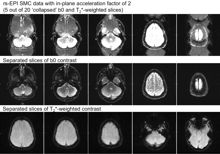
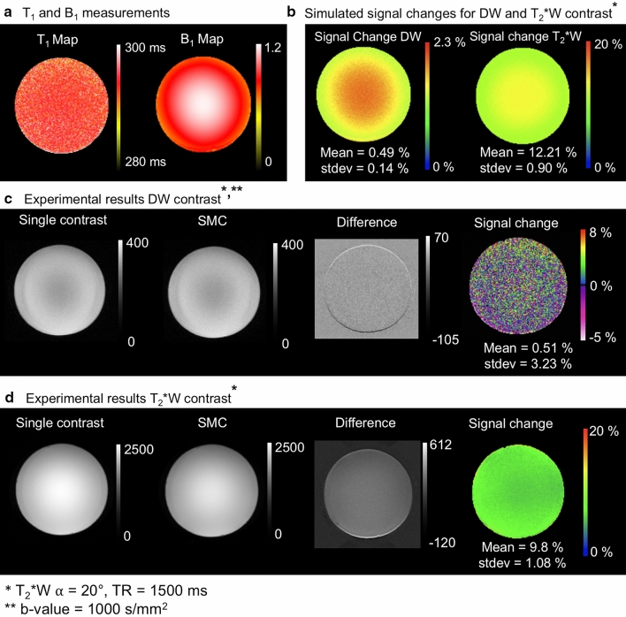
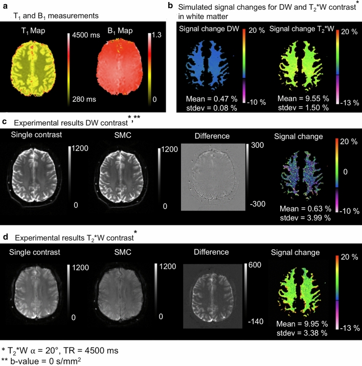
Results: The results demonstrated that simultaneous acquisition of two contrasts (here diffusion-weighted imaging and T2*-weighting) with SMC imaging is feasible with robust separation of contrasts and minimal effect on image quality.

Discussion: The simultaneous acquisition of multiple contrasts reduces the overall examination time and there is an inherent registration between contrasts. By using the results of this study to control saturation effects in SMC, the method enables rapid acquisition of distortion-matched and well-registered diffusion-weighted and T2*-weighted imaging, which could support rapid diagnosis and treatment of acute stroke.

Keywords: Diffusion; Echo-planar imaging; Stroke.


Conflict of interest statement

The authors have no conflicts of interest to declare.

Figures

Fig. 1
Fig. 1
Pulse sequence diagram of the rs-EPI sequence for SMC imaging. Data are acquired at two slice positions at the same time with one slice (A) generating DW-while the other slice (B) provides T2*W image contrast. Firstly, slice A is excited and DW preparation is applied. Then slice B is excited before both signals are read out simultaneously using rs-EPI with a variable amplitude encoding gradient in the readout (GRO) direction (labelled with arrows) and a blipped phase-encoding gradient (GPE). A blipped-CAIPIRINHA gradient scheme along the slice-select (Gslice) direction is used in conjunction with receiver phase modulation to shift the T2*W image by half a FOV in the phase-encoding direction. Finally, an RF refocusing pulse is applied to slice A only to generate a 2D navigator signal for phase correction. Note that a binomial water-excitation pulse was used in this study for the T2*W excitation
Fig. 2
Fig. 2
Data from five out of 20 measured SMC slices. The first row shows the collapsed SMC data with an in-plane acceleration factor of 2; The slices from the DW acquisition (with b = 0 s/mm2) are at the center of the FOV while the T2*W slices are shifted by FOV/2 in the anterior–posterior direction. The middle row shows the DWI data and the bottom row shows the T2*W data
Fig. 3
Fig. 3
a Measured T1 and B1 maps in the spherical water-based phantom, b Simulated magnetization signal changes [%] with Bloch equations on the basis of the T1 and B1 map, c DW acquisition without and with simultaneous acquisition of T2*W contrast, the corresponding difference image and signal-change map, d T2*W contrast without and with simultaneous acquisition of the DW contrast, the corresponding difference image and signal-change map. The difference images show evidence of an uncorrected frequency drift during measurement in the outer ring. These areas of high differences are masked out in the saturation maps. Positive values [%] of signal change means smaller grey values in the SMC images than in the single-contrast images and vice versa
Fig. 4
Fig. 4
Signal change result for slice iteration scheme A: a Measured T1 and B1 maps in vivo, b Simulated magnetization signal change [%] with Bloch equations on the basis of the T1 and B1 map in white matter, c DW acquisition without and with simultaneous acquisition of T2*W contrast, the corresponding difference image and signal change map, d T2*W contrast without and with simultaneous acquisition of the DW contrast, the corresponding difference image and signal change map. Positive values [%] of signal change means smaller grey values in the SMC images than in the single-contrast images and vice versa
Fig. 5
Fig. 5
Signal change results for slice iteration scheme B: a Measured T1 and B1 maps in vivo, b Simulated magnetization signal change [%] with Bloch equations on the basis of the T1 and B1 map in white matter, c DW acquisition without and with simultaneous acquisition of T2*W contrast, the corresponding difference image and signal change map, d T2*W contrast without and with simultaneous acquisition of the DW contrast, the corresponding difference image and signal change map. Positive values [%] of signal change means smaller grey values in the SMC images than in the single-contrast images and vice versa
Fig. 6
Fig. 6
a Comparison between simulated and measured signal change with and without split slice-GRAPPA reconstruction in the spherical phantom. b The table shows the mean signal change in terms of signal saturation and standard deviations for both contrasts in the phantom between three repeated measurements. c Comparison between simulated and measured signal change in vivo in grey and white matter (GM, WM) respectively
Fig. 7
Fig. 7
a Difference images between rs-EPI single-contrast (DW trace-weighted and T2*W) and rs-EPI SMC (separated trace-weighted and T2*W contrast) in-vivo central slice out of 20 slices. b Histograms of all slices in all subjects of the masked difference image
Fig. 8
Fig. 8
a The structural similarity index measure (SSIM) maps of the central slice of the calculated trace-weighted data for TR = 4500 ms and TR = 2250 ms. b Respective scatter plots of the gray level distributions of single-contrast and SMC data in the central slice
Fig. 9
Fig. 9
Comparison between the registered TrW single-contrast and central SMC slices of subject 2 with the corresponding SSIM map for TR and TR/2. The close-up image allows better interpretation of the correlation between high and low SSIM values and structural changes between the single-contrast and SMC slices
Fig. 10
Fig. 10
Contrast comparison in the central slice in one of four subjects. a Comparison of signal profile between single-contrast and SMC trace-weighted image. b Comparison of contrast between veins and tissue in T2*W contrast
Fig. 11
Fig. 11
Comparison between an T2*-weighted SMC, 2D Flash and GRE-EPI (both product) measurement in terms of scan time and visual vein-tissue contrast

Similar articles

Cited by

References

    1. Souza SP, Szumowski J, Dumoulin CL, Plewes DP, Glover G. SIMA. J Comput Assist Tomogr. 1988;12(6):1026–1030. doi: 10.1097/00004728-198811000-00021. - DOI - PubMed
    1. Müller S. Multifrequency selective rf pulses for multislice MR imaging. Magn Reson Med. 1988;6(3):364–371. doi: 10.1002/mrm.1910060315. - DOI - PubMed
    1. Weaver JB. Simultaneous multislice acquisition of MR images. Magn Reson Med. 1988;8(3):275–284. doi: 10.1002/mrm.1910080305. - DOI - PubMed
    1. Glover GH. Phase-offset multiplanar (POMP) volume imaging: a new technique. J Magn Reson Imaging. 1991;1(4):457–461. doi: 10.1002/jmri.1880010410. - DOI - PubMed
    1. Larkman DJ, Hajnal JV, Herlihy AH, Coutts GA, Young IR, Ehnholm G. Use of multicoil arrays for separation of signal from multiple slices simultaneously excited. J Magn Reson Imaging. 2001;13:313–317. doi: 10.1002/1522-2586(200102)13:2<313::aid-jmri1045>3.0.co;2-w. - DOI - PubMed

MeSH terms

LinkOut - more resources