. 2021 Dec 3;374(6572):1227-1237.
doi: 10.1126/science.abi7495. Epub 2021 Dec 2.

Fumarate is a terminal electron acceptor in the mammalian electron transport chain

Affiliations

Fumarate is a terminal electron acceptor in the mammalian electron transport chain

Jessica B Spinelli et al. Science. .

Abstract

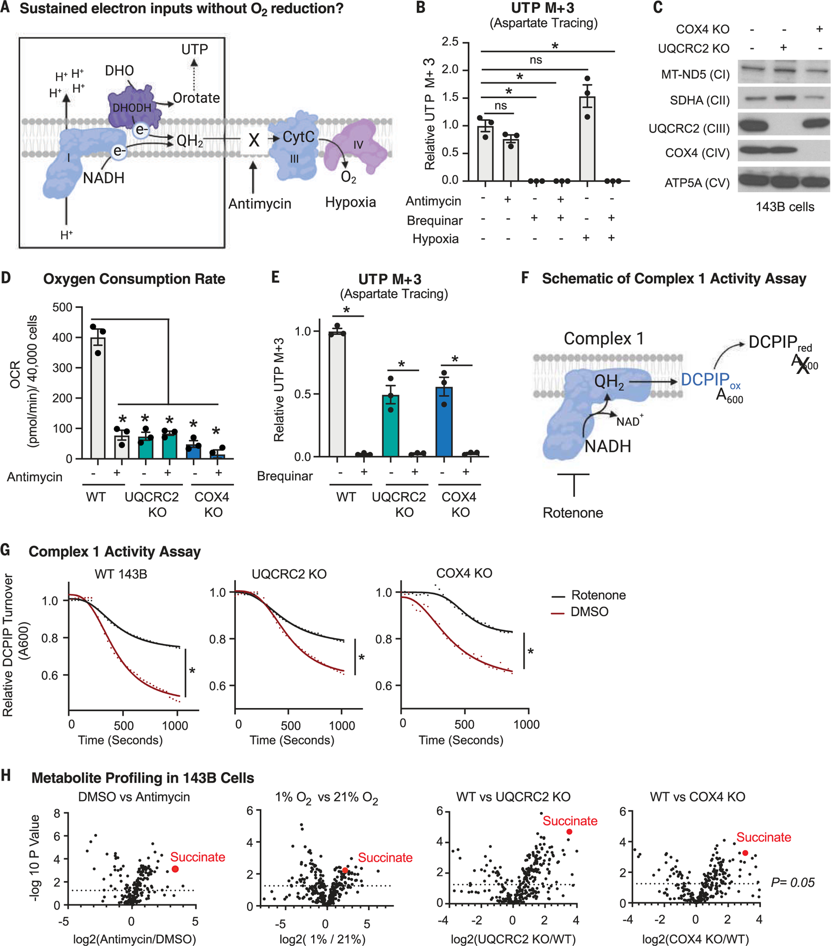
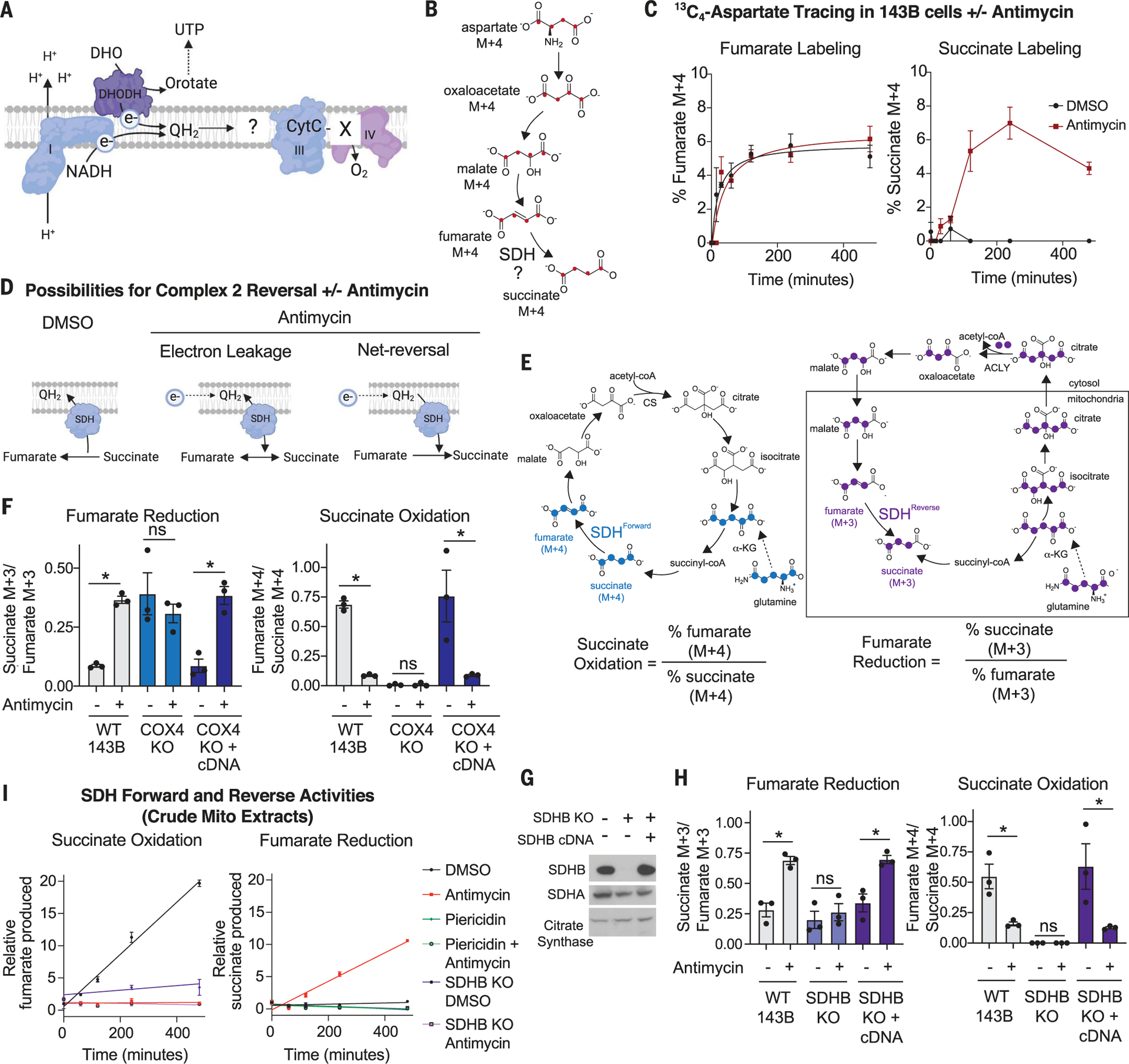
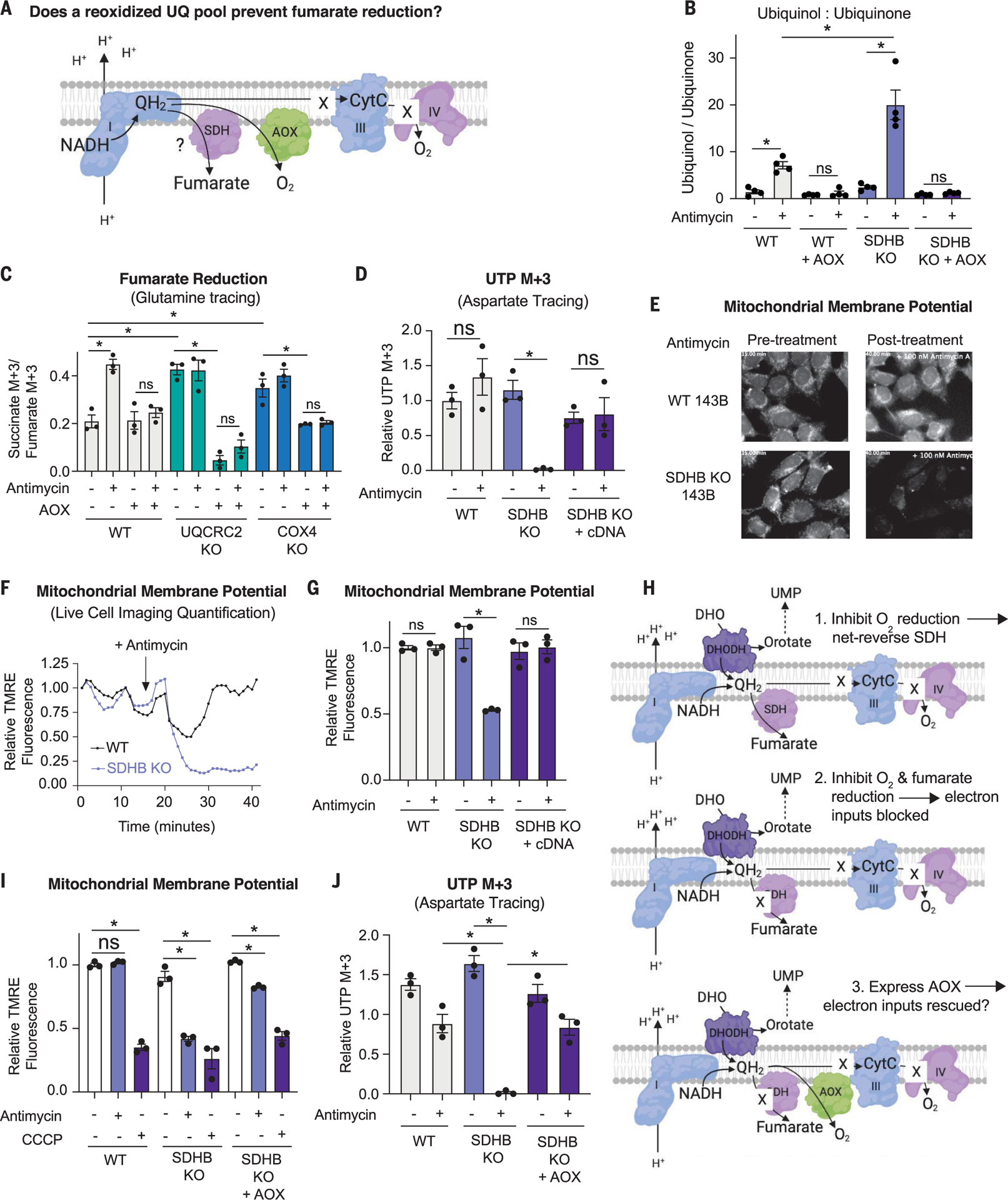
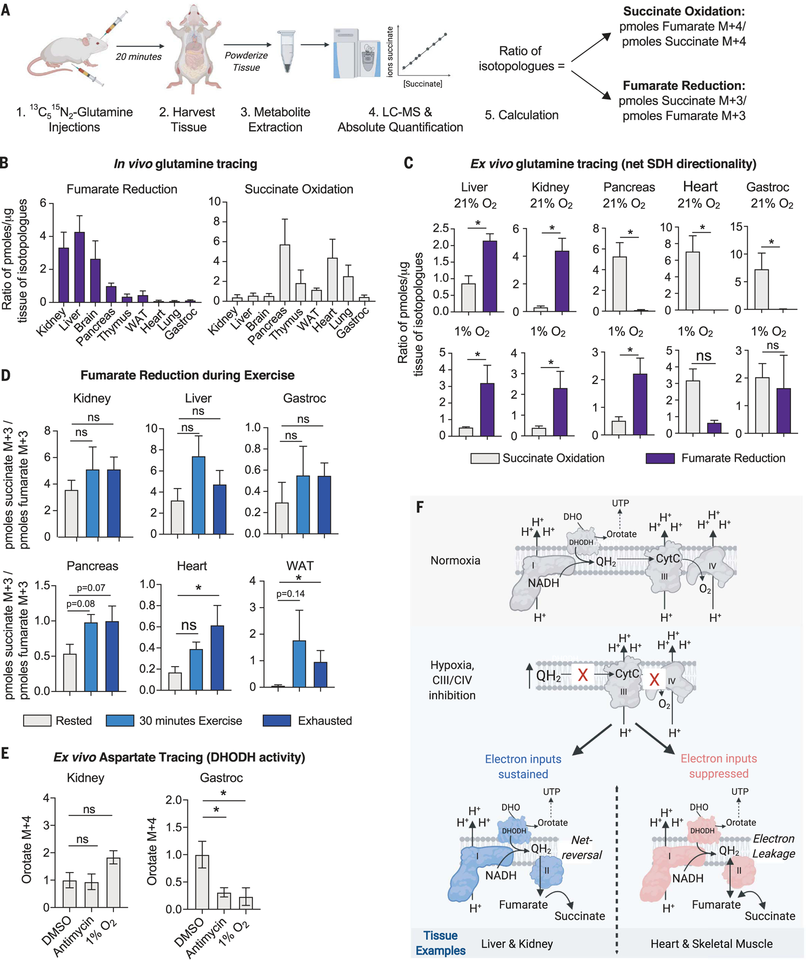
For electrons to continuously enter and flow through the mitochondrial electron transport chain (ETC), they must ultimately land on a terminal electron acceptor (TEA), which is known to be oxygen in mammals. Paradoxically, we find that complex I and dihydroorotate dehydrogenase (DHODH) can still deposit electrons into the ETC when oxygen reduction is impeded. Cells lacking oxygen reduction accumulate ubiquinol, driving the succinate dehydrogenase (SDH) complex in reverse to enable electron deposition onto fumarate. Upon inhibition of oxygen reduction, fumarate reduction sustains DHODH and complex I activities. Mouse tissues display varying capacities to use fumarate as a TEA, most of which net reverse the SDH complex under hypoxia. Thus, we delineate a circuit of electron flow in the mammalian ETC that maintains mitochondrial functions under oxygen limitation.


Conflict of interest statement

Competing interests: All authors declare that they have no competing interests.

Figures

Fig. 1.
Fig. 1.. Cells deficient in O2 reduction retain the capacity to input electrons into the ETC.
(A) Schematic depicting the electron transport chain (ETC) and the deposition of electrons (e−) onto a terminal electron acceptor. CytC, cytochrome c. (B) DHODH activity as determined by stable isotope tracing with 10 mM 13C4-aspartate, which generates 13C3-UTP if DHODH is active. Tracing was performed for 8 hours in 143B cells treated with vehicle [dimethyl sulfoxide (DMSO)], 500 nM antimycin, 2 μM brequinar, or in combination or cultured in 1% O2 for 24 hours (mean ± SEM, n = 3 biological replicates per condition). *P < 0.05. P values were calculated using a two-way analysis of variance (ANOVA). (C) Immunoblot analyses of mitochondrial proteins in wild-type (WT), UQCRC2 (complex III) knockout (KO), and COX4 (complex IV) KO 143B cells. (D) O2 consumption rate (OCR) of WT, UQCRC2 KO, and COX4 KO 143B cells treated with DMSO or 500 nM antimycin for 1 hour (mean ± SEM, n = 3 biological replicates per condition). *P < 0.05. P values were calculated using a two-way ANOVA. (E) DHODH activity as determined using stable isotope tracing of 10 mM 13C4- aspartate, which generates 13C3-UTP if DHODH is active. Tracing was performed for 8 hours in WT, UQCRC2 KO, and COX4 KO 143B cells treated with DMSO or 2 μM brequinar (mean ± SEM, n = 3 biological replicates per condition). *P < 0.05. P values were calculated using a parametric t test. (F) Schematic depicting the complex I activity assay on purified mitochondria. NADH initiates the reaction, and the absorbance (A600) of the oxidized electron acceptor 2,6-dichlorophenolindophenol (DCPIP) is measured over time. (G) Complex I activity in mitochondria purified from WT, UQCRC2 KO, and COX4 KO 143B cells in the presence or absence of 1 μM rotenone (complex I inhibitor). *P < 0.05. P values were calculated using an extra sum of squares F test in GraphPad Prism. (H) Polar metabolite profiling of 143B cells treated with DMSO versus 500 nM antimycin for 8 hours, grown in 21% versus 1% O2, WT versus UQCRC2 KO 143B cells, or WT versus COX4 KO 143B cells, n = 3 biological replicates per condition. P values were calculated using a parametric t test.
Fig. 2.
Fig. 2.. Upon inhibition of O2 reduction, fumarate accepts electrons through net reversal of the SDH complex.
(A) Schematic depicting the question “what is the fate of electrons in the ETC when O2 cannot be reduced?” (B) Schematic showing the expected isotopologues produced during 13C4-aspartate tracing if succinate is generated from fumarate. (C) Percent labeled fumarate and succinate from a stable isotope tracing experiment using 3 mM 13C4-aspartate. WT 143B cells were treated with DMSO or 500 nM antimycin for the indicated times (mean ± SEM, n = 3 biological replicates per time point). (D) Schematic depicting the reduction of fumarate from either electron leakage onto fumarate or net reversal of SDH upon antimycin treatment. (E) Schematic demonstrating the expected isotopologues of TCA cycle metabolites produced during 13C515N2-glutamine tracing. The forward direction of the SDH reaction can be monitored with the ratio of percent labeled fumarate M + 4 to percent labeled succinate M + 4. The reverse direction of the SDH reaction can be monitored with the ratio of percent labeled succinate M + 3 to percent labeled fumarate M + 3. ACLY, ATP citrate lyase; CoA, coenzyme A; CS, citrate synthase; α-KG, α-ketoglutarate. (F) Fumarate reduction and succinate oxidation as determined using stable isotope tracing of 2 mM 13C515N2-glutamine. Tracing was performed for 8 hours in WT, COX4 KO, and COX4 KO 143B cells expressing the COX4 cDNA and treated with DMSO or 100 nM antimycin for 8 hours (mean ± SEM, n = 3 biological replicates per condition). ns, not significant. (G) Immunoblot analyses for indicated proteins in SDHB KO and SDHB cDNA addback 143B cells. (H) Fumarate reduction and succinate oxidation as determined using stable isotope tracing of 2 mM 13C515N2-glutamine. Tracing was performed for 8 hours in WT, SDHB KO, and SDHB KO 143B cells expressing the SDHB cDNA and treated with DMSO or 100 nM antimycin for 8 hours (mean ± SEM, n = 3 biological replicates per condition). (I) SDH activity in purified mitochondria from WT and SDHB KO 143B cells. The succinate oxidation reaction was initiated by adding 10 mM succinate and monitored by the production of fumarate over time. The fumarate reduction reaction was initiated with 10 mM fumarate and 1 mM NADH and monitored through the production of succinate over time; 1 μM antimycin and 1 μM piericidin were included as indicated (mean ± SEM, n = 3 biological replicates per time point). Data points were fitted using linear regression. *P < 0.05 for all experiments. P values were calculated using an unpaired parametric t test.
Fig. 3.
Fig. 3.. Fumarate reduction is required to maintain nucleotide biosynthesis and the mitochondrial membrane potential in cells deficient in O2 reduction.
(A) Schematic depicting the potential impact of alternative oxidase (AOX) on the accumulation of QH2 in cells deficient for complex III or IV activity and the consequences for fumarate reduction. (B) Ratio of ion counts of ubiquinol to ubiquinone as measured by LC-MS on mitochondria isolated from WT and SDHB KO 143B cells expressing or not expressing AOX and treated with DMSO or 500 nM antimycin for 3 hours (mean ± SEM, n = 4 biological replicates per condition). *P < 0.05. P values were calculated using a two-way ANOVA. (C) Relative fumarate reduction as determined using stable isotope tracing of 2 mM 13C515N2-glutamine and the ratio of percent succinate M + 3 to percent fumarate M + 3, representing fumarate reduction in a stable isotope tracing experiment using 2 mM 13C515N2-glutamine. Tracing was performed for 8 hours in WT, UQCRC2 KO, and COX4 KO 143B cells expressing or not expressing AOX and treated with DMSO or 500 nM antimycin (mean ± SEM, n = 3 biological replicates per condition). *P < 0.05. P values were calculated using a two-way ANOVA. (D) DHODH activity as measured by stable isotope tracing with 10 mM 13C4- aspartate, which generates 13C3-UTP if DHODH is active. Tracing was for 8 hours in WT, SDHB KO, and KO 143B cells with the SDHB cDNA expressed and treated with DMSO or 500 nM antimycin (mean ± SEM, n = 3 biological replicates per condition). *P < 0.05. P values were calculated using a parametric t test. (E) First and last images from a live-cell imaging video of WT and SDHB KO 143B cells treated with DMSO or 100 nM antimycin. (F) Quantification of the mitochondrial membrane potential using live-cell imaging of WT and SDHB KO 143B cells treated with 100 nM antimycin, which was added at the time point indicated with the arrow. (G) Mitochondrial membrane potential of WT, SDHB KO, and SDHB KO cells expressing the SDHB cDNA treated with either DMSO or 500 nM antimycin for 1 hour (mean ± SEM, n = 3 biological replicates per condition). *P < 0.05. P values were calculated using a parametric t test. (H) Schematic depicting the hypothesis that expression of AOX will rescue complex I and DHODH activities in SDH KO cells treated with antimycin. UMP, uridine 5′-monophosphate. (I) Mitochondrial membrane potential in WT, SDHB KO, and SDHB KO 143B cells expressing AOX and treated with DMSO, 500 nM antimycin for 1 hour (mean ± SEM, n = 3 biological replicates per condition). *P < 0.05. P values were calculated using a two-way ANOVA. (J) DHODH activity as measured via stable isotope tracing with 10 mM 13C4- aspartate, which generates 13C3-UTP if DHODH is active. Tracing was performed for 8 hours in WT, SDHB KO, and SDHB KO 143B cells expressing AOX and treated with DMSO or 500 nM antimycin (mean ± SEM, n = 3 per biological replicates condition). *P < 0.05. P values were calculated using a two-way ANOVA.
Fig. 4.
Fig. 4.. Fumarate reduction supports mitochondrial functions in tissues capable of net reversal of the SDH reaction.
(A) Depiction of the workflow for the in vivo 13C515N2-glutamine stable isotope tracing experiment to measure the net directionality of the SDH complex in tissues. (B) In vivo stable isotope tracing of 13C515N2-glutamine in indicated tissues. Mice were euthanized 20 min after retroorbital and intraperitoneal injections. Succinate oxidation was calculated as the ratio of picomoles fumarate M + 4 to picomoles succinate M + 4. Fumarate reduction was calculated as the ratio of picomoles succinate M + 3 to picomoles fumarate M + 3 (mean ± SEM, n = 4 per condition). (C) Tissue-autonomous succinate oxidation or fumarate reduction as determined with ex vivo 2 mM 13C515N2-glutamine stable isotope tracing for 24 hours in indicated tissues kept in a tissue culture incubator at 21% O2 or a hypoxia incubator (1% O2). Succinate oxidation and fumarate reduction were calculated as described in (B) (mean ± SEM, n = 4 per condition). *P < 0.05. P values were calculated using a parametric t test. (D) In vivo 13C515N2-glutamine tracing in female mice 12 weeks old through intraperitoneal and intramuscular injections. Mice were rested, exercised for 30 min, or exercised until exhaustion for ~1.5 hours and then injected with 13C515N2-glutamine. The rested mice were euthanized 15 min after injection with no exercise, and the exercised mice continued to run on the treadmill for 15 min before being euthanized. Tissues were harvested for metabolite isolation and mass spectrometry. Absolute quantification was performed to calculate the concentration of succinate M + 3, succinate M + 4, fumarate M + 3, and fumarate M + 4 in picomoles per microgram tissue protein. The reported ratio representing fumarate reduction was calculated by the picomoles succinate M + 3 per microgram tissue protein to the picomoles fumarate M + 3 per microgram tissue protein. The reported ratio representing succinate oxidation was calculated by the picomoles fumarate M + 4 per microgram tissue protein to the picomoles succinate M + 4 per microgram tissue protein. Data represent the mean ± SEM, n = 5 mice per time point. *P < 0.05. P values were calculated using a two-way ANOVA. (E) Ex vivo 3 mM 13C4-aspartate stable isotope tracing for 16 hours in indicated tissues kept in an incubator at 21 or 1% O2 or treated with 2 μM antimycin. Orotate M + 4 levels reflect DHODH activity (mean ± SEM, n = 4 biological replicates per condition). *P < 0.05. P values were calculated using a two-way ANOVA. (F) Model in which net reversal of SDH supports certain mitochondrial functions in tissues under conditions that reduce electron transfer to O2.

Comment in

  • Short-circuiting respiration.
    Baksh SC, Finley LWS. Baksh SC, et al. Science. 2021 Dec 3;374(6572):1196-1197. doi: 10.1126/science.abm8098. Epub 2021 Dec 2. Science. 2021. PMID: 34855498

References

    1. Chandel NS et al., J. Biol. Chem 275, 25130–25138 (2000). - PubMed
    1. Titov DV et al., Science 352, 231–235 (2016). - PMC - PubMed
    1. Nakagawa T et al., Nature 434, 652–658 (2005). - PubMed
    1. Spinelli JB, Haigis MC, Nat. Cell Biol 20, 745–754 (2018). - PMC - PubMed
    1. Zhang H, Menzies KJ, Auwerx J, Development 145, dev143420 (2018). - PMC - PubMed

Publication types

MeSH terms