. 2022 Jan 21;375(6578):eabl6251.
doi: 10.1126/science.abl6251. Epub 2022 Jan 21.

Structural basis for continued antibody evasion by the SARS-CoV-2 receptor binding domain

Affiliations

Structural basis for continued antibody evasion by the SARS-CoV-2 receptor binding domain

Katherine G Nabel et al. Science. .

Erratum in

Abstract

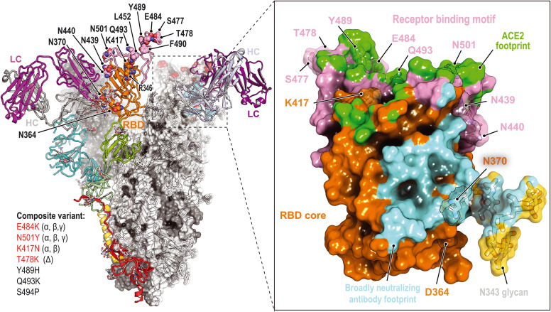
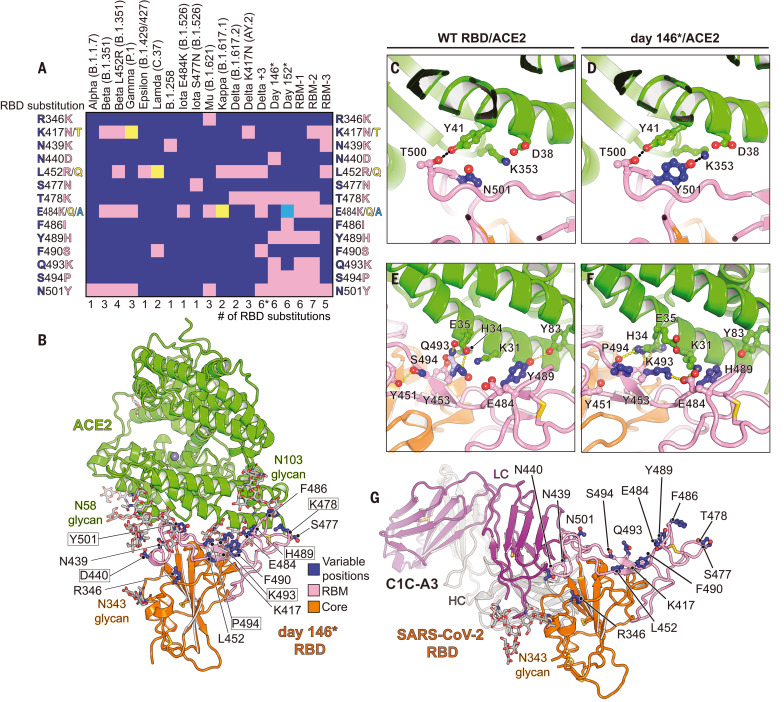
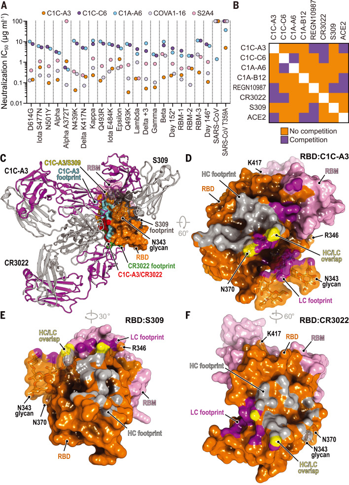
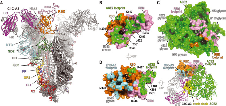
Many studies have examined the impact of severe acute respiratory syndrome coronavirus 2 (SARS-CoV-2) variants on neutralizing antibody activity after they have become dominant strains. Here, we evaluate the consequences of further viral evolution. We demonstrate mechanisms through which the SARS-CoV-2 receptor binding domain (RBD) can tolerate large numbers of simultaneous antibody escape mutations and show that pseudotypes containing up to seven mutations, as opposed to the one to three found in previously studied variants of concern, are more resistant to neutralization by therapeutic antibodies and serum from vaccine recipients. We identify an antibody that binds the RBD core to neutralize pseudotypes for all tested variants but show that the RBD can acquire an N-linked glycan to escape neutralization. Our findings portend continued emergence of escape variants as SARS-CoV-2 adapts to humans.


Figures

None
Immune escape at the SARS-CoV-2 spike protein RBD.
Structural plasticity accommodates the accumulation of composite substitutions in the RBD ACE2 binding site and allows the RBD to adeptly escape therapeutic antibodies. Cross-neutralizing antibodies bind the RBD core, but acquisition of an N-linked glycan at RBD residue Asn370 (N370) drives further neutralization escape. Single-letter abbreviations for the amino acid residues are as follows: D, Asp; E, Glu; F, Phe; H, His; K, Lys; L, Leu; N, Asn; P, Pro; Q, Gln; R, Arg; S, Ser; T, Thr; and Y, Tyr. LC, light chain; HC, heavy chain.
Fig. 1.
Fig. 1.. Structure of intrahost evolved RBD bound to human ACE2.
(A) Key RBD substitutions discussed in the text and the SARS-CoV-2 variants that contain them. Single-letter abbreviations for the amino acid residues are as follows: A, Ala; C, Cys; D, Asp; E, Glu; F, Phe; G, Gly; H, His; I, Ile; K, Lys; L, Leu; M, Met; N, Asn; P, Pro; Q, Gln; R, Arg; S, Ser; T, Thr; V, Val; W, Trp; and Y, Tyr. (B) Day 146* RBD–ACE2 ectodomain x-ray crystal structure. RBD residues that are mutated in variants discussed in the text are shown. Boxed residues are mutated in the day 146* RBD as compared with the Wuhan-Hu-1 (wild-type) SARS-CoV-2 RBD. The Delta +3 variant contains an additional RBD mutation that is not shown in the schematic diagram (see table S2). (C) Wild-type RBD–ACE2 contacts near N501RBD [Protein Data Bank (PDB) ID 6M0J] (2). (D) Day 146* RBD contacts near Y501RBD. (E) Wild-type SARS-CoV-2 RBD–ACE2 interactions near Q493RBD. (F) Day 146* RBD interactions near K493RBD. (G) Cryo-EM structure of the SARS-CoV-2 RBD bound to the C1C-A3 antibody Fab. RBD residues discussed in the text are labeled. LC, light chain; HC, heavy chain.
Fig. 2.
Fig. 2.. Neutralization escape from therapeutic antibodies and mRNA vaccine–elicited serum.
(A) Summary of neutralization IC50 values for lentivirus pseudotypes with the indicated monoclonal antibodies. (B) Tabulated IC50 values for lentivirus pseudotypes with the indicated monoclonal antibodies and an ACE2-Fc fusion protein (ACE2). (C) Mean ID50 neutralization titers for the indicated variant pseudotypes at the time of the second immunization but before vaccination (“dose 1”), or 28 days after second immunization (“dose 2”) with mRNA-1273 or BNT162b2. The fold change of the mean ID50 neutralization titer with respect to D614GS pseudotype is shown in each panel. Each experiment was performed twice independently in triplicate (n = 6). Wilcoxon matched-pairs signed rank test; ****P < 0.0001. (D) Tabulated fold change of mean ID50 neutralization titers for the indicated pseudotypes as compared with D614GS pseudotype.
Movie 1.
Movie 1.. Antibody footprints on an evolving SARS-CoV-2 RBD.
Antibodies are classified according to Barnes et al. (36). PDB IDs are listed in parentheses. Key RBD residues discussed in the main text are highlighted.
Fig. 3.
Fig. 3.. Neutralization of SARS-CoV-2 variants by an RBD core–targeting antibody.
(A) Summary of neutralization IC50 values for pseudotypes and the indicated antibodies. (B) Summary of the results of BLI-based competition assays. (C) Superposition of the CR3022 (PDB ID 6W41) (55) and S309 (PDB ID 6WPS) (44) structures onto the C1C-A3–bound RBD structure. Antibody Fabs are shown as ribbon diagrams, and the RBD is shown in surface representation. Antibody footprints are shown on the RBD surface. (D) RBD footprint of C1C-A3. (E) RBD footprint of S309 (PDB ID 6WPS) (44). (F) RBD footprint of CR3022 (PDB ID 6W41) (55). In panels (D) to (F), key RBD residues discussed in the main text are highlighted.
Fig. 4.
Fig. 4.. Structural basis for C1C-A3 neutralization.
(A) Cryo-EM structure of the C1C-A3–Fab SARS-CoV-2 spike protein complex. Two of the three spike protein protomers are shown in surface representation. One protomer is shown as a ribbon diagram with labeled subdomains. The trimer model shown was generated by superposition of an RBD–C1C-A3 Fab model generated by subparticle classification of the RBD region onto the coordinates of the trimeric spike protein–C1C-A3 Fab complex (see materials and methods). SD1, subdomain 1; SD2, subdomain 2; FP, fusion peptide; HR1, heptad repeat 1; CD, connector domain; S2, additional portions of S2 subunit. (B) Surface representation of the SARS-CoV-2 day 146* RBD showing the ACE2 footprint, including surfaces contacted by ACE2 N-linked glycans. Key RBD positions discussed in the text are labeled. (C) Surface representation of ACE2, showing the day 146* RBD and RBM footprints. (D) Surface representation of the RBD highlighting C1C-A3 Fab and ACE2 footprints. (E) Overlay of the C1C-A3 Fab–RBD complex with the day 146* RBD–ACE2 complex. Atoms within 1.54 Å of each other are shown in yellow surface representation to highlight steric clashes. Key RBD residues discussed in the text are labeled in (B) and (D).
Fig. 5.
Fig. 5.. Structural basis for immune evasion of a RBD core–targeting antibody.
(A and B) C1C-A3 antibody contacts with the SARS-CoV-2 RBD core. (C) C1C-A3 contacts with the N343RBD glycan with structural superposition of the SARS-CoV RBD (PDB ID 6NB6) (78). N-linked glycans found at N330RBD and N357RBD in SARS-CoV and the analogous N343RBD and N370RBD positions in SARS-CoV-2 are highlighted. (D) Superposition of the C1C-A3 Fab–SARS-CoV-2 RBD structure with the SARS-CoV RBD (PDB ID 6NB6) (78) showing that a glycan attached at SARS-CoV N357RBD may interfere with antibody binding. The SARS-CoV-2 RBD is not shown for clarity. (E) Superposition of the C1C-A3–SARS-CoV-2 RBD with the RaTG13 virus RBD (PDB ID 7CN4) (79) showing that a glycan attached at RaTG13 virus N370RBD would be more readily accommodated because the helix that contains it would be rotated away from the Fab. The SARS-CoV-2 RBD is omitted for clarity. (F) Sequence alignment of the RBD core region contacted by C1C-A3. SARS-CoV-2 numbering is shown at the top of the alignment, and SARS-CoV numbering is shown at the bottom. Circles indicate antibody contacts. (G) C1C-A3 neutralization curves for the indicated lentivirus pseudotypes. Data are plotted as the mean ± standard deviation of the mean. The experiment was performed twice in triplicate (n = 6). For some data points, error bars are smaller than symbols. (H) Tabulated neutralization IC50 values for the indicated pseudotypes.

References

    1. Harvey W. T., Carabelli A. M., Jackson B., Gupta R. K., Thomson E. C., Harrison E. M., Ludden C., Reeve R., Rambaut A., Peacock S. J., Robertson D. L.; COVID-19 Genomics UK (COG-UK) Consortium , SARS-CoV-2 variants, spike mutations and immune escape. Nat. Rev. Microbiol. 19, 409–424 (2021). 10.1038/s41579-021-00573-0 - DOI - PMC - PubMed
    1. Lan J., Ge J., Yu J., Shan S., Zhou H., Fan S., Zhang Q., Shi X., Wang Q., Zhang L., Wang X., Structure of the SARS-CoV-2 spike receptor-binding domain bound to the ACE2 receptor. Nature 581, 215–220 (2020). 10.1038/s41586-020-2180-5 - DOI - PubMed
    1. Shang J., Ye G., Shi K., Wan Y., Luo C., Aihara H., Geng Q., Auerbach A., Li F., Structural basis of receptor recognition by SARS-CoV-2. Nature 581, 221–224 (2020). 10.1038/s41586-020-2179-y - DOI - PMC - PubMed
    1. Piccoli L., Park Y.-J., Tortorici M. A., Czudnochowski N., Walls A. C., Beltramello M., Silacci-Fregni C., Pinto D., Rosen L. E., Bowen J. E., Acton O. J., Jaconi S., Guarino B., Minola A., Zatta F., Sprugasci N., Bassi J., Peter A., De Marco A., Nix J. C., Mele F., Jovic S., Rodriguez B. F., Gupta S. V., Jin F., Piumatti G., Lo Presti G., Pellanda A. F., Biggiogero M., Tarkowski M., Pizzuto M. S., Cameroni E., Havenar-Daughton C., Smithey M., Hong D., Lepori V., Albanese E., Ceschi A., Bernasconi E., Elzi L., Ferrari P., Garzoni C., Riva A., Snell G., Sallusto F., Fink K., Virgin H. W., Lanzavecchia A., Corti D., Veesler D., Mapping neutralizing and immunodominant sites on the SARS-CoV-2 spike receptor-binding domain by structure-guided high-resolution serology. Cell 183, 1024–1042.e21 (2020). 10.1016/j.cell.2020.09.037 - DOI - PMC - PubMed
    1. Greaney A. J., Loes A. N., Gentles L. E., Crawford K. H. D., Starr T. N., Malone K. D., Chu H. Y., Bloom J. D., Antibodies elicited by mRNA-1273 vaccination bind more broadly to the receptor binding domain than do those from SARS-CoV-2 infection. Sci. Transl. Med. 13, eabi9915 (2021). 10.1126/scitranslmed.abi9915 - DOI - PMC - PubMed

Publication types

MeSH terms

Supplementary concepts

LinkOut - more resources