. 2022 Jan 14;375(6577):183-192.
doi: 10.1126/science.abm0811. Epub 2021 Dec 2.

Heterologous infection and vaccination shapes immunity against SARS-CoV-2 variants

Affiliations

Heterologous infection and vaccination shapes immunity against SARS-CoV-2 variants

Catherine J Reynolds et al. Science. .

Abstract

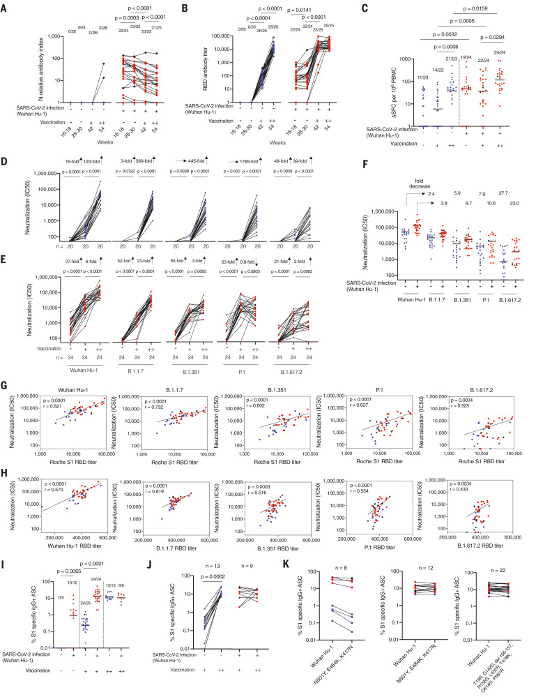
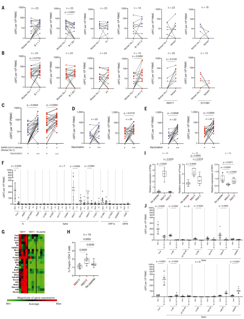
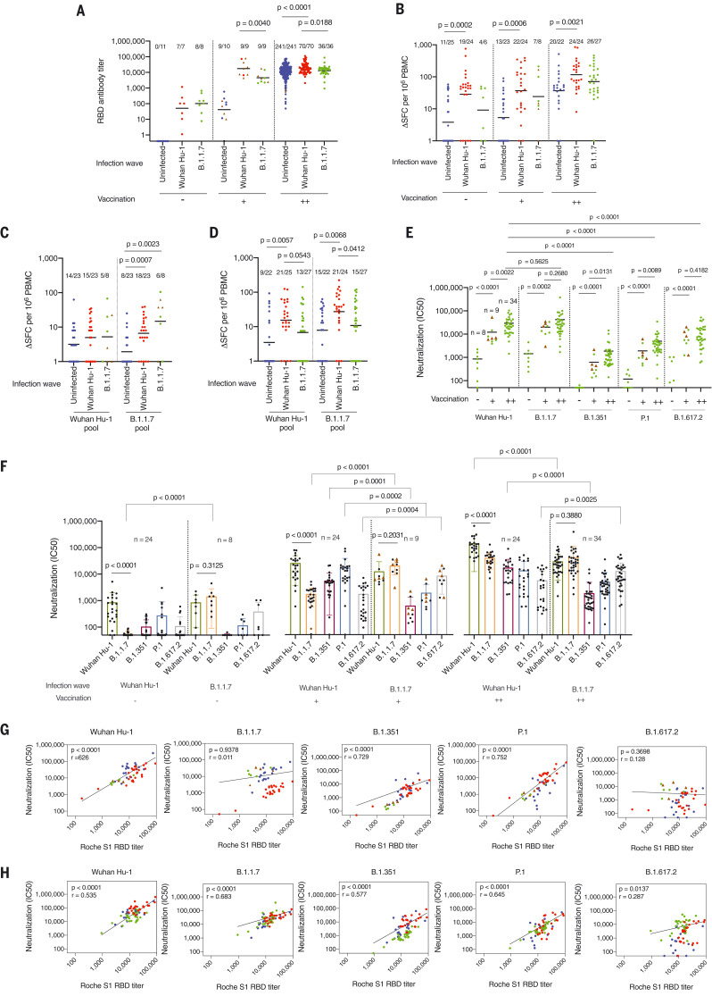
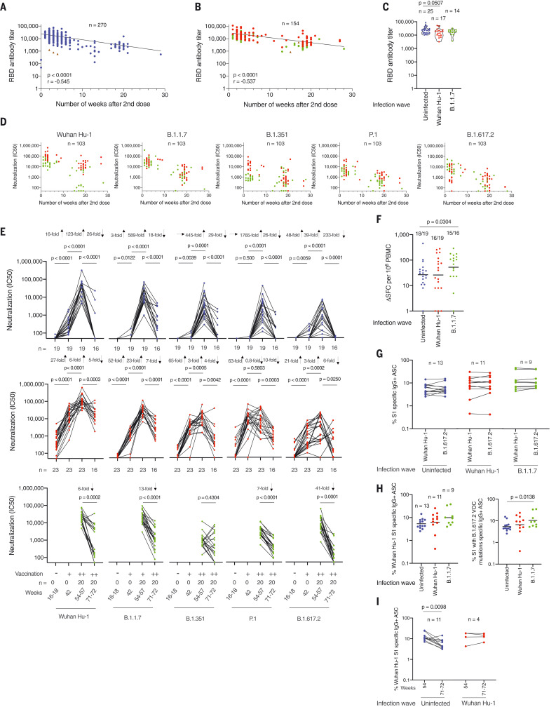
The impact of the initial severe acute respiratory syndrome coronavirus 2 (SARS-CoV-2) infecting strain on downstream immunity to heterologous variants of concern (VOCs) is unknown. Studying a longitudinal healthcare worker cohort, we found that after three antigen exposures (infection plus two vaccine doses), S1 antibody, memory B cells, and heterologous neutralization of B.1.351, P.1, and B.1.617.2 plateaued, whereas B.1.1.7 neutralization and spike T cell responses increased. Serology using the Wuhan Hu-1 spike receptor binding domain poorly predicted neutralizing immunity against VOCs. Neutralization potency against VOCs changed with heterologous virus encounter and number of antigen exposures. Neutralization potency fell differentially depending on targeted VOCs over the 5 months from the second vaccine dose. Heterologous combinations of spike encountered during infection and vaccination shape subsequent cross-protection against VOC, with implications for future-proof next-generation vaccines.


Figures

Fig. 1.
Fig. 1.. T and B cell immunity to infection with Wuhan Hu-1 SARS-CoV-2 and after one and two doses of BNT162b2 vaccine in infection naïve and previously infected HCWs.
(A) N Ab and (B) S1 RBD serum Ab titers measured by ECLIA in HCWs with (red, n = 25) and without (blue, n = 26) laboratory-confirmed SARS-CoV-2 infection at 16 to 18 weeks after the start of the first UK epidemic wave and 2 to 3 weeks after first (+, 42 weeks) and second (++, 54 weeks) dose of BNT162b2 vaccine. In all figures, HCWs without laboratory-confirmed infection are shown in blue and HCWs with laboratory-confirmed infection are shown in red. HCWs with new infection or reinfection after 30 weeks are shown in black. (C) Magnitude of T cell responses to spike MEP pool in HCWs with (red, n = 24) and without (blue, n = 23) laboratory-confirmed SARS-CoV-2 infection 16 to 18 weeks after the start of the first UK epidemic wave and 2 to 3 weeks after first (+, 42 weeks) and second (++, 54 weeks) dose of vaccine. Neutralizing Ab titer (IC50) against authentic Wuhan Hu-1 live virus and B.1.1.7, B.1.351, P.1, and B.1.617.2 VOCs is plotted longitudinally for (D) SARS-CoV-2 infection naïve (blue, n = 20) and (E) previously Wuhan Hu-1 infected HCWs (red, n = 24) unvaccinated and after the first and second dose of BNT162b2 vaccine. (F) IC50 nAb against Wuhan Hu-1 and VOCs after two-dose BNT162b2 vaccination in previously infected HCWs (red, n = 24) and infection-naïve HCWs (blue, n = 20). (G) Correlation between Roche S1 RBD Ab titer and nAb (IC50) against SARS-CoV-2 authentic Wuhan Hu-1 live virus and the B.1.1.7, B.1.351, P.1, and B.1.617.2 VOCs. (H) Correlation between Wuhan Hu-1, VOC RBD, and corresponding live virus in two-dose BNT162b2 vaccinated previously infected (red, n = 24) and SARS-CoV-2 infection-naïve (blue, n = 20) HCWs. (I) Percentage of S1-specific IgG+ antibody-secreting cells (ASCs) in SARS-CoV-2 infection naïve and previously infected HCWs at 16 to 18 weeks and after first- and second-dose BNT162b2 vaccination. (J) Percentage of S1-specific IgG+ ASCs plotted pairwise after first- and second-dose vaccination for HCWs with (red, n = 9) and without (blue, n = 13) prior SARS-CoV-2 infection. (K) Left, percentage of IgG+ ASCs specific for Wuhan Hu-1 S1 protein or spike protein containing the mutations N501Y, E484K, and K417N in HCWs with (red) and without (blue) prior SARS-CoV-2 infection after first-dose vaccination (n = 4 per group). Middle and right panels, percentage of IgG+ ASCs specific for Wuhan Hu-1 S1 or S1 spike protein containing N501Y, E484K, and K417N (B.1.351 VOC) mutations or S1 spike protein containing T19R, G142D, del 156-157, R158G, L452R, T478K, D614G, and P681R (B.1.617.2 VOC) mutations after second-dose BNT162b2 vaccination (middle panel, n = 6 per group; right panel, infection naïve HCWs are indicated in blue, n = 13, and previously Wuhan Hu-1 infected HCWs are indicated in red, n = 9). In (A), (B), (D), (E), and (H), Wilcoxon matched-pairs signed rank test was used; in (C) and (I), Mann-Whitney U test was used; and in (G), Spearman’s rank correlation was used. New infection (in the infection-naïve group, n = 2) and reinfection (in the previously infected group, n = 2) data points shown in black were excluded from statistical analysis. SFCs, spot-forming cells.
Fig. 2.
Fig. 2.. T cell responses to Wuhan Hu-1 and VOC peptide pools after one- and two-dose BNT162b2 vaccination in infection-naïve and previously infected HCWs.
Magnitude of T cell responses to B.1.1.7, B.1.351, P.1, and B.1.617.2 variant specific peptide pools and to the matched sequence peptide pools from Wuhan Hu-1 as well as to individual wild-type and variant N501Y and D1118H peptides in (A) SARS-Cov-2 infection-naïve (blue) HCWs and (B) previously infected (red) HCWs who had received two doses of BNT162b2 vaccine. In all figures, HCWs without labaoratory-confirmed infection are shown in blue, HCWs with laboratory-confirmed infection are shown in red, and HCWs with new infection or reinfection after 30 weeks are shown in black. (C to E) Magnitude of T cell responses to spike MEP (C), Wuhan Hu-1 (D), and B.1.1.7 variant (E) peptide pools after one (+) and two (++) doses of BNT162b2 vaccine in infection-naïve HCWs (blue) and previously infected HCWs (red). (F) Magnitude of T cell response to wild-type Wuhan Hu-1 and P.1 variant peptide pools and individual peptides in Wuhan Hu-1 peptide pool immunized HLA-DRB1*04:01 transgenic mice (n = 7). (G) Heatmap showing relative gene expression of T cell activation markers in draining lymph node (DLN) cells from Wuhan Hu-1 N501Y peptide–primed DRB1*04:01 transgenic mice (n = 4) stimulated for 24 h in vitro with 10 μg/ml of wild-type Wuhan Hu–1 or variant peptide. Genes shown in red are significantly different (P < 0.05) between no peptide control and Wuhan Hu-1 or variant peptide stimulated cells with a fold change greater than 1.5. (H) Percentage of Foxp3+ CD4 T cells by flow cytometry in DLN cells from Wuhan Hu-1 N501Y peptide-primed DRB1*04:01 transgenic mice (n = 10) stimulated for 24 h in vitro with 10 mg/ml of wild-type or variant peptide. (I) Relative gene expression of ifng, foxp3, and irf4 in DLN cells from Wuhan Hu-1 N501Y peptide-primed DRB1*0401 transgenic mice (n = 10) stimulated for 24 h in vitro with 10 μg/ml of Wuhan Hu-1 or variant peptide. (J) Magnitude of T cell response to wild-type Wuhan Hu-1 and B.1.617.2 variant peptide pools and individual peptides in Wuhan Hu-1 peptide pool immunized (top panel, n = 6) or B.1.617.2 peptide pool immunized (bottom panel, n = 6) HLA-DRB1*04:01 transgenic mice. In (A) to (E), (I), and (J), Wilcoxon matched-pairs signed rank test was used. New infection (in the infection-naïve group, n = 2) and reinfection (in the previously infected group, n = 2) data points shown in black were excluded from statistical analysis. In (F) and (G), Mann-Whitney U test was used.
Fig. 3.
Fig. 3.. T cell and B cell immunity and neutralization hierarchy after heterologous exposure through infection with the B.1.1.7 VOC during the second UK wave and in the context of single- and two-dose vaccination.
(A) RBD Ab titers at 54 to 57 weeks after the start of study recruitment in March 2020 in HCWs who were not infected with SARS-CoV-2 (blue, n = 256), those infected during the first UK wave by the Wuhan Hu-1 strain (red, n = 86), and those infected during the second UK wave by the B.1.1.7 (green, n = 53). (B) Magnitude of T cell response to Wuhan Hu-1 spike MEP peptide pool. Data are plotted according to whether individuals were unvaccinated (–) or had received one (+) or two (++) doses of BNT162b2 vaccine. (C and D) Magnitude of T cell response to B.1.1.7 peptide pools (Wuhan Hu-1 or B.1.1.7 variant peptides) after first dose (C) or second dose (D) of vaccine. (E and F) Neutralizing Ab titers (IC50) against authentic Wuhan Hu-1 live virus and B.1.1.7, B.1.351, P.1, or B.1.617.2 VOCs in HCWs infected with SARS-CoV-2 during the B.1.1.7 wave (E) and during the Wuhan Hu-1 first UK wave (F), plotted according to whether individuals were unvaccinated (–, Wuhan Hu-1 wave, n = 24; B.1.1.7 wave, n = 8) or had received one dose (+, Wuhan Hu-1 wave n = 24, B.1.1.7 wave n = 9) or two doses (++, Wuhan Hu-1 wave n = 24, B.1.1.7 wave n = 34) of vaccine. (G and H) Correlation between Roche S1 RBD Ab titer and nAb (IC50) against authentic Wuhan Hu-1 live virus and B.1.1.7, B.1.351, P.1, and B.1.617.2 VOC in one-dose (G) and two-dose (H) BNT162b2 vaccinated HCWs previously infected by Wuhan Hu-1 (red, n = 23) or B.1.1.7 infected (green, one dose n = 9, two doses n = 31) and two-dose vaccinated infection-naïve (blue, n = 19) HCWs at 54 to 57 weeks after initial study recruitment. In all graphs, individuals who received the ChAdOx1 nCoV-19 vaccine are marked as brown triangles. In (A) to (E), Mann-Whitney U test was used; in (F), Mann-Whitney U test or Wilcoxon matched-pairs signed rank test was used; and in (G) and (H), Spearman’s rank correlation was used.
Fig. 4.
Fig. 4.. Differential impact of heterologous exposure through B.1.1.7 infection on neutralization of other VOC and durability of the immune response.
(A) S1 RBD Ab titers in HCWs with no previous SARS-CoV-2 infection (blue) and two-dose vaccination (n = 270). (B) S1 RBD Ab titers in HCWs with a history of SARS-CoV-2 infection (Wuhan Hu-1, red; B.1.17, green) and two-dose vaccination (n = 154), plotted by the number of weeks that serum was sampled after the second vaccine dose. HCWs who received the ChAdOx1 nCoV-19 vaccine are indicated by brown triangles. (C) S1 RBD Ab titers in infection-naïve HCWs (blue, n = 25) or those with a history of SARS-CoV-2 infection with the Wuhan Hu-1 strain (red, n = 17) or B.1.17 VOCs (green, n = 14) and three-dose vaccination sampled between 10 d and 7 weeks after the third vaccine dose. (D) Neutralizing Ab titers (IC50) against authentic Wuhan Hu-1 live virus and B.1.1.7, B.1.351, P.1, and B.1.617.2 VOC in HCWs with a history of previous SARS-CoV-2 infection who had received two vaccine doses, plotted by the number of weeks that serum was sampled after the second vaccine dose. (E) Neutralizing Ab titer (IC50) against authentic Wuhan Hu-1 live virus and B.1.1.7, B.1.351, P.1, and B.1.617.2 VOCs plotted longitudinally 20 d after the first (+) and 20 days after the second (++) dose and then 21 weeks after the second BNT162b2 vaccination. Shown are SARS-CoV-2 infection-naïve HCWs (blue, upper panel), Wuhan Hu-1 previously infected HCWs (red, middle panel), and B.1.1.7–infected HCWs (green, lower panel). (F) Magnitude of T cell response to spike MEP peptide pool at 71 to 72 weeks after initial study recruitment in double-vaccinated infection-naïve HCWs (blue, n = 19) and HCWs with a history of SARS-CoV-2 infection by Wuhan Hu-1 (red, n = 19) or B.1.1.7 (green, n = 16). (G) Percentage of IgG+ ASCs specific for Wuhan Hu-1 S1 or S1 protein containing T19R, G142D, del 156-157, R158G, L452R, T478K, D614G, and P681R (B.1.617.2 VOC) mutations at 71 to 72 weeks after initial study recruitment in double-vaccinated infection-naïve (blue, n = 13) HCWs and those with a history of SARS-CoV-2 infection with the Wuhan Hu-1 strain (red, n = 11) and B.1.1.7 VOC (green, n = 9) 21 weeks after the second vaccine dose. (H) Percentage of IgG+ ASCs specific for Wuhan Hu-1 S1 protein (left panel) or S1 protein containing T19R, G142D, del 156-157, R158G, L452R, T478K, D614G, and P681R (B.1.617.2 VOC) mutations (right panel) at 71 to 72 weeks after initial study recruitment in double-vaccinated infection-naïve HCWs (blue, n = 13), previously Wuhan Hu-1–infected HCWs (red, n = 11), and B.1.1.7–infected HCWs (green, n = 9). (I) Percentage of IgG+ ASCs specific for Wuhan Hu-1 S1 protein in double-vaccinated HCWs with (red, n = 4) or without (blue, n = 11) a history of Wuhan Hu-1 infection at 54 and 71 to 72 weeks after initial study recruitment. In (A) and (B), Spearman’s rank correlation was used; in (C), (F), and (H), Mann-Whitney U test was used; and in (E) and (I), Wilcoxon matched-pairs signed rank test was used.

Comment in

References

    1. Collier D. A., Ferreira I. A. T. M., Kotagiri P., Datir R. P., Lim E. Y., Touizer E., Meng B., Abdullahi A., CITIID-NIHR BioResource COVID-19 Collaboration, Elmer A., Kingston N., Graves B., Le Gresley E., Caputo D., Bergamaschi L., Smith K. G. C., Bradley J. R., Ceron-Gutierrez L., Cortes-Acevedo P., Barcenas-Morales G., Linterman M. A., McCoy L. E., Davis C., Thomson E., Lyons P. A., McKinney E., Doffinger R., Wills M., Gupta R. K., Age-related immune response heterogeneity to SARS-CoV-2 vaccine BNT162b2. Nature 596, 417–422 (2021). 10.1038/s41586-021-03739-1 - DOI - PMC - PubMed
    1. Wall E. C., Wu M., Harvey R., Kelly G., Warchal S., Sawyer C., Daniels R., Hobson P., Hatipoglu E., Ngai Y., Hussain S., Nicod J., Goldstone R., Ambrose K., Hindmarsh S., Beale R., Riddell A., Gamblin S., Howell M., Kassiotis G., Libri V., Williams B., Swanton C., Gandhi S., Bauer D. L., Neutralising antibody activity against SARS-CoV-2 VOCs B.1.617.2 and B.1.351 by BNT162b2 vaccination. Lancet 397, 2331–2333 (2021). 10.1016/S0140-6736(21)01290-3 - DOI - PMC - PubMed
    1. Planas D., Veyer D., Baidaliuk A., Staropoli I., Guivel-Benhassine F., Rajah M. M., Planchais C., Porrot F., Robillard N., Puech J., Prot M., Gallais F., Gantner P., Velay A., Le Guen J., Kassis-Chikhani N., Edriss D., Belec L., Seve A., Courtellemont L., Péré H., Hocqueloux L., Fafi-Kremer S., Prazuck T., Mouquet H., Bruel T., Simon-Lorière E., Rey F. A., Schwartz O., Reduced sensitivity of SARS-CoV-2 variant Delta to antibody neutralization. Nature 596, 276–280 (2021). 10.1038/s41586-021-03777-9 - DOI - PubMed
    1. Harvey W. T., Carabelli A. M., Jackson B., Gupta R. K., Thomson E. C., Harrison E. M., Ludden C., Reeve R., Rambaut A., Peacock S. J., Robertson D. L.; COVID-19 Genomics UK (COG-UK) Consortium , SARS-CoV-2 variants, spike mutations and immune escape. Nat. Rev. Microbiol. 19, 409–424 (2021). 10.1038/s41579-021-00573-0 - DOI - PMC - PubMed
    1. Treibel T. A., Manisty C., Burton M., McKnight Á., Lambourne J., Augusto J. B., Couto-Parada X., Cutino-Moguel T., Noursadeghi M., Moon J. C., COVID-19: PCR screening of asymptomatic health-care workers at London hospital. Lancet 395, 1608–1610 (2020). 10.1016/S0140-6736(20)31100-4 - DOI - PMC - PubMed

Publication types

MeSH terms

Supplementary concepts