. 2021 Dec 2;16(12):e0260719.
doi: 10.1371/journal.pone.0260719. eCollection 2021.

The efficacy and safety of pinocembrin in a sheep model of bleomycin-induced pulmonary fibrosis

Affiliations

The efficacy and safety of pinocembrin in a sheep model of bleomycin-induced pulmonary fibrosis

Habtamu B Derseh et al. PLoS One. .

Abstract

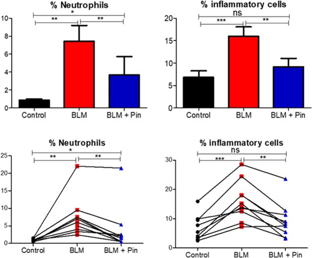
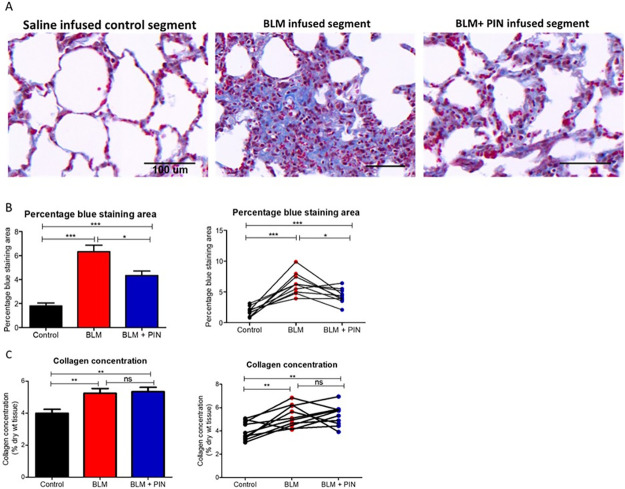
The primary flavonoid, pinocembrin, is thought to have a variety of medical uses which relate to its reported anti-oxidant, anti-inflammatory, anti-microbial and anti-cancer properties. Some studies have reported that this flavonoid has anti-fibrotic activities. In this study, we investigated whether pinocembrin would impede fibrosis, dampen inflammation and improve lung function in a large animal model of pulmonary fibrosis. Fibrosis was induced in two localized lung segments in each of the 10 sheep participating in the study. This was achieved via two infusions of bleomycin delivered bronchoscopically at a two-week interval. Another lung segment in the same sheep was left untreated, and was used as a healthy control. The animals were kept for a little over 5 weeks after the final infusion of bleomycin. Pinocembrin, isolated from Eucalyptus leaves, was administered to one of the two bleomycin damaged lung segments at a dose of 7 mg. This dose was given once-weekly over 4-weeks, starting one week after the final bleomycin infusion. Lung compliance (as a measure of stiffness) was significantly improved after four weekly administrations of pinocembrin to bleomycin-damaged lung segments. There were significantly lower numbers of neutrophils and inflammatory cells in the bronchoalveolar lavage of bleomycin-infused lung segments that were treated with pinocembrin. Compared to bleomycin damaged lung segments without drug treatment, pinocembrin administration was associated with significantly lower numbers of immuno-positive CD8+ and CD4+ T cells in the lung parenchyma. Histopathology scoring data showed that pinocembrin treatment was associated with significant improvement in inflammation and overall pathology scores. Hydroxy proline analysis showed that the administration of pinocembrin did not reduce the increased collagen content that was induced by bleomycin in this model. Analyses of Masson's Trichrome stained sections showed that pinocembrin treatment significantly reduced the connective tissue content in lung segments exposed to bleomycin when compared to bleomycin-infused lungs that did not receive pinocembrin. The striking anti-inflammatory and modest anti-fibrotic remodelling effects of pinocembrin administration were likely linked to the compound's ability to improve lung pathology and functional compliance in this animal model of pulmonary fibrosis.


Conflict of interest statement

We have read the journal’s policy and the authors of this manuscript have the following competing interests: AC is the CEO of Gretals Australia Pty Ltd. Gretals Australia Pty Ltd has an interest in the development of pinocembrin for lung diseases. This does not alter our adherence to PLOS ONE policies on sharing data and materials.

Figures

Fig 1
Fig 1. Experimental design.
Locations of spatially separate lung-segments, which received the different treatments. Note that the treatments to either the left or right caudal segments were randomized, so that half (n = 5 of the sheep) received the treatments as shown above, while for the other (n = 5 sheep), the treatments in the caudal segments were reversed. Note that the rest of the lung was available for healthy respiration.
Fig 2
Fig 2. Study timeline for bleomycin challenges, bioactive compound treatments, lung function assessments, and sampling time.
Fig 3
Fig 3. Lung function in the differentially treated lung segments as assessed at week 11 of the study.
The differentially treated lung segments were the right medial (RM) lung-segments which were left untreated for healthy lung controls (Control), the right caudal (RC) and the left caudal (LC) lung-segments which were either infused with bleomycin without drug treatment (BLM), or infused with bleomycin and received 4 once-weekly doses of pinocembrin (BLM + PIN). Part A shows mean data for Cseg (n = 10), which is a measure for how easy it is to inflate the lung segment. Part B shows individual sheep data. Part C shows percent change of Cseg at week 11 from baseline values taken at week 0 at the beginning of the study. Significance was determined using paired t-tests, *p<0.05, ***p<0.001, n = 10 sheep.
Fig 4
Fig 4. Neutrophils and inflammatory cells recovered from the bronchoalveolar lavage (BAL) fluid of the differentially treated lung-segments at week 12.
The differentially treated lung segments were the right medial (RM) lung-segments which were left untreated for healthy lung controls (Control), the right caudal (RC) and the left caudal (LC) lung-segments which were either infused with bleomycin without drug treatment (BLM) or infused with bleomycin and received 4 once-weekly doses of pinocembrin (BLM + PIN). The left panels show neutrophil data, and the right panels show inflammatory cell data, which included the sum of the percentages of neutrophils, lymphocytes, and eosinophils. The top panels show mean data for ten sheep. The bottom panel shows individual sheep data. Significance was determined using paired t-tests, *p<0.05, **p<0.01, ***p<0.001, n = 10 sheep.
Fig 5
Fig 5. The effect of pinocembrin on lung parenchymal T cells.
(A) representative photomicrographs showing CD8+ and CD4+ T cells (arrows show examples of immuno-positive cells) in the lung parenchyma sampled from the differentially treated lung-segments at week 12. (B) graphs showing the average number of positive cells per field at 400x magnification for each differentially treated segment. The differentially treated lung segments were the right medial (RM) lung-segments which were left untreated for healthy lung controls (Control), the right caudal (RC) and the left caudal (LC) lung-segments which were either infused with bleomycin without drug treatment (BLM) or infused with bleomycin and received 4 once-weekly doses of pinocembrin (BLM + PIN). The left panels show mean lung segment data and the right panels show individual sheep data. Significance was determined using paired t-tests, **p<0.01, ***p<0.001, n = 10 sheep. Scale bars = 100 μm.
Fig 6
Fig 6. Histopathology scoring data as assessed on histological H+E-stained sections sampled at post-mortem from the differentially treated lung-segments.
The differentially treated lung segments were the right medial (RM) lung-segments which were left untreated for healthy lung controls (Control), the right caudal (RC) and the left caudal (LC) lung-segments which were either infused with bleomycin without drug treatment (BLM) or infused with bleomycin and received 4 once-weekly doses of pinocembrin (BLM + PIN). The top panels show mean scoring data for ten sheep. The bottom panels show individual sheep data. Significance was determined using paired t-tests, *p<0.05, **p<0.01, n = 10 sheep. Scoring criteria is described in the Materials and methods.
Fig 7
Fig 7. The effects of pinocembrin on connective tissue content.
(A) Shows representative photomicrographs of Masson’s trichrome stained lung parenchyma from the differentially treated lung-segments. Masson’s Trichrome stains most connective tissues blue. The stained connective tissue includes collagen and other extracellular matrix proteins associated with fibrosis. The scale bar in each image indicates a length of 100 μm. (B) shows data for percentage of Masson’s trichrome stained lung tissue sections. (C) shows data for the hydroxyproline assay to determine collagen content after four once-weekly treatments with pinocembrin. For each sheep, the differentially treated lung segments were the right medial (RM) lung-segments which were left untreated for healthy lung controls (Control), the right caudal (RC) and the left caudal (LC) lung-segments which were either infused with bleomycin without drug treatment (BLM) or infused with bleomycin and received 4 once-weekly doses of pinocembrin (BLM + PIN). In the graphs B and C, the left panels show mean data. The right panels show individual sheep data. Significance was determined using paired t-tests, *p<0.05, **p<0.01, ***p<0.001, n = 10 sheep.

References

    1. El-Demerdash AA, Menze ET, Esmat A, Tadros MG, Elsherbiny DA. Protective and therapeutic effects of the flavonoid "pinocembrin" in indomethacin-induced acute gastric ulcer in rats: impact of anti-oxidant, anti-inflammatory, and anti-apoptotic mechanisms. Naunyn-Schmiedeberg’s archives of pharmacology. 2021;394(7):1411–24. Epub 2021/02/28. doi: 10.1007/s00210-021-02067-5 . - DOI - PubMed
    1. Gan W, Li X, Cui Y, Xiao T, Liu R, Wang M, et al.. Pinocembrin relieves lipopolysaccharide and bleomycin induced lung inflammation via inhibiting TLR4-NF-kappaB-NLRP3 inflammasome signaling pathway. International immunopharmacology. 2021;90:107230. Epub 2020/12/09. doi: 10.1016/j.intimp.2020.107230 . - DOI - PubMed
    1. Yang X, Wang X, Chen XY, Ji HY, Zhang Y, Liu AJ. Pinocembrin(-)Lecithin Complex: Characterization, Solubilization, and Antioxidant Activities. Biomolecules. 2018;8(2). Epub 2018/06/21. doi: 10.3390/biom8020041 . - DOI - PMC - PubMed
    1. Pei B, Sun J. Pinocembrin alleviates cognition deficits by inhibiting inflammation in diabetic mice. J Neuroimmunol. 2018;314:42–9. Epub 2017/11/19. doi: 10.1016/j.jneuroim.2017.11.006 . - DOI - PubMed
    1. Giri SS, Sen SS, Sukumaran V, Park SC. Pinocembrin attenuates lipopolysaccharide-induced inflammatory responses in Labeo rohita macrophages via the suppression of the NF-kappaB signalling pathway. Fish Shellfish Immunol. 2016;56:459–66. Epub 2016/08/06. doi: 10.1016/j.fsi.2016.07.038 . - DOI - PubMed

Publication types

MeSH terms