. 2021 Dec 2;16(12):e0260705.
doi: 10.1371/journal.pone.0260705. eCollection 2021.

A translational rat model for ex vivo lung perfusion of pre-injured lungs after brain death

Affiliations

A translational rat model for ex vivo lung perfusion of pre-injured lungs after brain death

Judith E van Zanden et al. PLoS One. .

Abstract

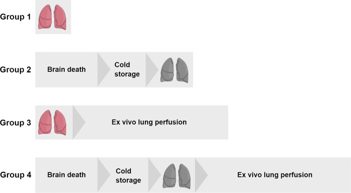
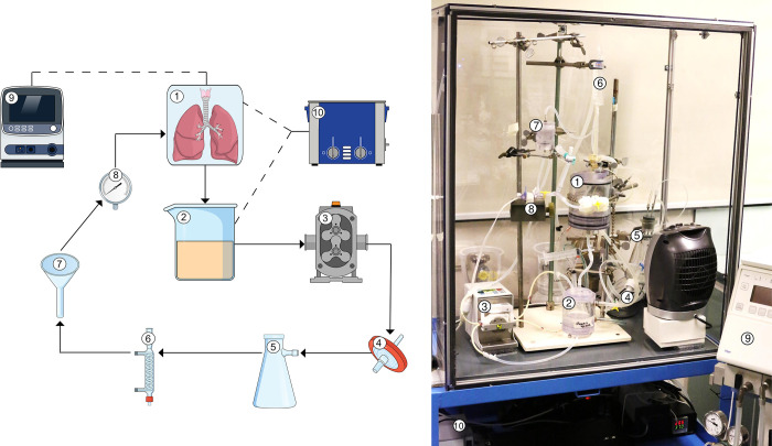
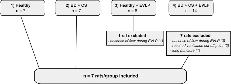
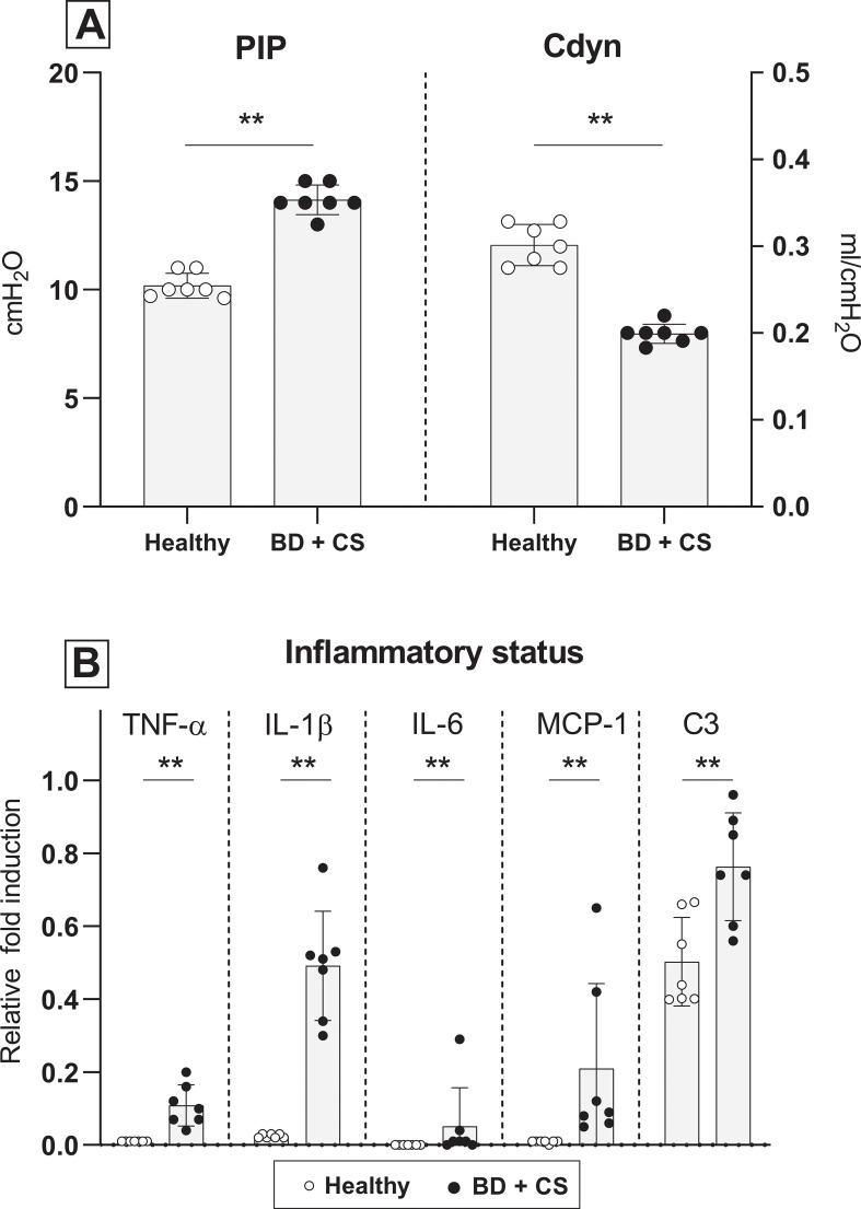
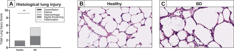
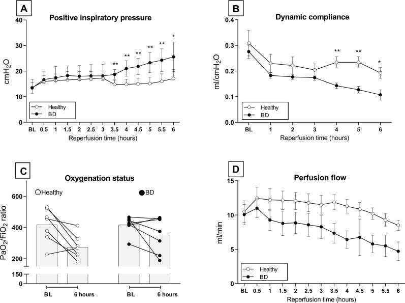
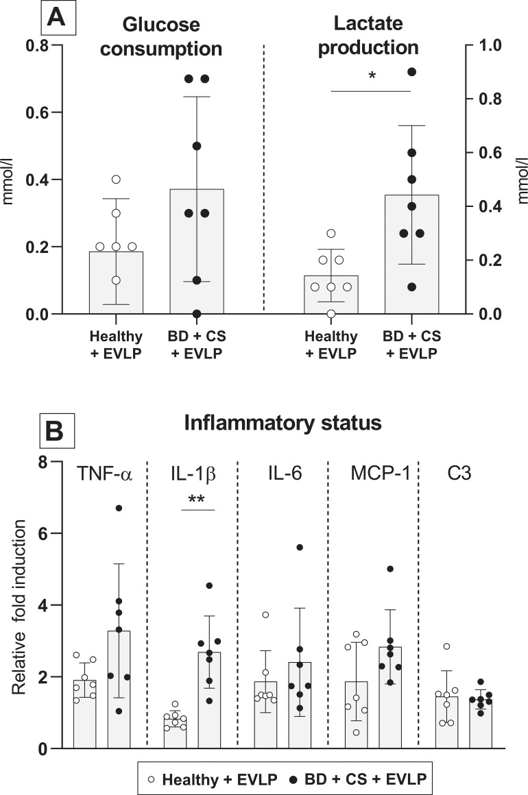
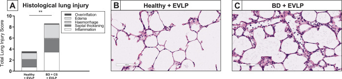
The process of brain death (BD) detrimentally affects donor lung quality. Ex vivo lung perfusion (EVLP) is a technique originally designed to evaluate marginal donor lungs. Nowadays, its potential as a treatment platform to repair damaged donor lungs is increasingly studied in experimental models. Rat models for EVLP have been described in literature before, yet the pathophysiology of BD was not included in these protocols and prolonged perfusion over 3 hours without anti-inflammatory additives was not achieved. We aimed to establish a model for prolonged EVLP of rat lungs from brain-dead donors, to provide a reliable platform for future experimental studies. Rat lungs were randomly assigned to one of four experimental groups (n = 7/group): 1) healthy, directly procured lungs, 2) lungs procured from rats subjected to 3 hours of BD and 1 hour cold storage (CS), 3) healthy, directly procured lungs subjected to 6 hours EVLP and 4), lungs procured from rats subjected to 3 hours of BD, 1 hour CS and 6 hours EVLP. Lungs from brain-dead rats showed deteriorated ventilation parameters and augmented lung damage when compared to healthy controls, in accordance with the pathophysiology of BD. Subsequent ex vivo perfusion for 6 hours was achieved, both for lungs of healthy donor rats as for pre-injured donor lungs from brain-dead rats. The worsened quality of lungs from brain-dead donors was evident during EVLP as well, as corroborated by deteriorated ventilation performance, increased lactate production and augmented inflammatory status during EVLP. In conclusion, we established a stable model for prolonged EVLP of pre-injured lungs from brain-dead donor rats. In this report we describe tips and pitfalls in the establishment of the rat EVLP model, to enhance reproducibility by other researchers.


Conflict of interest statement

The authors have declared that no competing interests exist.

Figures

Fig 1
Fig 1. Experimental outline of the study.
Lungs from donor rats were randomly assigned to one of four experimental groups (n = 7/group): 1) healthy, directly procured lungs, 2) lungs procured from rats subjected to 3 hours of brain death (BD) and 1 hour cold storage (CS), 3) healthy, directly procured lungs subjected to 6 hours ex vivo lung perfusion (EVLP) and 4), lungs procured from rats subjected to 3 hours of BD, 1 hour CS and 6 hours EVLP.
Fig 2
Fig 2. Schematic overview of the rat ex vivo lung perfusion model.
Lungs from healthy or brain-dead donor rats were subjected to 6 hours ex vivo lung perfusion (EVLP). The EVLP circuit consisted of a (1) graft humidity chamber, (2) reservoir, (3) roller pump, (4) leukocyte filter, (5) deoxygenator, (6) heat exchanger, (7) funnel, (8) pressure sensor, (9) ventilator, and a (10) water bath.
Fig 3
Fig 3. Experimental groups with number of rats utilized and rationale for exclusion.
Lungs from donor rats were randomly assigned to one of four experimental groups (n = 7/group): 1) healthy, directly procured lungs, 2) lungs procured from rats subjected to 3 hours of brain death (BD) and 1 hour cold storage (CS), 3) healthy, directly procured lungs subjected to 6 hours ex vivo lung perfusion (EVLP) and 4), lungs procured from rats subjected to 3 hours of BD, 1 hour CS and 6 hours EVLP. In total, 38 rats were utilized for the establishment of a stable BD and EVLP protocol, of which 10 rats were excluded. Eventually, 7 rats per group were included in the final protocol.
Fig 4
Fig 4. Ventilation parameters and inflammatory status after lung procurement.
Lungs were procured from either healthy rats (experimental group 1) or rats subjected to 3 hours of brain death (BD, experimental group 2). (A) Pulmonary Inspiratory Pressure (PIP) required to maintain tidal volume at 7 ml/kg of bodyweight and dynamic compliance (Cdyn) of healthy donor lungs 1) versus donor lungs subjected to 3 hours of BD, at time of lung procurement. (B) Pro-inflammatory gene expressions of TNF-α, IL-1β, IL-6, MCP-1 and C3 of healthy donor lungs versus donor lungs subjected to 3 hours of BD. ** p<0.01 in healthy donor lungs versus lungs from brain-dead donors.
Fig 5
Fig 5. Lung morphology after lung procurement.
Lungs were procured from either healthy rats (experimental group 1) or rats subjected to 3 hours of brain death (BD, experimental group 2). Histological lung injury was scored after staining for hematoxylin and eosin (H&E). (A) Quantification of lung injury scores in H&E-stained lung slides. (B-C) Representative H&E-stained slices of healthy donor lungs and lungs from brain-dead donors. ** p<0.01 in healthy donor lungs versus lungs from brain-dead donors.
Fig 6
Fig 6. Ventilation and perfusion performance during ex vivo lung perfusion.
Lungs from healthy donor rats or rats subjected to 3 hours of brain death (BD) and 1 hour cold storage (CS) were ex vivo perfused for 6 hours (EVLP, experimental group 3 and 4). (A) Positive Inspiratory Pressure (PIP) required to maintain tidal volume at 7 ml/kg of bodyweight over time, during EVLP. (B) Dynamic compliance (Cdyn) of donor lungs over time, during EVLP. (C) Oxygenation capacity of donor lungs as reflected by PaO2/FiO2 ratio. (D) Perfusion flow of donor lungs over time, during EVLP. * p<0.05 in healthy donor lungs versus lungs from brain-dead donors subjected to EVLP. ** p<0.01 in healthy donor lungs versus lungs from brain-dead donors subjected to EVLP.
Fig 7
Fig 7. Metabolic profile and inflammatory status after ex vivo lung perfusion.
Lungs from healthy donor rats or rats subjected to 3 hours of brain death (BD) and 1 hour cold storage (CS) were ex vivo perfused for 6 hours (EVLP, experimental group 3 and 4). (A) Cumulative glucose consumption of healthy donor lungs versus lungs from brain-dead rats, during EVLP. (B) Cumulative lactate production of healthy donor lungs versus lungs from brain-dead rats, during EVLP. (C) Pro-inflammatory gene expressions of TNF-α, IL-1β, IL-6, MCP-1 and C3 in donor lungs, after 6 hours of EVLP. * p<0.05 in healthy donor lungs versus lungs from brain-dead donors subjected to EVLP. ** p<0.01 in healthy donor lungs versus lungs from brain-dead donors subjected to EVLP.
Fig 8
Fig 8. Lung morphology after ex vivo lung perfusion.
Lungs from healthy donor rats or rats subjected to 3 hours of brain death (BD) and 1 hour cold storage (CS) were ex vivo perfused for 6 hours (EVLP, experimental group 3 and 4). Histological lung injury was scored after staining for hematoxylin and eosin (H&E). (A) Quantification of lung injury scores after EVLP in H&E-stained lung slides. (B-C) Representative H&E-stained slices of healthy donor lungs and lungs from brain-dead donors, after EVLP. ** p<0.01 in healthy donor lungs versus lungs from brain-dead donors subjected to EVLP.

Similar articles

Cited by

References

    1. Avlonitis VS, Fisher AJ, Kirby JA, et al.. Pulmonary transplantation: The role of brain death in donor lung injury. Transplantation 2003; 75: 1928–1933. doi: 10.1097/01.TP.0000066351.87480.9E - DOI - PubMed
    1. Van Raemdonck D, Neyrinck A, Verleden GM, et al.. Lung Donor Selection and Management. Proc Am Thorac Soc 2009; 6: 28–38. doi: 10.1513/pats.200808-098GO - DOI - PubMed
    1. Somers J, Ruttens D, Verleden SE, et al.. A decade of extended-criteria lung donors in a single center: Was it justified? Transpl Int 2015; 28: 170–179. doi: 10.1111/tri.12470 - DOI - PubMed
    1. Cypel M, Yeung JC, Donahoe L, et al.. Normothermic ex vivo lung perfusion: Does the indication impact organ utilization and patient outcomes after transplantation? J Thorac Cardiovasc Surg 2020; 159: 346–355.e1. - PubMed
    1. Hsin M, Au T. Ex vivo lung perfusion: A potential platform for molecular diagnosis and ex vivo organ repair. J Thorac Dis 2018; 10: S1871–S1883. doi: 10.21037/jtd.2018.04.119 - DOI - PMC - PubMed

Publication types