. 2022 Feb 16;23(4):e202100604.
doi: 10.1002/cbic.202100604. Epub 2021 Dec 16.

A Conformationally Stable Acyclic β-Hairpin Scaffold Tolerating the Incorporation of Poorly β-Sheet-Prone Amino Acids

Affiliations

A Conformationally Stable Acyclic β-Hairpin Scaffold Tolerating the Incorporation of Poorly β-Sheet-Prone Amino Acids

Vesna Stanojlovic et al. Chembiochem. .

Abstract

The β-hairpin is a structural element of native proteins, but it is also a useful artificial scaffold for finding lead compounds to convert into peptidomimetics or non-peptide structures for drug discovery. Since linear peptides are synthetically more easily accessible than cyclic ones, but are structurally less well-defined, we propose XWXWXpPXK(/R)X(R) as an acyclic but still rigid β-hairpin scaffold that is robust enough to accommodate different types of side chains, regardless of the secondary-structure propensity of the X residues. The high conformational stability of the scaffold results from tight contacts between cross-strand cationic and aromatic side chains, combined with the strong tendency of the d-Pro-l-Pro dipeptide to induce a type II' β-turn. To demonstrate the robustness of the scaffold, we elucidated the NMR structures and performed molecular dynamics (MD) simulations of a series of peptides displaying mainly non-β-branched, poorly β-sheet-prone residues at the X positions. Both the NMR and MD data confirm that our acyclic β-hairpin scaffold is highly versatile as regards the amino-acid composition of the β-sheet face opposite to the cationic-aromatic one.

Keywords: CH−π interactions; NMR spectroscopy; cation−π interactions; circular dichroism; hairpins.


Conflict of interest statement

The authors declare no conflict of interest.

Figures

Figure 1
Figure 1
Overview of the synthetic peptides of this work obtained by modifications of the Trp K pocket β‐hairpin published by Riemen and Waters. The residues expected to be involved in π−π, cation/CH−π, and amide−π cross‐strand interactions are in the cells highlighted with black borders. All peptides are N‐terminally acetylated and C‐terminally amidated (βA=β‐alanine; Ln=norleucine; O=ornithine; p=d‐proline).
Figure 2
Figure 2
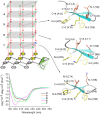
Impact of the β‐turn motif on the formation of a β‐hairpin based on the Trp K pocket. (a) CD spectra of peptide 1 at 58 μM (top), and gramicidin S‐like CD spectra of peptide 2 at 69 μM (bottom). (b) 1H NMR spectra of peptides 1 and 2 measured at 298 K in H2O/D2O with amide proton assignments and 3 J HNHA scalar coupling constants. Values in brackets are estimates and could not be exactly extracted due to overlap. One of the ten low‐energy NMR solution structures of peptide 2 in water is shown as an example (the residue number is based on Figure 7, vide infra. The residues on the cationic−aromatic face are shown in yellow; the residues on the opposite face are shown in coral). The two Trp side chains are both found in the g+ rotamer (χ1≈−60°), so the aromatic rings are tilted towards the N‐terminus, thus exposing the two Hβ atoms towards the next residue.
Figure 3
Figure 3
Versatility of the hairpin scaffold WXWXpPXKX for the assembly of different amino acids on the β‐sheet plate. Peptide concentrations were in the range of 36–61 μM. One of the ten low‐energy NMR solution structures of peptides 57 in water is shown as an example (the residue number is based on Figure 7, vide infra. The residues on the cationic−aromatic face are shown in yellow; the residues on the opposite face are shown in coral).
Figure 4
Figure 4
CD spectra of peptide 8 at 52 μM. One of the ten low‐energy NMR solution structures in water is shown as an example (the residue number is based on Figure 7, vide infra. The residues on the cationic−aromatic face are shown in yellow; the residues on the opposite face are shown in coral).
Figure 5
Figure 5
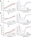
Comparison of the thermal stability of the peptide hairpins based on the Trp K/R (a/b) pocket, and aromatic/basic side‐chain zipper (c). In the panels on the left, the temperature dependence of the minima is shown. In the panels on the right, the CD curves before and after heating as well as after cooling are shown. Peptide concentrations were in the range of 31–68 μM.
Figure 6
Figure 6
CD and NMR spectra of peptides 911 containing the aromatic/basic side‐chain zipper. (a) CD spectra measured with peptide concentrations of 67 μM (9), 39 μM (10), 70 μM (11). (b) 1H NMR spectra of peptides 911 measured at 298 K in H2O/D2O with amide proton assignments and 3 J HNHA scalar coupling constants. Values in brackets are estimates and could not be exactly extracted due to overlap. One of the ten low‐energy NMR solution structures in water is shown as an example for each peptide (the residue number is based on Figure 7, vide infra. The residues on the cationic−aromatic face are shown in yellow; the residues on the opposite face are shown in coral).
Figure 7
Figure 7
Backbone chemical shift deviations from random‐coil values. (a) Hα chemical shift deviations. Norleucine is indicated with Ln and d‐Pro with p. Protons displaying values larger than 0.1 (dashed line) are shown with a more intense color. The Hα protons of the residues indicated with an asterisk experience a strong upfield chemical shift due to ring currents of the Trp residues counteracting the downfield contribution of the peptide backbone in β‐strand conformation. (b) HN chemical shift deviations. All amide chemical shifts with a deviation larger than 0.3 ppm (intense colors, above the dashed line) are involved in H‐bonds. (c) Chemical shift deviations of Cα and Cβ. Here [(Cα‐ Cα(r.c.))‐(Cβ‐Cβ(r.c.))] is plotted according to Marsh at al. (no smoothening was applied).
Figure 8
Figure 8
Chemical shift deviations from random‐coil values[ 31 , 33 ] of the Lys (a) or Arg (b and c) side chain involved in cation/CH−π interactions. Protons displaying deviations larger than −0.4 ppm (dashed line) are shown with a more intense color.
Figure 9
Figure 9
The versatile face of the β‐hairpin scaffold XWXWXpPXK(/R)X(R). The displayed side chains in the different peptides are represented by circles (for peptide 11 the ensemble of ten low‐energy NMR solution structures is also shown. The residue number is based on Figure 7, vide supra).

Similar articles

Cited by

References

    1. Sibanda B. L., Thornton J. M., Nature 1985, 316, 170–174. - PubMed
    1. Iuchi S., in Zinc Finger Proteins: From Atomic Contact to Cellular Function (Eds.: luchi S., Kuldell N.), Landes Bioscience/Eurekah.com and Kluwer Academic/Plenum Publishers, 2005, pp. 7–13.
    1. Blanco F. J., Rivas G., Serrano L., Nat. Struct. Biol. 1994, 1, 584–590. - PubMed
    1. Franklin M. C., Carey K. D., Vajdos F. F., Leahy D. J., de Vos A. M., Sliwkowski M. X., Cancer Cell 2004, 5, 317–328. - PubMed
    1. None

Publication types

MeSH terms

LinkOut - more resources