Review
. 2022 Jul;59(4):528-545.
doi: 10.1177/03009858211057197. Epub 2021 Dec 2.

Hamster models of COVID-19 pneumonia reviewed: How human can they be?

Affiliations
Review

Hamster models of COVID-19 pneumonia reviewed: How human can they be?

Achim D Gruber et al. Vet Pathol. 2022 Jul.

Abstract

The dramatic global consequences of the coronavirus disease 2019 (COVID-19) pandemic soon fueled quests for a suitable model that would facilitate the development and testing of therapies and vaccines. In contrast to other rodents, hamsters are naturally susceptible to infection with severe acute respiratory syndrome coronavirus 2 (SARS-CoV-2), and the Syrian hamster (Mesocricetus auratus) rapidly developed into a popular model. It recapitulates many characteristic features as seen in patients with a moderate, self-limiting course of the disease such as specific patterns of respiratory tract inflammation, vascular endothelialitis, and age dependence. Among 4 other hamster species examined, the Roborovski dwarf hamster (Phodopus roborovskii) more closely mimics the disease in highly susceptible patients with frequent lethal outcome, including devastating diffuse alveolar damage and coagulopathy. Thus, different hamster species are available to mimic different courses of the wide spectrum of COVID-19 manifestations in humans. On the other hand, fewer diagnostic tools and information on immune functions and molecular pathways are available than in mice, which limits mechanistic studies and inference to humans in several aspects. Still, under pandemic conditions with high pressure on progress in both basic and clinically oriented research, the Syrian hamster has turned into the leading non-transgenic model at an unprecedented pace, currently used in innumerable studies that all aim to combat the impact of the virus with its new variants of concern. As in other models, its strength rests upon a solid understanding of its similarities to and differences from the human disease, which we review here.

Keywords: COVID-19; animal model; hamster; histopathology; pathology; preclinical research; review; severe acute respiratory syndrome coronavirus 2; vaccines.


Conflict of interest statement

Declaration of Conflicting Interests: The author(s) declared no potential conflicts of interest with respect to the research, authorship, and/or publication of this article.

Figures

Figures 1–9.
Figures 1–9.
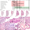
SARS-CoV-2 infection, lung, Syrian hamster. In situ hybridization (ISH) for viral RNA (red chromogen, blue hemalaun counterstain). Figure 1. At 2 days post infection (dpi), ISH localizes the virus only in bronchial epithelium throughout the entire left lung lobe. Figure 2. At 3 dpi, viral infection spreads to the respiratory parenchyma in a patchy pattern and the bronchial epithelium is still strongly infected. Figure 3. At 5 dpi, the infection turns into a purely parenchymal pattern with less signal. Figure 4. Viral RNA was no longer detected at 14 dpi. Figure 5. At 3 dpi, there is almost complete infection of the alveolar lining cells in affected areas as well as patchy infection of bronchial epithelial cells (asterisk). #: a blood vessel with virus-negative endothelial cells. Figures 6–9. Viral RNA is localized in the cytosol of bronchial epithelial cells (Fig. 6, arrow), alveolar epithelial cells (AEC) type-I (Fig. 7, arrow), AEC-II (Fig. 8, arrow), and alveolar macrophages (Fig. 9, arrow). Methods were described earlier. ,
Figures 10–24.
Figures 10–24.
SARS-CoV-2 infection, lung, Syrian hamster. Figure 10. Characteristic lesions can be divided into a phase of tissue damage and inflammation (red box) followed by regeneration (green box). Fibrosis is not a consistently reported feature in this model. Gray boxes show the time points for which histological analyses are available, and the remaining time points represent estimates. Asterisk, infection with SARS-CoV-2. BE, bronchial epithelial cells. AEC-II, alveolar epithelial type II cells. Figures 11–16. Overview of time-dependent microscopic lesions of the entire left lung (hematoxylin and eosin, HE). Figure 11. Uninfected control, 3 days after transnasal application of 30 µl of Dulbecco’s Modified Eagle Medium (DMEM). Figures 12, 13. Early bronchointerstitial pneumonia at 2 and 3 days post infection (dpi) begins to spread into alveoli surrounding the hilus. Figure 14. Severe diffuse interstitial pneumonia peaks at 5 dpi with little residual bronchitis. Figure 15. At 7 dpi, lesions regress with a patchy distribution over the entire lobe. Figure 16. After 14 days, almost complete recovery is visible with only little interstitial pneumonia left in a small proportion of hamsters. Figure 17. At 3 dpi, bronchi have epithelial necrosis with intraluminal neutrophils and cellular debris. Inset: Necrosis (arrow) of a BE cell. HE. Figure 18. Hyperplasia of BE at 5 dpi (double-headed arrow). HE. Figure 19. Early infiltration of neutrophils in interalveolar septa, with alveolar edema (asterisks) at 2 dpi. HE. Figure 20. At 3 dpi, interalveolar septa and alveolar spaces are densely infiltrated by macrophages and neutrophils. HE. Figure 21. Multifocal necrosis of alveolar epithelial cells at 3 dpi. HE. Figure 22. At 5 dpi, AEC-II are hyperplastic with occasional bizarre multinucleated syncytia (inset, arrow). HE. Figure 23. At 7 dpi, most alveoli are cleared but marked interstitial pneumonia remains. HE. Figure 24. At 5 dpi, endothelialitis is present in a medium-sized blood vessel. Inset: endothelialitis, with patches of elevated endothelial cells (arrows) and subendothelial infiltration with mostly monomorphonuclear cells but without thrombosis.
Figure 25.
Figure 25.
Cellular and molecular pathogenesis in the alveolus with a selection of relevant immune mediators and other activated genes as detected by temporal and cell type–specific multi-omics analyses from lungs of severe acute respiratory syndrome coronavirus 2 (SARS-CoV-2)-infected Syrian hamsters at 2 to 3 days (left half, red background) and 5 to 7 days post infection (dpi; right half, green background). Initially, the virus spreads through the upper airways where it infects and multiplies in airway epithelial cells. After entering the alveolar space, it infects both alveolar epithelial type-I (AEC-I) and type-II cells (AEC-II) with effective replication more likely in AEC-II. At 2 and 3 dpi, both types of AEC synthesize and secrete primarily chemokines such as CCL3 and CXCL10 as well as other cytokines that attract neutrophils and macrophages into the interalveolar wall and the alveolar space and activate them. Activated and dysfunctional endothelial and myeloid cells as well as necrosis of AEC-I and also some AEC-II result in leakage of the alveolar wall with exudation of protein-rich edema, together resulting in diffuse alveolar damage. Adjacent vascular endothelial cells are also activated by immune mediators from AEC and macrophages and upregulate, among other factors, vascular adhesion molecules such as ICAM-1 and VCAM-1 that attract leukocytes to the alveolar wall and promote their diapedesis. CD8+ lymphocytes are most effective in eliminating SARS-CoV-2-infected AEC-II. At 5 to 7 dpi, macrophages in the alveolar space carry the highest virus burden, likely due to their phagocytic activity. Proliferating AEC-II result in a cobblestone-like appearance of the alveolar lining and together with restored endothelial cells terminate the leakage of the alveolar wall. Concomitant expression of the viral spike protein, its receptor ACE2, and the required proteases result in fusion of adjacent AEC-II, forming large, bizarre, multinucleated syncytia. Proliferating AEC-II differentiate into AEC-I during the subsequent days and largely restore the structure of the alveolar lining until 14 dpi. A selection of upregulated immune mediators, mostly chemokines and other cytokines as revealed by multi-omics analyses, are given in white boxes for the different cell types and time points. Gene symbols in italics follow the nomenclature of the murine genome.
Figures 26–34.
Figures 26–34.
SARS-CoV-2 infection with the standard dose of 105 plaque forming units, lung, Roborovski dwarf hamster. Figure 26. Roborovski dwarf hamsters developed drastic bronchial, alveolar, and interstitial lesions at 2 and 3 days post infection (dpi). At 3 dpi, they reached humane end points of the experimental protocol and were euthanized (compare with Fig. 10). Red box, phase of tissue damage and inflammation; green box, phase of regeneration; gray box, time points for which histological analyses are available; the remaining time points represent estimates. Asterisk, infection with SARS-CoV-2. †, death. BE, bronchial epithelial cells. AEC-II, alveolar epithelial type II cells. Figure 27. Overview of left lung lobe at 3 dpi with almost diffuse consolidation. Hematoxylin and eosin stain (HE). Figure 28. Marked bronchointerstitial pneumonia with additional consolidation of the alveolar parenchyma at 3 dpi. HE. Figure 29. Marked alveolar edema and fibrin (asterisks) with formation of hyaline membranes (arrows) at 3 dpi. HE. Inset: Periodic acid–Schiff reaction (PAS). Figure 30. Diffuse alveolar damage including necrosis of AEC-I, fibrin deposition, and alveolar edema at 3 dpi. HE. Figure 31. Hyaline thrombi in pulmonary capillaries (small arrows) at 3 dpi. HE. Figure 32. Hyaline thrombi stain positive with the PAS reaction (small arrows). Figure 33. Interalveolar septa and alveolar spaces are densely infiltrated by macrophages and neutrophils with necrosis of AEC (inset, arrows) at 3 dpi. HE. Figure 34. Also at 3 dpi, there is some necrosis and flattening of BE with simultaneous mild and patchy hyperplasia with increased mitotic activity (arrows). HE.
Figure 35.
Figure 35.
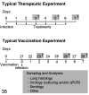
Proposed time lines for typical therapeutic and vaccination experiments in Syrian hamsters with SARS-CoV-2 infection. In therapeutic interventions, the first week following infection is the most useful time frame to observe the clinical outcome and take samples for virological and histological examination. In vaccination experiments, 21 days following vaccination is a typical time point for challenge infection that allows the vaccine to have elicited effective immune responses. The week after challenge infection is a useful time frame to examine clinical efficacy and to sample animals to assess other parameters of vaccination success. Indicates recommended sampling time points.

Similar articles

Cited by

References

    1. Abdelnabi R, Boudewijns R, Foo CS, et al. Comparative infectivity and virulence of emerging SARS-CoV-2 variants in Syrian hamsters. EBioMedicine. 2021;68:103403. - PMC - PubMed
    1. Ackermann M, Anders HJ, Bilyy R, et al. Patients with COVID-19: in the dark-NETs of neutrophils. Cell Death Differ. Published online May 24, 2021. doi:10.1038/s41418-021-00805-z - PMC - PubMed
    1. Ackermann M, Verleden SE, Kuehnel M, et al. Pulmonary vascular endothelialitis, thrombosis, and angiogenesis in COVID-19. N Engl J Med. 2020;383(2):120–128. - PMC - PubMed
    1. Agwa SHA, Kamel MM, Elghazaly H, et al. Association between interferon-lambda-3 rs12979860, TLL1 rs17047200 and DDR1 rs4618569 variant polymorphisms with the course and outcome of SARS-CoV-2 patients. Genes (Basel). 2021;12(6):830. - PMC - PubMed
    1. Allnoch L, Beythien G, Leitzen E, et al. Vascular inflammation is associated with loss of aquaporin 1 expression on endothelial cells and increased fluid leakage in SARS-CoV-2 infected golden Syrian hamsters. Viruses. 2021;13(4):639. - PMC - PubMed

Publication types