Sellar, suprasellar, and parasellar masses: Imaging features and neurosurgical approaches
- PMID: 34856828
- PMCID: PMC9244752
- DOI: 10.1177/19714009211055195
Sellar, suprasellar, and parasellar masses: Imaging features and neurosurgical approaches
Abstract
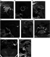
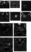
The sellar, suprasellar, and parasellar space contain a vast array of pathologies, including neoplastic, congenital, vascular, inflammatory, and infectious etiologies. Symptoms, if present, include a combination of headache, eye pain, ophthalmoplegia, visual field deficits, cranial neuropathy, and endocrine manifestations. A special focus is paid to key features on CT and MRI that can help in differentiating different pathologies. While most lesions ultimately require histopathologic evaluation, expert knowledge of skull base anatomy in combination with awareness of key imaging features can be useful in limiting the differential diagnosis and guiding management. Surgical techniques, including endoscopic endonasal and transcranial neurosurgical approaches are described in detail.
Keywords: Cavernous sinus; parasellar mass; sellar mass; skull base; suprasellar mass.
Conflict of interest statement
Figures


References
-
- Guerrero-Pérez F, Marengo AP, Vidal N, et al. Primary tumors of the posterior pituitary: A systematic review. Rev Endocr Metab Disord 2019; 20(2): 219–238. - PubMed
-
- Chin BM, Orlandi RR, Wiggins RH, 3rd. Evaluation of the sellar and parasellar regions. Magn Reson Imaging Clin North America 2012; 20(3): 515–543. - PubMed
-
- Rao VJ, James RA, Mitra D. Imaging characteristics of common suprasellar lesions with emphasis on MRI findings. Clin Radiol 2008; 63(8): 939–947. - PubMed
-
- Gao R, Isoda H, Tanaka T, et al. Dynamic gadolinium-enhanced MR imaging of pituitary adenomas: usefulness of sequential sagittal and coronal plane images. Eur J Radiol 2001; 39(3): 139–146. - PubMed
MeSH terms
LinkOut - more resources
Full Text Sources
Medical

