. 2022 Apr;149(4):1329-1339.
doi: 10.1016/j.jaci.2021.10.004. Epub 2021 Nov 29.

Transcriptomic characterization of prurigo nodularis and the therapeutic response to nemolizumab

Affiliations

Transcriptomic characterization of prurigo nodularis and the therapeutic response to nemolizumab

Lam C Tsoi et al. J Allergy Clin Immunol. 2022 Apr.

Abstract

Background: Prurigo nodularis (PN) is a debilitating, difficult-to-treat, intensely pruritic, chronic inflammatory skin disease characterized by hyperkeratotic skin nodules. The pathogenesis of PN is not well understood but is believed to involve cross talk between sensory nerve fibers, immune cells, and the epidermis. It is centered around the neuroimmune cytokine IL-31, driving an intractable itch-scratch cycle.

Objective: We sought to provide a comprehensive view of the transcriptomic changes in PN skin and characterize the mechanism of action of the anti-IL-31 receptor inhibitor nemolizumab.

Method: RNA sequencing of biopsy samples obtained from a cohort of patients treated with the anti-IL-31 receptor inhibitor nemolizumab and taken at baseline and week 12. Generation and integration of patient data with RNA-Seq data generated from reconstructed human epidermis stimulated with IL-31 and other proinflammatory cytokines.

Results: Our results demonstrate that nemolizumab effectively decreases IL-31 responses in PN skin, leading to effective suppression of downstream inflammatory responses including TH2/IL-13 and TH17/IL-17 responses. This is accompanied by decreased keratinocyte proliferation and normalization of epidermal differentiation and function. Furthermore, our results demonstrate how transcriptomic changes associated with nemolizumab treatment correlate with improvement in lesions, pruritus, stabilization of extracellular matrix remodeling, and processes associated with cutaneous nerve function.

Conclusion: These data demonstrate a broad response to IL-31 receptor inhibition with nemolizumab and confirm the critical upstream role of IL-31 in PN pathogenesis.

Keywords: Prurigo nodularis; T(H)17; T(H)2; mechanism; nemolizumab.


Figures

Figure 1.
Figure 1.. PN is characterized by immune activation and abnormal keratinocyte differentiation.
Number of differentially expressed genes (DEGs) in PN lesional vs. non-lesional skin (n=62, FC≥2 or FC≤0.5, FDR≤0.1) (A). Enriched GO categories in PN lesional skin (−log (FDR) values are shown) (B). Literature-based gene network obtained from the top 1,000 DEGs in PN skin, generated using Genomatix Pathway System (GePS, www.Genomatix.de). The picture displays the top connected genes co-cited in PubMed abstracts in the same sentence linked to a function word (most relevant genes/interactions). The color represents fold change: orange represent the genes that are increased, and green represent the genes that are decreased in PN lesional vs. non-lesional skin. Critical nodes included the proliferation markers Ki67 (MKI67), IL-1 family members IL36G and IL1A, and CXCL8 and CDKN1A (C). Number of genes and within cluster correlation in the modules identified from the weighted gene co-expression gene networks (WCGNA) analysis of non-lesional and lesional PN skin (D). Top functions enriched in key co-expression modules (module #5, #6 and #8) from PN skin. The observed-expected enrichment is shown on the x-axis (E).
Figure 2.
Figure 2.. Enriched transcriptomic cellular signatures and overlap with psoriasis and AD.
Cell type inference analysis on non-lesional (NL) and lesional (L) PN skin samples using xCell. The color spectrum in the heatmap represents relative cellular signature across samples. The bar on the left side shows statistical difference in the enrichment between lesional and non-lesional PN skin with the colors representing different p-value thresholds (A). Comparison of PN associated DEGs against DEGs in Psoriasis (Pso) and atopic dermatitis (AD) for increased and decreased DEGs (B). Correlation analysis between the effect sizes in PN lesion versus those in psoriasis (Pso) and atopic dermatitis (AD). Spearman’s rank-order correlation was included. The genes significant in the x-axis, y-axis, and both axes are colored in red/blue/purple respectively (C).
Figure 3.
Figure 3.. Transcriptomic changes associated with the anti-IL31R inhibitor nemolizumab
Principal component analyses (PCA) of the transcriptomic data from PN biopsies prior to and after 12-weeks of prospective placebo controlled, double blinded clinical trial with the anti-IL-31R inhibitor nemolizumab. Different colors represent different treatment groups, with lesional samples shown as triangles, and non-lesional skin shown as circles (A). Heatmap showing 2-way clustering (using genes differentially expressed between non-lesional vs lesional skin at baseline) for all samples (B).
Figure 4.
Figure 4.. Effect of nemolizumab on PN associated transcriptomic changes.
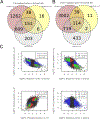
3-way Venn diagram of increased and decreased DEGs in PN skin and overlap with DEGs (compared to baseline) in the nemolizumab and placebo cohorts (A/B). Correlation analyses between different groups (PN baseline vs. placebo and nemolizumab DEGs) (Spearman’s rank-order correlation) (C).
Figure 5.
Figure 5.. Nemolizumab treatment leads to normalized epidermal differentiation and decreased IL-31/Th2 responses in PN skin
Nemolizumab treatment led to decrease in IL-31 and IL-13 responses in PN skin compared to placebo, along with decreased expression of IL-17A response genes. Color spectrum represents -log10 p-value of the hypergeometric enrichment test (A). Immune cell signature was tested against genes that are i) up-regulated in baseline non-lesional vs lesional PN skin; ii) down-regulated in the placebo baseline vs week 12; iii) down-regulated in the nemolizumab group baseline vs week 12 comparisons. Color spectrum represents -log10 p-value of the hypergeometric enrichment test (B). Cross comparing transcriptomic responses in PN skin against cellular signatures obtained from single-cell data of healthy epidermis demonstrated that nemolizumab treatment led to normalization of epidermal gene expression related to the differentiated layer (KRT10+) of the epidermis, corresponding to normalization of epidermal differentiation. The different nomenclatures correspond to the different layers of the epidermis with “basal” corresponding to KRT5+ basal cells, KRT10+ “differentiated” corresponding to the spinous layer, and “keratinized” corresponding to the granular layers (FLG+) (C). Transcription Factor Binding Site (TFBS) of PN associated DEGs that greater normalization amongst nemolizumab down-regulated genes compared to placebo. The axes represent -log10 (p) values, and the color spectrum reflects enrichment p-value for TFBS results for up-regulated genes in the baseline non-lesional vs. lesional skin comparison (D).
Figure 6.
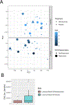
Figure 6.. Nemolizumab driven decrease in IGA scoring was accompanied by tighter clustering of PN samples on PCA analyses after 12 weeks of treatment.
The IGA value was superimposed on the PCA coordinates from the transcriptomic data. Nemolizumab group is shown as circles, whereas placebo group is shown as triangles. Baseline (top) and week 12 of treatment (bottom) are shown. The responder represents patients achieved almost clear skin by week 18 (i.e. IGA=0 or 1), and are showed in larger circles or triangles (A). Nemolizumab treatment led to tighter clustering of PN samples on PCA analyses compared to biopsy samples from the placebo treated cohort (B).

Comment in

References

    1. Huang AH, Williams KA, Kwatra SG. Prurigo nodularis: Epidemiology and clinical features. Journal of the American Academy of Dermatology. 2020;83(6):1559–65. - PubMed
    1. Weigelt N, Metze D, Stander S. Prurigo nodularis: systematic analysis of 58 histological criteria in 136 patients. J Cutan Pathol. 2010;37(5):578–86. - PubMed
    1. Johansson O, Liang Y, Marcusson JA, Reimert CM. Eosinophil cationic protein- and eosinophil-derived neurotoxin/eosinophil protein X-immunoreactive eosinophils in prurigo nodularis. Arch Dermatol Res. 2000;292(8):371–8. - PubMed
    1. Liang Y, Marcusson JA, Jacobi HH, Haak-Frendscho M, Johansson O. Histamine-containing mast cells and their relationship to NGFr-immunoreactive nerves in prurigo nodularis: a reappraisal. J Cutan Pathol. 1998;25(4):189–98. - PubMed
    1. Sonkoly E, Muller A, Lauerma AI, Pivarcsi A, Soto H, Kemeny L, et al. IL-31: a new link between T cells and pruritus in atopic skin inflammation. J Allergy Clin Immunol. 2006;117(2):411–7. - PubMed

Publication types