. 2022 Mar 1;71(3):440-452.
doi: 10.2337/db21-0638.

The Role of TRAPγ/SSR3 in Preproinsulin Translocation Into the Endoplasmic Reticulum

Affiliations

The Role of TRAPγ/SSR3 in Preproinsulin Translocation Into the Endoplasmic Reticulum

Xiaoxi Xu et al. Diabetes. .

Abstract

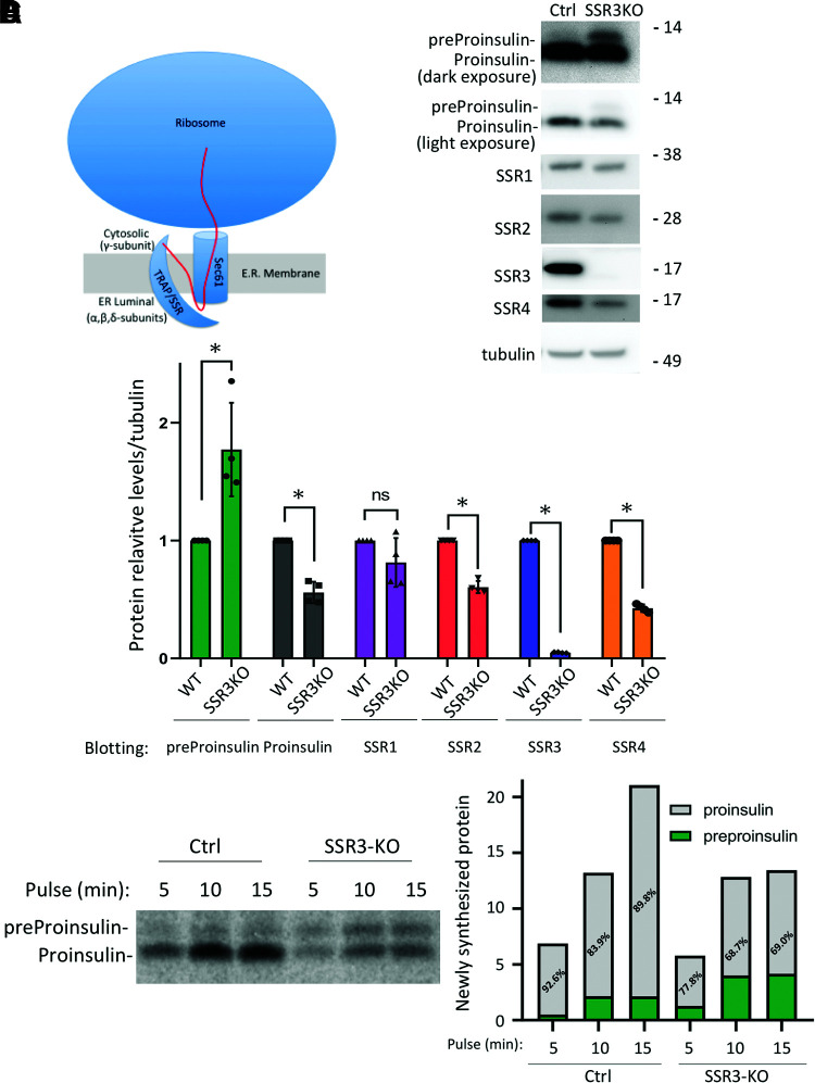
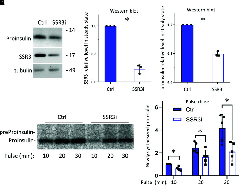
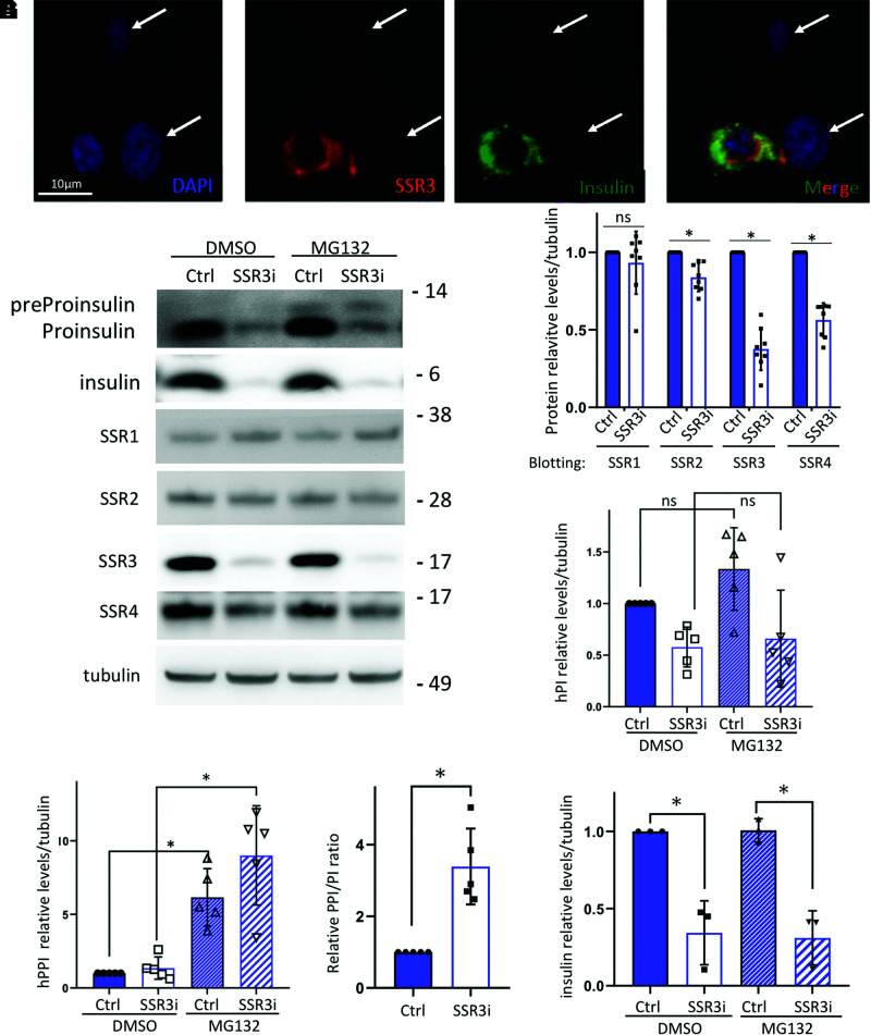
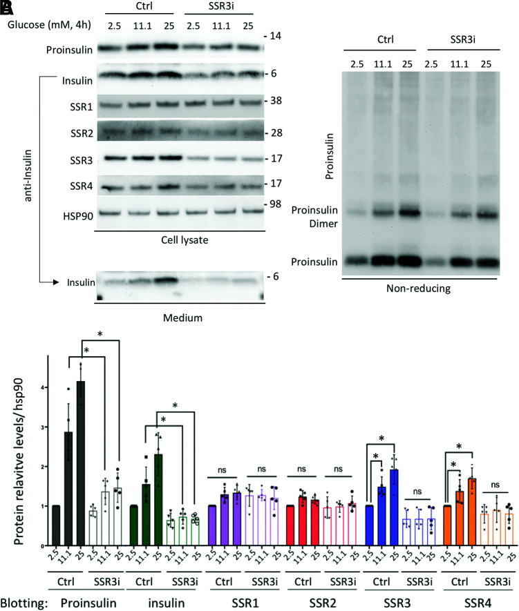
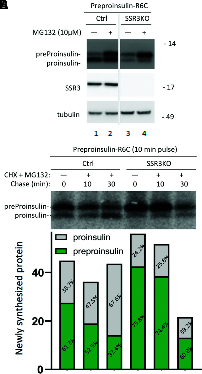
In the endoplasmic reticulum (ER), the translocation-associated protein complex (TRAP), also called signal sequence receptor (SSR), includes four integral membrane proteins TRAPα/SSR1, TRAPβ/SSR2, and TRAPδ/SSR4 with the bulk of their extramembranous portions primarily in the ER lumen, whereas the extramembranous portion of TRAPγ/SSR3 is primarily cytosolic. Individually diminished expression of either TRAPα/SSR1, TRAPβ/SSR2, or TRAPδ/SSR4 mRNA is known in each case to lower TRAPα/SSR1 protein levels, leading to impaired proinsulin biosynthesis, whereas forced expression of TRAPα/SSR1 at least partially suppresses the proinsulin biosynthetic defect. Here, we report that diminished TRAPγ/SSR3 expression in pancreatic β-cells leaves TRAPα/SSR1 levels unaffected while nevertheless inhibiting cotranslational and posttranslational translocation of preproinsulin into the ER. Crucially, acute exposure to high glucose leads to a rapid upregulation of both TRAPγ/SSR3 and proinsulin protein without change in the respective mRNA levels, as observed in cultured rodent β-cell lines and confirmed in human islets. Strikingly, pancreatic β-cells with suppressed TRAPγ/SSR3 expression are blocked in glucose-dependent upregulation of proinsulin (or insulin) biosynthesis. Most remarkably, overexpression of TRAPγ/SSR3 in control β-cells raises proinsulin levels, even without boosting extracellular glucose. The data suggest the possibility that TRAPγ/SSR3 may fulfill a rate-limiting function in preproinsulin translocation across the ER membrane for proinsulin biosynthesis.


Figures

Figure 1
Figure 1
The TRAPγ/SSR3 subunit of the TRAP/SSR complex contributes to the efficiency of recombinant proinsulin biosynthesis. A: Schematic of the TRAP/SSR complex highlights the predominant cytosolic exposure of the TRAPγ/SSR3 subunit, including a hypothetical contact with a signal peptide N-terminus during polypeptide translocation (red line). B: Control (Ctrl) (wild-type [WT] 293T cells) and SSR3-KO 293T cells were transfected with untagged human preproinsulin plasmids. At 48 h posttransfection, cell lysates were analyzed by reducing SDS-PAGE gel and immunoblotting with antiproinsulin and anti-SSR1–4 antibodies as indicated. C: Quantitation (mean ± SD) of preproinsulin, proinsulin, and TRAP/SSR subunit protein levels from four independent experiments like that shown in B (normalized to tubulin). *P < 0.05 compared with Ctrl. D: Ctrl and SSR3-KO 293T cells were transfected with human preproinsulin plasmids. At 48 h posttransfection, the cells were pulse labeled with 35S-Met/Cys for the times indicated; this is a representative pulse labeling from three identical experiments. Cell lysates (normalized to trichloroacetic acid–precipitable counts) were subjected to immunoprecipitation with anti-insulin and analyzed by reducing SDS-PAGE and phosphorimaging; both the absolute and relative amounts of recovered preproinsulin and proinsulin are shown in the graph.
Figure 2
Figure 2
Effect of TRAPγ/SSR3 deficiency on endogenous proinsulin biosynthesis in β-cells. A: INS1E cells were transfected with scrambled oligo or TRAPγ/SSR3-KD (SSR3i). At 72 h posttransfection, cells were lysed and analyzed by reducing SDS-PAGE and immunoblotted with antiproinsulin and anti-SSR3 antibodies. B and C: Quantitation (mean ± SD) of TRAPγ/SSR3 protein and proinsulin protein from three independent experiments (normalized to tubulin). D: At 72 h posttransfection, control (Ctrl) and TRAPγ/SSR3-KD (SSR3i) INS1E cells were pulse labeled with 35S-Met/Cys for the indicated times. Cell lysates (normalized to trichloroacetic acid–precipitable counts) were immunoprecipitated with anti-insulin and analyzed by reducing SDS-PAGE and phosphorimaging. E: Quantitation (mean ± SD) of newly synthesized proinsulin from five independent experiments like that shown in D, with the proinsulin band from Ctrl INS1E cells at 10 min labeling set to 1. *P < 0.05 compared with Ctrl.
Figure 3
Figure 3
TRAPγ/SSR3 deficiency results in a decrease in the steady-state level of stored, processed insulin in β-cells. INS832/13 cells were transfected with TRAPγ/SSR3-targeted siRNA. A: Immunofluorescence with anti-TRAPγ/SSR3 (red) and anti-insulin (green) in INS832/13 cells at 72 h posttransfection. Nuclei were counterstained with DAPI (blue). B: Cells transfected as in A were treated with or without MG132 (10 μmol/L) for 4 h. Cells were lysed and analyzed by reducing SDS-PAGE and immunoblotting with either anti-human-(pre)proinsulin or a battery of antibodies, as indicated. C: Quantitation (mean ± SD, n = 8 independent experiments) of each of the TRAP/SSR subunits in control (Ctrl) cells and TRAPγ/SSR3-KD (SSR3i). D: Quantitation of human proinsulin levels from five independent experiments like that shown in B. E: Quantitation of human preproinsulin levels from five independent experiments like that shown in B. F: Human preproinsulin-to-proinsulin ratios in MG132-treated INS832/13 cells were calculated from the experiments shown in D and E (n = 5). G: Quantitation (mean ± SD) of insulin protein levels from three independent experiments like that shown in B. *P < 0.05 compared with Ctrl. h, human; PI, proinsulin; PPI, preproinsulin.
Figure 4
Figure 4
The ability of β-cells to acutely increase proinsulin (and insulin) level in response to high-glucose stimulation depends on TRAPγ/SSR3. A: INS1E cells were transfected with scrambled oligo or TRAPγ/SSR3-KD (SSR3i). At 72 h posttransfection, cells were preincubated in RPMI medium containing 2.5 mmol/L glucose for 1.5 h followed by a 4-h incubation at either 2.5, 11.1, or 25 mmol/L glucose, respectively. Cells were lysed and analyzed by SDS-PAGE under reducing conditions (left) or nonreducing conditions (right) for immunoblotting with the antibodies indicated. The 4-h media were collected and similarly analyzed by SDS-PAGE and immunoblotting with anti-insulin (bottom left). B: Quantitation (mean ± SD) of proinsulin, insulin, and each TRAP/SSR subunit from five independent experiments. *P < 0.05. Ctrl, control.
Figure 5
Figure 5
TRAPγ/SSR3 re-expression rescues proinsulin protein levels in TRAPγ/SSR3-KO β-cells. TRAPγ/SSR3-KO INS832/13 cells were transfected with empty vector (−) or plasmids encoding Flag-tagged TRAPγ/SSR3 cDNA (+). Wild-type (WT) INS832/13 cells were also transfected with empty vector as control. A: At 48 h posttransfection, cells were lysed and analyzed by reducing SDS-PAGE and immunoblotting for anti-rodent proinsulin and the additionally indicated antibodies. B: Quantitation (mean ± SD) of proinsulin and each TRAP/SSR subunit from four independent experiments like that shown in A; expression levels in WT control cells were set to 1.0. C: TRAPγ/SSR3-KO INS832/13 cells transfected as in A were examined by immunofluorescence with anticalnexin (red), anti-Flag (Flag-SSR3, purple), and anti-rodent proinsulin (green); a merged anti-Flag/antiproinsulin image is shown. D: Quantitation (mean ± SD) of rodent proinsulin–positive TRAPγ/SSR3-KO cells that either re-express or do not re-express TRAPγ/SSR3 protein from three independent experiments like that shown in C. E: Quantitation (mean ± SD) of insulin-positive TRAPγ/SSR3-KO cells that either re-express or do not re-express TRAPγ/SSR3 protein from four independent experiments. *P < 0.05.
Figure 6
Figure 6
Overexpression of TRAPγ/SSR3 in wild-type INS832/13 cells increases the steady-state level of proinsulin. A: Wild-type INS832/13 cells were transfected with plasmids encoding empty vector (−) or Flag-tagged TRAPγ/SSR3 cDNA (+). At 48 h posttransfection, cells were lysed and analyzed in duplicate by reducing SDS-PAGE and immunoblotting with the indicated antibodies. B: Quantitation (mean ± SD) of rodent proinsulin (CCI-17 antibody) and human proinsulin (human C-A junction antibody) and each TRAP/SSR subunit as indicated (n = 4 independent experiments). C: Wild-type INS832/13 cells were transfected with plasmids encoding empty vector or each individual Flag-tagged TRAP/SSR subunit and processed as in A. The red arrows highlight expression of the Flag-tagged constructs. D: Quantitation (mean ± SD) showing increased expression of each of the TRAP/SSR subunits (top) and the impact of that overexpression on rodent proinsulin (bottom left) and human proinsulin (bottom right) levels from five independent experiments. E: Wild-type 293T cells were transfected with human wild-type preproinsulin plasmid plus plasmid encoding empty vector or each individual Flag-tagged TRAP/SSR subunit and processed as in A. F: Quantitation (mean ± SD) of individually overexpressed TRAP/SSR subunits (left) and recombinant human proinsulin (right) from four independent experiments. G: Human islets were preincubated for 90 min in RPMI medium containing 2.5 mmol/L glucose and 0.1% BSA without serum. Aliquots of 130 human islet equivalents were sedimented at 1,000g, and the preincubation medium was removed, followed by a 2-h incubation in 500 μL of fresh medium bearing either 2.5 or 25 mmol/L glucose in the absence or presence of CHX (100 μg/mL). Human islets were then lysed in RIPA buffer; lysates normalized to total protein were analyzed by SDS-PAGE and immunoblotting for the indicated proteins (tubulin was used as a loading control to confirm proper normalization of samples). *P < 0.05. EV, empty vector; PI, proinsulin.
Figure 7
Figure 7
Effects of TRAPγ/SSR3 on the posttranslational translocation of mutant preproinsulin-R6C. Control (Ctrl) 293T cells and SSR3-KO 293T cells were transfected to express untagged human preproinsulin-R6C. A: At 48 h posttransfection, cells were treated with or without MG132 (10 μmol/L) for 2 h. The cells were lysed and analyzed by reducing SDS-PAGE and immunoblotting with the indicated antibodies. B: After transfection as in A, cells were pulse labeled with 35S-Met/Cys for 10 min followed by a 0-, 10-, or 30-min chase in the presence of CHX (10 μg/mL) and MG132 (10 μmol/L). Cell lysates (normalized to trichloroacetic acid–precipitable counts) were subjected to immunoprecipitation with anti-insulin and analyzed by reducing SDS-PAGE and phosphorimaging; both the absolute and the relative amounts of recovered preproinsulin and proinsulin are shown below each lane of the gel.

Similar articles

Cited by

References

    1. Mahajan A, Taliun D, Thurner M, et al. . Fine-mapping type 2 diabetes loci to single-variant resolution using high-density imputation and islet-specific epigenome maps. Nat Genet 2018;50:1505–1513 - PMC - PubMed
    1. Mahajan A, Go MJ, Zhang W, et al. .; DIAbetes Genetics Replication And Meta-analysis (DIAGRAM) Consortium; Asian Genetic Epidemiology Network Type 2 Diabetes (AGEN-T2D) Consortium; South Asian Type 2 Diabetes (SAT2D) Consortium; Mexican American Type 2 Diabetes (MAT2D) Consortium; Type 2 Diabetes Genetic Exploration by Next-generation sequencing in multi-Ethnic Samples (T2D-GENES) Consortium . Genome-wide trans-ancestry meta-analysis provides insight into the genetic architecture of type 2 diabetes susceptibility. Nat Genet 2014;46:234–244 - PMC - PubMed
    1. Li X, Itani OA, Haataja L, et al. . Requirement for translocon-associated protein (TRAP) α in insulin biogenesis. Sci Adv 2019;5:eaax0292. - PMC - PubMed
    1. Kriegler T, Kiburg G, Hessa T. Translocon-associated protein complex (TRAP) is crucial for co-translational translocation of pre-proinsulin. J Mol Biol 2020;432:166694. - PubMed
    1. Schuit FC, In’t Veld PA, Pipeleers DG. Glucose stimulates proinsulin biosynthesis by a dose-dependent recruitment of pancreatic beta cells. Proc Natl Acad Sci U S A 1988;85:3865–3869 - PMC - PubMed

Publication types

MeSH terms